
Ketoacidosis think lab test, instrumental, differential diagnosis,treatment
"diabetic ketoacidosis" AND management
diabetic ketoacidosis DKA management flowchart treatment algorithm
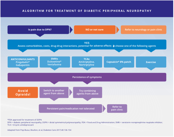
Summary : This flowchart presents an algorithm for the treatment of diabetic peripheral neuropathy (DPN), guiding clinicians through decision-making steps for pain management, agent selection, and escalation of care based on symptom persistence and medication tolerance. flowchart: # Nodes : • "Is pain due to DPN?" (hexagon) • "NO or not sure" (rectangle) • "Refer to neurology or pain clinic" (rectangle) • "YES" (rectangle) • "Assess comorbidities, costs, drug-drug interactions, potential for adverse effects ➔ choose one of the following agents" (rectangle) • "ANTICONVULSANTS Pregabalin* Gabapentin" (rectangle) • "SNRIs Duloxetine* Venlafaxine" (rectangle) • "TCAs Amitriptyline, Nortriptyline" (rectangle) • "Capsaicin* 8% patch" (rectangle) • "Exercise" (rectangle) • "Persistence of symptoms" (rectangle) • "Avoid Opioids!" (rectangle, orange) • "Switch to another agent from above" (rectangle) • "Try combining agents from above" (rectangle) • "Persistent pain/medication not tolerated" (rectangle) • "Refer to pain clinic" (rectangle) # Connectors : • Arrow from "Is pain due to DPN?" to "NO or not sure" and "YES". • Arrow from "NO or not sure" to "Refer to neurology or pain clinic". • Arrow from "YES" to "Assess comorbidities..." and then to five agent options: "ANTICONVULSANTS", "SNRIs", "TCAs", "Capsaicin* 8% patch", "Exercise". • Arrows from all five agent options to "Persistence of symptoms". • Arrow from "Persistence of symptoms" to "Avoid Opioids!", "Switch to another agent from above", and "Try combining agents from above". • Arrow from "Switch to another agent from above" and "Try combining agents from above" to "Persistent pain/medication not tolerated". • Arrow from "Persistent pain/medication not tolerated" to "Refer to pain clinic". # Layout : • Top-down, stepwise progression. • Initial decision node at top, branching to referral or treatment assessment. • Five parallel agent options in the middle. • Symptom persistence leads to further branching for agent switching, combination, or referral. • Orange highlight for "Avoid Opioids!" as a warning. # Additional Information : • Footnotes: *FDA approved for treatment of DSPN. • Abbreviations: DPN = diabetic peripheral neuropathy; DSPN = distal symmetrical polyneuropathy; FDA = Food and Drug Administration; SNRI = serotonin-norepinephrine reuptake inhibitor; TCA = tricyclic antidepressant. • Source: Adapted from Pop-Busui, Boulton, et al, Diabetes Care 2017;40:136-154. # Analysis : • The algorithm prioritizes non-opioid agents for pain management in DPN, with anticonvulsants, SNRIs, TCAs, topical capsaicin, and exercise as first-line options. • If symptoms persist, clinicians are advised to avoid opioids, switch agents, or combine therapies. • Persistent pain or intolerance to medication prompts referral to a pain clinic. • The flowchart emphasizes assessment of comorbidities and adverse effects before agent selection, and provides clear escalation steps for refractory cases.
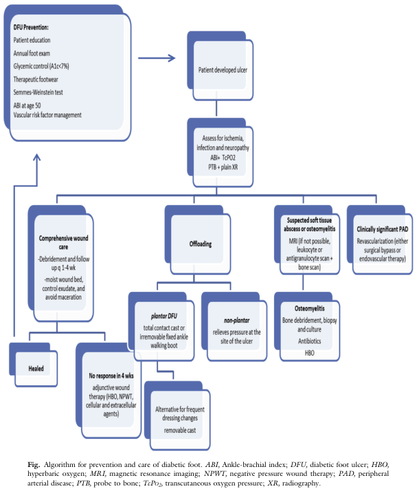
Summary : This flowchart presents a comprehensive algorithm for the prevention and care of diabetic foot, including screening, ulcer management, wound care, offloading, infection and ischemia assessment, and advanced therapies. flowchart: # Nodes : • DFU Prevention (rounded rectangle): Patient education, annual foot exam, glycemic control (A1c<7%), therapeutic footwear, Semmes-Weinstein test, ABI at age 50, vascular risk factor management. • Patient developed ulcer (rectangle). • Assess for ischemia, infection and neuropathy (rectangle): ABI + TcPO2, PTB + plain XR. • Comprehensive wound care (rectangle): Debridement and follow up q 1-4 wk, moist wound bed, control exudate, avoid maceration. • Offloading (rectangle). • Suspected soft tissue abscess or osteomyelitis (rectangle): MRI (if not possible, leukocyte or antigranulocyte scan + bone scan). • Clinically significant PAD (rectangle): Revascularization (either surgical bypass or endovascular therapy). • Healed (rectangle). • No response in 4 wks (rectangle): Adjunctive wound therapy (HBO, NPWT, cellular and extracellular agents). • plantar DFU (rectangle): Total contact cast or irremovable fixed ankle walking boot. • non-plantar (rectangle): Relieves pressure at the site of the ulcer. • Alternative for frequent dressing changes (rectangle): Removable cast. • Osteomyelitis (rectangle): Bone debridement, biopsy and culture, antibiotics, HBO. # Connectors : • Arrow from DFU Prevention to Patient developed ulcer. • Arrow from Patient developed ulcer to Assess for ischemia, infection and neuropathy. • From Assess for ischemia, infection and neuropathy, three branches: – To Comprehensive wound care. – To Offloading. – To Suspected soft tissue abscess or osteomyelitis and Clinically significant PAD. • Comprehensive wound care leads to Healed or, if no response in 4 wks, to adjunctive wound therapy. • Offloading splits into plantar DFU (total contact cast or irremovable boot) and non-plantar (relieves pressure at ulcer site), with alternative for frequent dressing changes (removable cast). • Suspected soft tissue abscess or osteomyelitis leads to Osteomyelitis (bone debridement, biopsy, antibiotics, HBO). • Clinically significant PAD leads to revascularization. • All branches ultimately aim for healing. # Layout : • Top-down flow, starting with prevention, then ulcer development, assessment, and branching into wound care, offloading, infection/ischemia management. • Multiple parallel branches for different clinical scenarios. • Merges at points where healing is achieved or advanced therapies are considered. # Analysis : • The flowchart emphasizes early prevention and regular screening to reduce diabetic foot ulcer (DFU) risk. • Once an ulcer develops, a systematic assessment for ischemia, infection, and neuropathy guides further management. • Wound care and offloading are central, with specific strategies for plantar and non-plantar ulcers. • Advanced imaging and therapies are reserved for cases with suspected infection or poor response to standard care. • The algorithm integrates vascular assessment and revascularization for patients with peripheral arterial disease (PAD). • The workflow is designed to optimize healing and minimize complications through stepwise escalation of care.
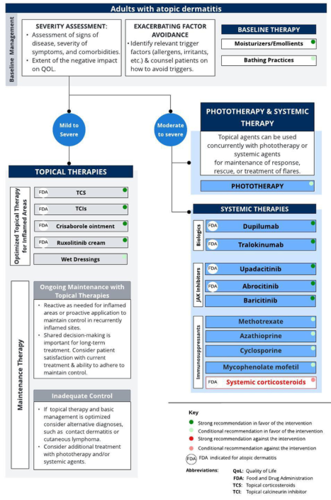
Summary : This flowchart provides a comprehensive management algorithm for adults with atopic dermatitis, detailing baseline management, topical therapies, phototherapy, and systemic therapies, including FDA-approved and recommended treatments, maintenance strategies, and escalation steps for inadequate control. flowchart: # Baseline Management : • Severity Assessment: Assessment of signs of disease, severity of symptoms, comorbidities, and impact on quality of life (QOL). • Exacerbating Factor Avoidance: Identify trigger factors (allergens, irritants, etc.) and counsel patients on avoidance. • Baseline Therapy: Moisturizers/Emollients (strong recommendation), Bathing Practices (conditional recommendation). # Initial Pathways : • Mild to Severe: Proceed to Topical Therapies. • Moderate to Severe: Proceed to Phototherapy & Systemic Therapy. # Topical Therapies : ## Optimized Topical Therapy for Inflamed Areas : • TCS (Topical corticosteroids) (FDA, strong recommendation) • TCIs (Topical calcineurin inhibitors) (FDA, strong recommendation) • Crisaborole ointment (FDA, strong recommendation) • Ruxolitinib cream (FDA, strong recommendation) • Wet Dressings (strong recommendation) ## Ongoing Maintenance with Topical Therapies : • Reactive or proactive application for maintenance. • Shared decision-making for long-term treatment. • Consider patient satisfaction and adherence. ## Inadequate Control : • If topical therapy and basic management optimized, consider alternative diagnoses. • Consider additional treatment with phototherapy and/or systemic agents. # Phototherapy & Systemic Therapy : • Topical agents can be used concurrently with phototherapy or systemic agents for maintenance, rescue, or flares. # Phototherapy : • No specific agents listed; included as a treatment option for moderate to severe cases. # Systemic Therapies : ## Biologics : • Dupilumab (FDA, strong recommendation) • Tralokinumab (FDA, strong recommendation) ## JAK Inhibitors : • Upadacitinib (FDA, strong recommendation) • Abrocitinib (FDA, strong recommendation) • Baricitinib (strong recommendation) ## Immunosuppressants : • Methotrexate (strong recommendation) • Azathioprine (strong recommendation) • Cyclosporine (strong recommendation) • Mycophenolate mofetil (strong recommendation) • Systemic corticosteroids (FDA, strong recommendation against use) # Key : • Green circle: Strong recommendation in favor. • Yellow circle: Conditional recommendation in favor. • Red circle: Strong recommendation against. • Orange circle: Conditional recommendation against. • FDA: Indicated for atopic dermatitis. # Abbreviations : • QOL: Quality of Life • FDA: Food and Drug Administration • TCS: Topical corticosteroids • TCI: Topical calcineurin inhibitor # Layout : • The flowchart is organized from baseline management at the top, splitting into two main pathways (mild to severe and moderate to severe), with further branches into topical, phototherapy, and systemic therapies. • Maintenance and escalation steps are included for ongoing management and inadequate control. # Analysis : • The algorithm emphasizes starting with baseline management and topical therapies, escalating to phototherapy and systemic therapies for more severe or refractory cases. • Strong recommendations are visually highlighted for first-line agents, with systemic corticosteroids strongly discouraged. • FDA-approved options are clearly marked, supporting evidence-based decision-making. • The flowchart supports a stepwise, patient-centered approach, integrating maintenance and escalation strategies.
Ketoacidosis - Complete Clinical Overview
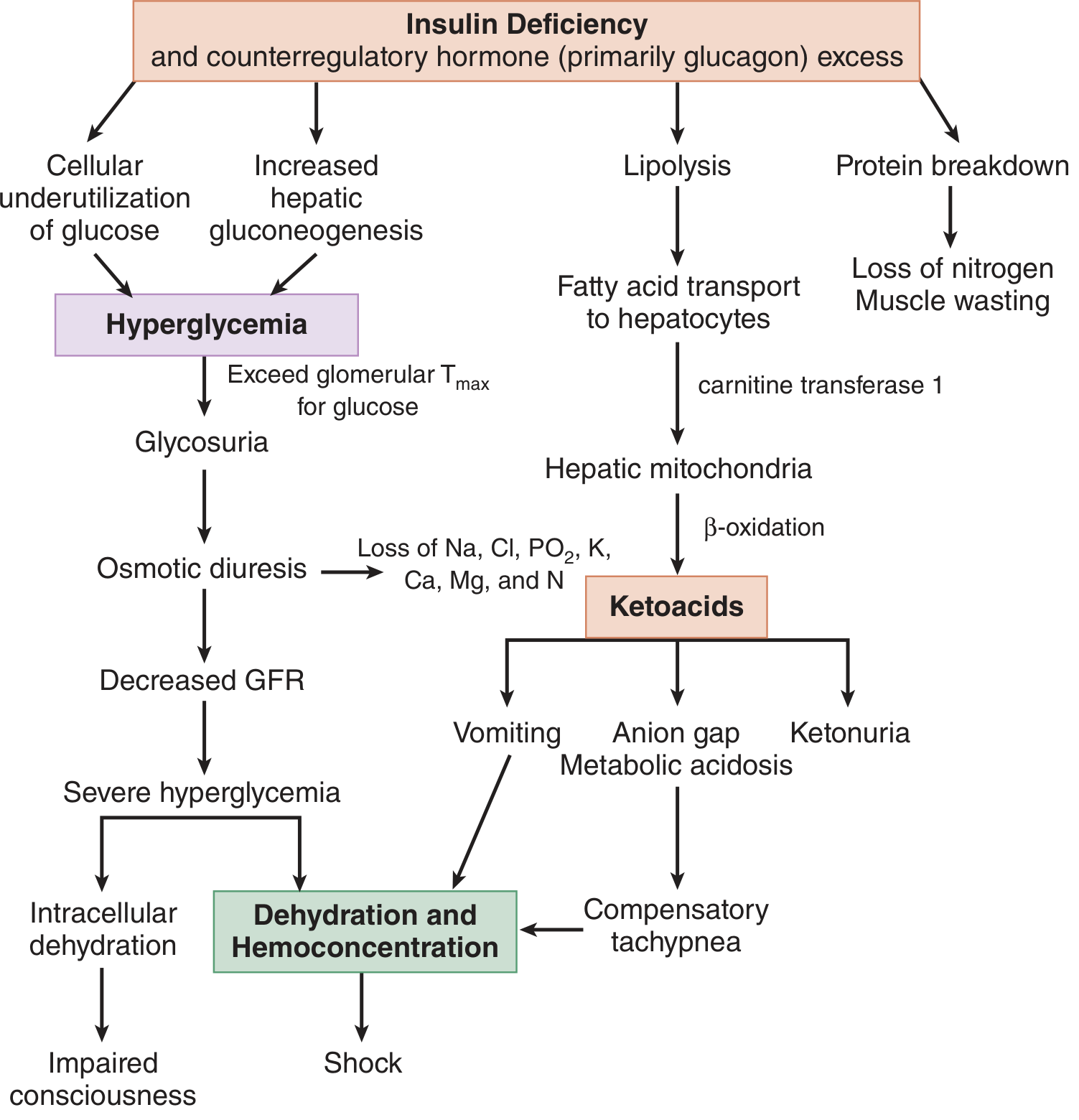
Pathophysiology in Brief
- Hyperglycemia - via increased gluconeogenesis, glycogenolysis, and impaired glucose uptake
- Ketogenesis - free fatty acids transported to liver undergo beta-oxidation → acetoacetate (AcAc) + beta-hydroxybutyrate (βHB) + acetone
- Osmotic diuresis - leads to profound dehydration + renal losses of Na, K, Cl, PO4, Mg, Ca
Lab Tests
Diagnostic Criteria for DKA
| Parameter | Mild DKA | Moderate DKA | Severe DKA |
|---|---|---|---|
| Blood glucose | >250 mg/dL (>13.9 mmol/L) | >250 mg/dL | >250 mg/dL |
| Arterial pH | 7.25-7.30 | 7.00-7.24 | <7.00 |
| Serum HCO3 | 15-18 mEq/L | 10-15 mEq/L | <10 mEq/L |
| Anion gap | >10 mEq/L | >12 mEq/L | >12 mEq/L |
| Mental status | Alert | Alert to drowsy | Stuporous/Comatose |
Essential Lab Panel
- Rapid bedside glucose (fingerstick)
- Serum electrolytes (Na, K, Cl, HCO3) - calculate anion gap: Na - (Cl + HCO3); normal 10-12 mEq/L
- BUN and creatinine (assess renal function and degree of dehydration)
- Venous blood gas (pH; correlates well with arterial pH - preferred as less invasive)
- CBC (look for leukocytosis - may reflect stress response even without infection)
- Urinalysis (glycosuria, ketonuria)
- Phosphate, magnesium, calcium
- ECG (assess for signs of hypo/hyperkalemia)
- Serum beta-hydroxybutyrate (βHB) - quantitative level >3 mmol/L confirms significant ketoacidosis; more reliable than urine nitroprusside test (which only detects AcAc, NOT βHB)
- Blood cultures, wound cultures if infection suspected
- Serum lactate (if hemodynamic instability or metformin use - risk of concurrent lactic acidosis)
- Serum osmolality (if altered consciousness; correlates better with mental status than pH)
- Serum lipase/amylase (both can be elevated in DKA itself; lipase more specific for pancreatitis)
- Arterial blood gas (if critically ill, respiratory compromise, or need to assess ventilation)
- Serum acetaminophen and toxicology screen (if toxic ingestion cannot be excluded)
- Troponin, chest X-ray, cardiac workup (if MI or PE suspected as precipitant)
- Urine dipstick ketones (nitroprusside reaction) detects only AcAc - βHB is NOT detected; test may appear falsely negative or weakly positive early in DKA when βHB predominates. As treatment progresses and the patient improves, βHB converts to AcAc, so "ketones appear to increase" on dipstick - this is normal, not worsening
- Captopril (ACE inhibitor with sulfhydryl group) can cause a false-positive urine nitroprusside test
- Serum K may appear normal or elevated at presentation due to acidosis-driven transcellular shift - total body K is always depleted. Serum K falls rapidly with insulin and fluid therapy
- A normal anion gap in DKA can occur if concurrent metabolic alkalosis (from vomiting) masks the acidosis, or if aggressive prior saline resuscitation has caused hyperchloremic acidosis
- Calculate delta gap (delta AG - delta HCO3): >+6 suggests concomitant metabolic alkalosis; <-6 suggests concomitant hyperchloremic acidosis
| DKA | HHS | |
|---|---|---|
| Glucose (mg/dL) | >350 | >700 |
| Sodium (mEq/L) | Low 130s | 140s |
| Potassium (mEq/L) | ~4.5-6.0 | ~5 |
| Bicarbonate (mEq/L) | <10 | >15 |
| BUN (mg/dL) | 25-50 | >50 |
| Serum ketones | Present | Absent |
- Water: 70-120 mL/kg
- Sodium: 8-10 mEq/kg
- Potassium: 5-7 mEq/kg
- Phosphorus: ~3 mEq/kg
Instrumental / Monitoring
- ECG - mandatory at presentation and with potassium changes: peaked T-waves (hyperkalemia) or U-waves, flat T-waves, prolonged QT (hypokalemia)
- Continuous cardiac monitoring - K shifts during treatment can cause arrhythmias
- Pulse oximetry - monitor oxygenation, especially if Kussmaul respirations are present
- Chest X-ray - rule out pulmonary infection, pulmonary edema (especially in children - cerebral/pulmonary edema risk during treatment)
- Brain CT or MRI - if altered consciousness does not improve with treatment; rule out cerebral edema (especially children) or cerebrovascular event
- Abdominal imaging (CT abdomen) - if severe abdominal pain persists after DKA correction; rule out pancreatitis, infarction, perforation
- Urine output monitoring - essential; Foley catheter in obtunded patients
Differential Diagnosis
High Anion Gap Metabolic Acidosis (MUDPILES/GOLD MARK)
| Condition | Key distinguishing features |
|---|---|
| Diabetic Ketoacidosis (DKA) | Glucose >250, known T1DM/T2DM, βHB >3 mmol/L, ± precipitant |
| Alcoholic Ketoacidosis (AKA) | Glucose low/normal, recent alcohol binge then starvation, βHB predominates (urine ketones may be negative/trace), HCO3 usually >18 mEq/L, mild ketosis (βHB <3 mEq/L) |
| Starvation Ketoacidosis | Mild, HCO3 rarely <18 mEq/L, no hyperglycemia, history of prolonged fasting |
| Lactic Acidosis | Elevated serum lactate >2 mmol/L; associated with hypoperfusion, sepsis, metformin toxicity; may co-exist with DKA |
| Uremia (Renal failure) | Elevated creatinine/BUN, no significant ketonemia |
| Salicylate toxicity | Respiratory alkalosis + AGMA; tinnitus, salicylate level elevated |
| Methanol / Ethylene glycol toxicity | Elevated osmol gap; methanol: visual symptoms; ethylene glycol: calcium oxalate crystals in urine; both cause severe AGMA |
| Isoniazid / Iron / Inborn errors | History, specific toxicology |
| Hyperglycemic Hyperosmolar State (HHS) | Glucose >700, serum osm >320, no significant acidosis, no ketones, older T2DM patients, more profound mental status change |
| Sepsis with AGMA | Fever, source of infection, lactic acidosis; note - sepsis can also trigger DKA |
| Acetaminophen toxicity | Elevated LFTs, aminotransferases, acetaminophen level; renal failure + AGMA |
Note: DKA should not exclude investigation for other simultaneous causes. Physiologic stress from sepsis, MI, or PE can precipitate DKA and both may coexist. The presence of DKA explains the ketosis and acid-base abnormality, but you must still look for the precipitant.
Treatment
Step 1 - Fluid Resuscitation (Priority #1)
- Normal saline (0.9% NaCl) is the standard first-line resuscitation fluid
- Initial bolus: 1-2 L IV over 1 hour in adults (10-20 mL/kg in children)
- Subsequent rate: 250-500 mL/hour, adjusted for hemodynamics and urine output
- Important caveat: Large volumes of 0.9% NaCl cause predictable hyperchloremic acidosis. Multiple studies show patients receiving balanced crystalloids (e.g., Plasmalyte-148) have shorter duration of acidosis, better bicarbonate levels, and better blood pressure profile compared to normal saline - Tintinalli's/Miller's both note this emerging evidence
Step 2 - Potassium Replacement (Do Before Insulin if K <3.5)
- Do NOT start insulin if serum K <3.5 mEq/L - insulin drives K further into cells → fatal arrhythmia
- K = 3.5-5.0 mEq/L: add 20-40 mEq KCl to each liter of IV fluid
- K <3.5 mEq/L: replace aggressively (20-40 mEq/hour IV) and delay insulin until K ≥3.5
- K >5.5 mEq/L: hold potassium supplementation, recheck every 2 hours
- Monitor K every 2-4 hours during treatment
Step 3 - Insulin
- Regular insulin: 0.1 units/kg/hour IV infusion (weight-based fixed rate)
- No bolus required if fluid resuscitation initiated first (ADA guideline)
- If no initial IV option: 0.1 units/kg SC or IM as bridge
- Add dextrose (D5W or D5 0.45% NaCl) to IV fluids - this allows continued insulin infusion to clear ketones without causing hypoglycemia
- Reduce insulin rate to 0.05 units/kg/hour once glucose <200 mg/dL
- Lispro 0.3 units/kg initial dose, then 0.1 units/kg/hour, OR
- 0.3 units/kg initial, then 0.2 units/kg every 2 hours until glucose <250 mg/dL
- Reserved for carefully selected mild-moderate cases; requires close monitoring
- A 2024 systematic review and meta-analysis (PMID 39090718) found comparable outcomes between SC insulin and continuous IV infusion for adult DKA; further supported by a 2026 meta-analysis of RCTs (PMID 41208563) showing early SC basal insulin co-administered with IV insulin safely reduces DKA recurrence
- Criteria for stopping IV insulin: glucose <200 mg/dL AND pH >7.3 AND HCO3 >15 mEq/L AND anion gap closed (≤12)
- Overlap IV and SC insulin by 2-4 hours to prevent rebound hyperglycemia
- New-onset diabetics: start 0.5-0.8 units/kg/day total daily dose
- Prefer long-acting (glargine) + rapid-acting (lispro/aspart) regimens over NPH + regular insulin
Step 4 - Electrolyte Management
- Serum phosphate appears normal at presentation but total body deficit exists
- IV phosphate only if serum PO4 <1.0 mg/dL (0.323 mmol/L)
- Give as IV K2PO4: 2.5-5 mg/kg if indicated
- No routine IV phosphate in ED - hypophosphatemia usually worsens 24-48 hours into therapy
- Monitor serum calcium if giving IV phosphate (risk of hypocalcemia)
- Not routinely recommended for most DKA (insulin and fluids correct acidosis)
- Consider NaHCO3 only if pH <6.9 (severe life-threatening acidosis) - 100 mEq over 2 hours
- Risks: paradoxical CNS acidosis, hypokalemia, cerebral edema in children
- Replace if <1.2 mEq/L or symptomatic (tetany, arrhythmia)
Step 5 - Treat the Precipitant
- Antibiotics for infection (common precipitant - fever absence doesn't exclude infection; hypothermia can occur due to peripheral vasodilation)
- Revascularization/anticoagulation for MI or PE
- Address medication errors or insulin pump failure
Step 6 - Monitoring Goals for Resolution
- Glucose every 1-2 hours during IV insulin
- Electrolytes (especially K) every 2-4 hours
- BG, pH, HCO3, anion gap every 2-4 hours
- Resolution criteria: BG <200 mg/dL + pH >7.3 + HCO3 >18 mEq/L + anion gap ≤12
Special Considerations
- Cerebral edema - rare but life-threatening, especially in children; avoid rapid rehydration; treat with mannitol or hypertonic saline if suspected
- DKA in pregnancy - can occur at lower glucose thresholds ("euglycemic DKA"); early fetal monitoring; associated with poor fetal outcomes per 2025 systematic review (PMID 40447103)
- SGLT-2 inhibitor-associated euglycemic DKA - glucose may be near-normal despite DKA; easy to miss; high clinical suspicion needed