
Define Nephrotic syndrome,it's epidermology, pathophysiology and treatment guidelines for it.
"nephrotic syndrome"[MeSH Terms] AND treatment
nephrotic syndrome pathophysiology glomerular basement membrane podocyte
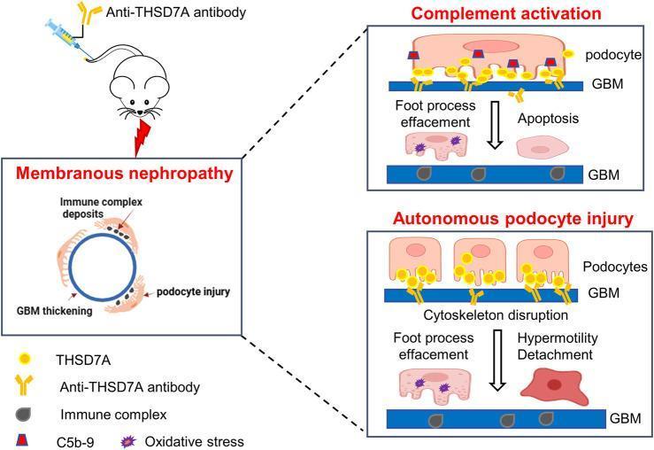
This pathophysiology diagram illustrates the dual pathomechanisms of THSD7A-associated Membranous Nephropathy (MN) based on a mouse model. The schematic shows a mouse being injected with anti-THSD7A antibodies, leading to MN characterized by subepithelial immune complex deposits, glomerular basement membrane (GBM) thickening, and podocyte injury. Two distinct pathways of injury are detailed: 1) Complement Activation: Anti-THSD7A antibodies (yellow) bind to THSD7A antigens on the podocyte surface, recruiting C5b-9 (red/blue markers) to form immune complexes. This leads to foot process effacement, oxidative stress, and podocyte apoptosis. 2) Autonomous Podocyte Injury: A complement-independent pathway where antibody binding directly causes cytoskeleton disruption. This results in foot process effacement, podocyte hypermotility, and eventual detachment from the GBM. The diagram serves as a model for primary MN, emphasizing both the immune-complex-mediated classical pathway and a direct, antibody-mediated cytotoxic effect that compromises the glomerular filtration barrier.
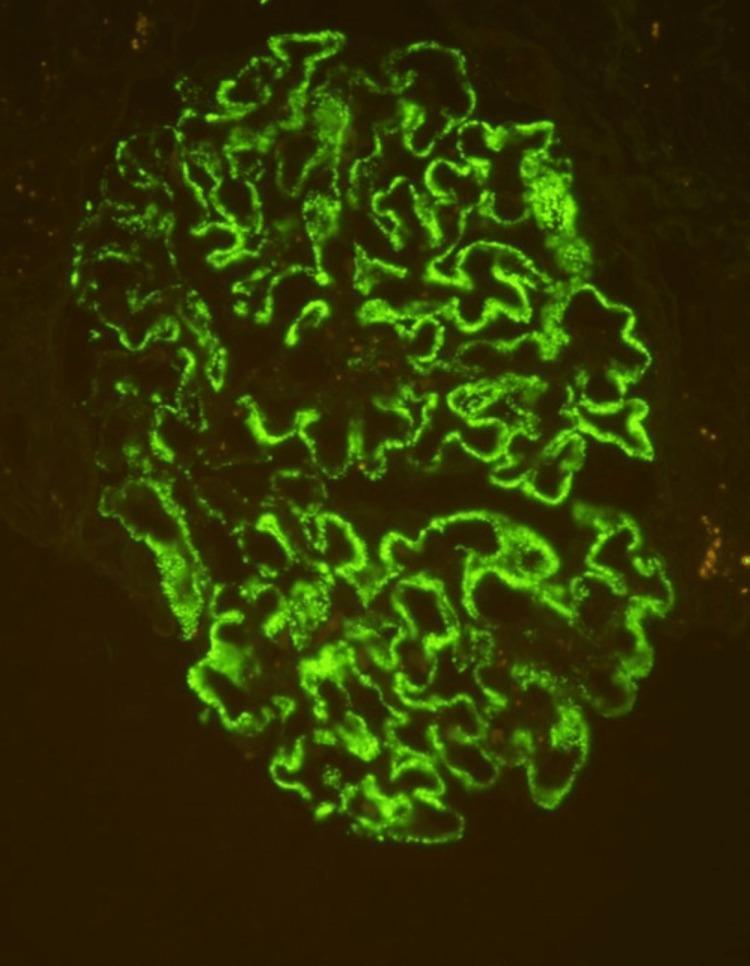
This diagnostic image is a high-power (400x) immunofluorescence micrograph of a human glomerulus from a renal biopsy specimen. The image demonstrates a diffuse, granular staining pattern of 3+ intensity, specifically localized along the glomerular capillary loops. The green fluorescent signal highlights the convoluted, interconnected morphology of the basement membrane area in a 'beaded' fashion, which is characteristic of immune complex deposition. Pathologically, this visual finding represents positive staining for phospholipase A2 receptor (PLA2R) antibodies, a definitive diagnostic marker for primary membranous nephropathy. The absence of mesangial or endocapillary staining emphasizes the subepithelial nature of the deposits. This specimen is critical for differentiating primary autoimmune-mediated glomerular disease from secondary causes in patients presenting with nephrotic syndrome.
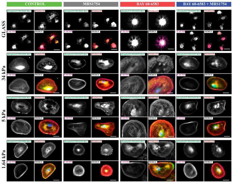
This composite of epifluorescence microscopy images illustrates the mechanobiological response of human podocytes to varying matrix stiffness (Glass, 34 kPa, 5 kPa, and 1.64 kPa) under A2B adenosine receptor (A2BAR) pharmacological modulation. The panel tracks four groups: Control, MRS1754 (A2BAR antagonist), BAY 60-6583 (A2BAR agonist), and a combined treatment. Immunofluorescent markers include phospho-S19 Myosin Light Chain (MLC) (green), Paxillin (red), and F-actin (phalloidin, magenta/white), with DAPI (blue) for nuclei. The images demonstrate that increasing matrix stiffness promotes the formation of prominent actin stress fibers and larger paxillin-positive focal adhesions. A2BAR stimulation by BAY 60-6583 enhances cell spreading and actomyosin contractility, evidenced by intensified phospho-S19 MLC and organized actin cables, particularly at intermediate stiffness (5-34 kPa). Conversely, MRS1754 antagonizes these effects, resulting in reduced cytoskeletal organization and attenuated focal adhesion development. This highlights the role of A2BAR signaling in regulating podocyte contractility and structural plasticity in response to the physical environment of the glomerular basement membrane.
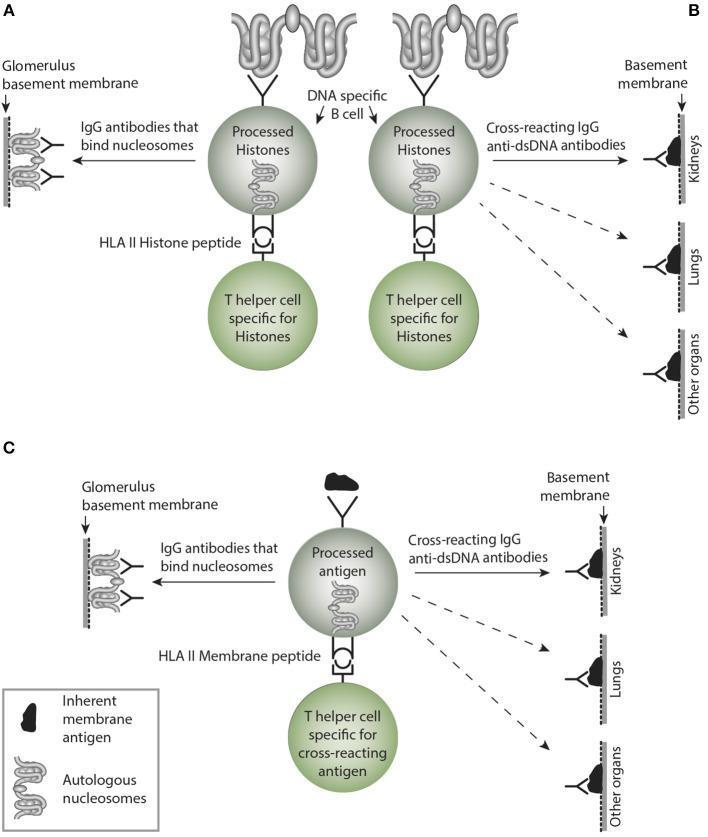
This medical illustration depicts the pathophysiology of autoimmune antibody responses involving nucleosomes and the glomerular basement membrane (GBM). The diagram is divided into three sections (A, B, and C). Section A shows a DNA-specific B cell presenting processed histones via HLA Class II molecules to a histone-specific T helper cell, leading to the production of IgG antibodies that bind nucleosomes and target autologous nucleosomes associated with the GBM. Section B illustrates a similar cellular interaction resulting in cross-reacting IgG anti-dsDNA antibodies that target the basement membranes of kidneys, lungs, and other organs. Section C demonstrates a B cell presenting processed inherent membrane antigens, resulting in both IgG antibodies that bind nucleosomes on the GBM and cross-reacting IgG anti-dsDNA antibodies affecting multiple organ systems. Key structures illustrated include nucleosomes, B cell receptors, HLA II peptides, T helper cells, and specific tissue targets like the kidney's glomerular basement membrane. This visual highlights the mechanisms of cross-reactivity and direct immune targeting in conditions such as systemic lupus erythematosus and Goodpasture-like syndromes.