
Teach Bell's palsy in detail
Reading File
Now I'll search the medical library for Bell's palsy comprehensively, running parallel searches.
Finding Sources
Finding Sources
Searching PubMed
"Bell palsy" treatment
Excellent - I have the key sources. Now I'll read the richest sections in parallel: Harrison's, Adams & Victor's, and Cummings (the specialist ENT reference).
Reading File
Reading File
Reading File
Rich content. Now let me read more from the key sources - the treatment sections, House-Brackmann grading, prognosis, and complications - plus fetch the MRI image.
Reading File
Reading File
Reading File
Loading Image
I now have comprehensive material from 3 major textbooks plus the MRI image. Let me read the remaining key sections on treatment (Cummings) and House-Brackmann grading, and electrodiagnostic testing.
Reading File
Finding Sources
Reading File
Reading File
I have comprehensive material from all major textbooks. Now let me compile the complete, detailed teaching on Bell's palsy.
Bell's Palsy - A Complete Teaching
1. Definition and Naming
Bell's palsy is idiopathic acute unilateral peripheral (lower motor neuron) facial nerve paralysis. The term was coined after Sir Charles Bell (1821), who first described the anatomy and function of the facial nerve. The diagnosis is one of exclusion with positive clinical criteria - the term should only be used when a diligent search for another cause is negative. As Cummings puts it: "All that palsies are not Bell."
Taverner's minimum diagnostic criteria:
- Paralysis or paresis of all muscle groups of one side of the face
- Sudden onset
- Absence of signs of CNS disease
- Absence of signs of ear or cerebellopontine angle disease
2. Epidemiology
- Annual incidence: 23-37 per 100,000 population (approximately 1 in 60 persons over a lifetime)
- Higher in those >65 years (59/100,000); lower in children <13 years (13/100,000)
- Equal sex ratio overall; slight female predominance in those <20 years; slight male predominance in those >40 years
- Left and right sides equally affected
- ~30% present with incomplete paralysis; ~70% with complete paralysis
- Bilateral paralysis: only 0.3% of cases (bilateral should prompt search for systemic disease)
- Recurrence: ~7-8% of cases (average 10-year interval between episodes)
- Family history: 8% of patients
- Risk factors: pregnancy (especially 3rd trimester, peripartum), diabetes mellitus, and possibly hypertension
3. Anatomy of the Facial Nerve (CN VII)
The facial nerve's unique anatomy explains why Bell's palsy produces such a distinctive constellation of symptoms:
Intracranial course:
- Nucleus in the caudal pons (facial motor nucleus)
- Loops around the abducens nucleus (forms the facial colliculus)
- Exits at the cerebellopontine angle (CPA)
Internal auditory canal: travels with CN VIII
Intratemporal (fallopian canal) segments:
- Labyrinthine segment - narrowest, most vulnerable segment; site of greatest nerve swelling and compression in Bell's palsy
- Geniculate ganglion - location of the HSV/VZV reactivation; gives off the greater superficial petrosal nerve (lacrimation, salivation)
- Tympanic (horizontal) segment
- Mastoid (vertical) segment - gives off the nerve to stapedius, then the chorda tympani (taste anterior 2/3 tongue; submandibular/sublingual salivation)
Extratemporal: exits stylomastoid foramen → parotid gland → five terminal branches: temporal, zygomatic, buccal, marginal mandibular, cervical
Clinical implication: Because the lesion in Bell's palsy is proximal (near the geniculate ganglion/labyrinthine segment), all functions of the nerve distal to this point can be affected: motor (all facial muscles), taste (chorda tympani), lacrimation (greater petrosal nerve), salivation, and the stapedius reflex (hyperacusis).
4. Etiology and Pathophysiology
The viral hypothesis (most widely accepted):
- HSV-1 reactivation from the geniculate ganglion is the leading cause. Murakami et al. (1996) using PCR detected HSV type 1 DNA in endoneurial fluid surrounding CN VII in 11 of 14 Bell's palsy cases
- VZV (varicella-zoster virus) reactivation accounts for up to one-third of cases (sometimes without skin vesicles - "zoster sine herpete")
- Other implicated agents: SARS-CoV-2, HIV (at seroconversion), EBV, CMV
Other proposed mechanisms:
- Microcirculatory failure of the vasa nervorum (dilated engorged veins, hemorrhage in the IAC - Fowler's autopsy findings)
- Ischemic neuropathy
- Autoimmune reaction
Final common pathway: Regardless of trigger, the nerve becomes edematous and swells within the bony fallopian canal (especially the tight labyrinthine segment at the meatal foramen). This causes compression and ischemia → demyelination → varying degrees of axonal degeneration.
Histopathology:
- Diffuse demyelination throughout the intratemporal course
- Lymphocytic infiltration and phagocytosis of myelin by macrophages
- Most severe changes at the labyrinthine segment and meatal foramen
- Engorged veins, hemorrhage within the IAC
- Osteoclastic bone resorption around the geniculate ganglion (in severe cases)
5. Clinical Features
Onset: Acute; ~50% reach maximum weakness within 48 hours; virtually all within 3-4 days. Progressive facial paralysis (>3 weeks to maximum) is NOT Bell's palsy - it mandates imaging to exclude neoplasm.
Motor: Unilateral weakness of ALL facial muscles (forehead + lower face) - this peripheral pattern distinguishes it from central (supranuclear) palsy where the forehead is relatively spared.
| Feature | Peripheral (Bell's) | Central (UMN) |
|---|---|---|
| Forehead | Affected (cannot wrinkle) | Spared (bilateral cortical input) |
| Eye closure | Incomplete (lagophthalmos) | Usually possible |
| Lower face | Weak | Weak |
| Bell's phenomenon | Present (eyeball rolls up on attempted closure) | Absent |
Associated features (by level of lesion):
- Retroauricular/post-auricular pain - present in ~50%; may precede paralysis by 1-2 days (common, can be intense)
- Taste impairment (chorda tympani involvement) - anterior 2/3 tongue; present in most but rarely persists beyond 2nd week
- Hyperacusis (stapedius muscle paralysis) - present in up to 30%; ipsilateral ear
- Decreased lacrimation (greater petrosal nerve) - dry eye; clinically important for eye care
- Numbness/fullness of the face - reported by patients; formal sensory testing usually normal (pure motor nerve)
- Mild CSF lymphocytosis - in a small number of cases
Bell's phenomenon: On attempted eye closure, the eyeball rolls upward (protective reflex); becomes visible because lid closure is incomplete (lagophthalmos). This is NOT pathological - it is a normal reflex unmasked by the palsy.
6. House-Brackmann Grading Scale
The standard clinical grading system endorsed by the American Academy of Otolaryngology - Head and Neck Surgery:
| Grade | Description | Function |
|---|---|---|
| I | Normal | 100% |
| II | Slight dysfunction - slight weakness on close inspection; complete eye closure with minimal effort; slight asymmetry | >80% |
| III | Moderate dysfunction - obvious but not disfiguring difference; complete eye closure with effort; some forehead movement | 50-80% |
| IV | Moderately severe - obvious weakness/disfigurement; incomplete eye closure; no forehead movement | 25-50% |
| V | Severe - only barely perceptible motion; incomplete eye closure; no forehead movement | <25% |
| VI | Total paralysis - no movement | 0% |
Limitation: A single global score mainly reflects eye function; regional scoring (forehead, eye, nose, mouth separately) is more precise.
7. Investigations
Diagnosis is clinical in typical cases. No routine workup is needed if:
- Typical presentation (acute-onset, unilateral, peripheral pattern)
- No risk factors for other causes
- No skin vesicles in the ear canal
- Normal neurologic exam (other than CN VII)
When to investigate further (atypical features):
- Bilateral palsy → Lyme titer, HIV, ACE, CXR (sarcoidosis), LP (Guillain-Barré)
- Slowly progressive → MRI (tumor)
- Recurrent palsy → consider Melkersson-Rosenthal syndrome
- Signs of herpes zoster → confirm Ramsay Hunt
Electrodiagnostic testing:
- Electroneuronography (ENoG): Evoked compound muscle action potential; compares affected to normal side. Used for prognosis in complete palsy (performed 4-14 days after onset)
-
90% degeneration on ENoG: poor prognosis; may consider surgical decompression
-
- Electromyography (EMG): Detects fibrillation potentials (denervation) or voluntary motor unit potentials (regeneration)
- Evidence of denervation after 10 days = axonal degeneration → expect 3+ months before recovery, may be incomplete
- Voluntary motor units = ongoing reinnervation, good sign
- Nerve conduction (blink reflex): Assesses conduction across the geniculate ganglion
MRI (Gadolinium-enhanced, fat suppressed):
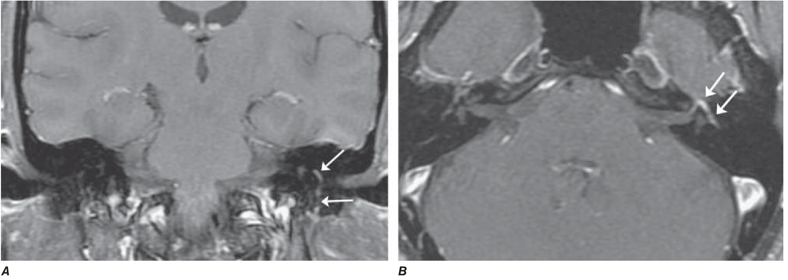
Gadolinium-enhanced T1 MRI showing diffuse smooth linear enhancement of the left facial nerve (arrows) involving the genu, tympanic, and mastoid segments within the temporal bone - consistent with Bell's palsy. Note: similar enhancement can occur in Lyme disease, sarcoidosis, and perineural malignant spread. - Harrison's Principles of Internal Medicine, 22e
8. Differential Diagnosis
| Condition | Key Distinguishing Features |
|---|---|
| Ramsay Hunt syndrome (herpes zoster oticus) | Vesicular eruption in external auditory canal/auricle; more severe palsy; pain; CN VIII involvement (vertigo, sensorineural hearing loss) |
| Lyme disease (Borrelia burgdorferi) | Tick bite/erythema migrans history; endemic area; can be bilateral; CSF pleocytosis |
| Sarcoidosis | Bilateral facial palsy (Heerfordt syndrome); uveitis, hilar adenopathy; elevated ACE |
| Acoustic neuroma | Progressive; CN VIII symptoms; cerebellopontine angle mass on MRI |
| Cholesteatoma / chronic otitis media | History of ear disease; conductive hearing loss; otoscopy findings |
| Parotid malignancy | Slowly progressive; palpable mass |
| Guillain-Barré syndrome | Bilateral facial palsy + ascending weakness; CSF albuminocytologic dissociation |
| Leprosy | Thickened nerves; endemic area; skin patches |
| Melkersson-Rosenthal syndrome | Recurrent facial palsy + facial (labial) edema + plicated tongue |
| Diabetes mellitus | Peripheral neuropathy context; metabolic |
| Central lesion (pontine stroke) | Ipsilateral CN VI palsy, gaze palsy, contralateral hemiplegia; forehead spared |
9. Treatment
A. Eye Care (IMMEDIATE - most important)
Corneal exposure from incomplete eye closure can cause keratitis and corneal ulceration:
- Artificial tears (lubricating eye drops) during the day
- Lubricating eye ointment at night
- Tape eyelid shut or moisture chamber goggles during sleep
- If lagophthalmos is severe: ophthalmology referral; consider temporary tarsorrhaphy
B. Corticosteroids (First-line treatment)
Most important pharmacologic intervention.
- Prednisone 60-80 mg/day for the first 5 days, then taper over 5 days (Harrison's) OR
- Prednisolone 25 mg twice daily for 10 days (Sullivan et al. RCT protocol)
- Must be started within 72 hours of onset for maximum benefit (ideally within 48 hours)
- Mechanism: reduces nerve edema/inflammation, decreases compression within the fallopian canal
- Evidence: Multiple RCTs (Sullivan 2007, Engstrom 2008) showed statistically significant benefit; NNT approximately 6
C. Antiviral Agents (Adjunct - contested)
- Acyclovir 400 mg 5× daily × 10 days OR Valacyclovir 1000 mg daily × 5-7 days (better bioavailability)
- Evidence is mixed:
- Major RCT (Sullivan et al.): antiviral alone = no benefit over placebo; combination = no significant additional benefit over steroids alone
- Hato et al.: suggested additive benefit of valacyclovir + prednisolone particularly in complete palsy
- Cochrane review: low-quality evidence for combination benefit; moderate-quality evidence for reduction of synkinesis with combination therapy
- Current consensus: Antivirals should be added if vesicular lesions are seen (ear canal, palate) - confirming HSV/VZV involvement. In pure Bell's palsy, steroids alone are standard; antivirals remain optional but are commonly co-prescribed in severe/complete palsy
D. Surgical Decompression
- Highly controversial and not routinely recommended
- Considered in severe cases with >90% degeneration on ENoG within 14 days, no recovery by 4-6 weeks
- Approach: middle cranial fossa decompression of the labyrinthine segment (highest-yield site)
- Risk: sensorineural hearing loss
- Most guidelines and systematic reviews do NOT support routine decompression
- Adams & Victor: "There is no evidence that surgical decompression of the facial nerve is effective, and it may be harmful."
E. Physical Therapy
- Facial exercises, neuromuscular re-education
- Most useful during recovery phase and for sequelae (synkinesis rehabilitation)
- 2025 systematic review (PMID 39647183) supports physical therapy for idiopathic facial paralysis
F. Surgical Rehabilitation (Late/Permanent Paralysis)
For patients with permanent paralysis:
- Static procedures: Gold weight implant (upper eyelid loading for lagophthalmos), facial slings
- Dynamic reinnervation: Hypoglossal-facial nerve anastomosis; cross-facial nerve graft
- Gracilis free muscle flap: For long-standing complete paralysis
- Botulinum toxin: For synkinesis management
10. Prognosis
- ~80-90% recover completely
- ~70% recover completely within 1-2 months
- ~85% achieve near-normal function
- Incomplete paralysis at presentation: 95-100% complete recovery (most favorable)
- Evidence of denervation on EMG after 10 days: long delay (3+ months) before recovery; may be incomplete; proceeds via axonal regeneration (up to 2 years)
- Most favorable prognostic sign: some voluntary motor recovery in the first 5-7 days; early return of taste (first week)
Poor prognostic factors:
- Complete paralysis at onset
- Age >60 years
- Diabetes mellitus / hypertension
- Hyperacusis + decreased tearing (high lesion level)
- Severe pain at onset
- Late onset of recovery
Recurrence: ~7-8%; average 10-year interval; more common with Lyme disease, sarcoidosis, or familial variety.
11. Sequelae
When recovery is incomplete:
- Synkinesis - aberrant regeneration; voluntary movement of one muscle group triggers involuntary movement in another (e.g., eye closes when smiling - "crocodile tears" variant); managed with botulinum toxin + physiotherapy
- Contracture - persistent hemifacial hypertonicity
- Crocodile tears (Bogorad syndrome) - gustatory lacrimation; aberrant regeneration of greater petrosal nerve fibers to lacrimal gland (tears when eating/salivating)
- Hemifacial spasm (rare late sequela)
12. Special Situations
Pregnancy
- 3× increased incidence; highest risk in 3rd trimester and first 2 weeks postpartum
- Managed identically to non-pregnant patients (corticosteroids are safe in pregnancy)
- Collaborative management with obstetrician
Children
- Less common; 8% of Bell's palsy patients are children
- Evidence for corticosteroids less clear in children (some studies show no benefit)
- Generally favorable prognosis
Bilateral Bell's Palsy (0.3% of cases)
- Must actively exclude: Lyme disease, Guillain-Barré syndrome, sarcoidosis, HIV, bilateral acoustic neuromas
13. Key Teaching Points Summary
| Point | Detail |
|---|---|
| Most common cause of peripheral CN VII palsy | Bell's palsy (HSV-1 reactivation) |
| Defines peripheral vs. central | Forehead involvement: peripheral affects forehead; central spares it |
| Most common site of nerve entrapment | Labyrinthine segment / meatal foramen |
| Mainstay of treatment | Prednisone (within 72 hours of onset) |
| Most favorable prognostic sign | Incomplete palsy at onset; early motor recovery (days 5-7) |
| Overall recovery rate | ~80-90% |
| Ramsay Hunt vs. Bell's | RHS has vesicles in ear canal, more severe, CN VIII involved |
| Bilateral palsy key DDx | Lyme disease #1 in endemic areas |
| Eye care urgency | Immediate - corneal exposure is the main short-term risk |
Sources:
- Harrison's Principles of Internal Medicine 22e, p. 3597-3598
- Adams and Victor's Principles of Neurology, 12th Ed., p. 1371-1372
- Cummings Otolaryngology Head and Neck Surgery (Bell's Palsy chapter)
- Scott-Brown's Otorhinolaryngology Head & Neck Surgery, Vol 2 (Grading Systems)
- Recent systematic reviews: physical therapy (PMID 39647183), photobiomodulation (PMID 39627559), laser therapy (PMID 39546047)
Teach bell's palsy to first year mbbs
Reading File
I have the full context from the previous detailed session. For a 1st year MBBS student, I need anatomy-focused content (their syllabus stage) with simple language. Let me pull the anatomy-specific content about the facial nerve course and Bell's phenomenon from the library.
Finding Sources
Finding Sources
Finding Sources
Reading File
Reading File
Loading Image
I have everything I need. Now I'll write a 1st year MBBS-level teaching - focused on anatomy, simple language, with mnemonics, clinical correlation, and exam-ready format.
Bell's Palsy for 1st Year MBBS
Level: Anatomy + basic Physiology foundation | Tone: Simple, story-like, exam-ready
🧠 Start With a Story
Imagine you wake up one morning and half your face feels "frozen." You can't close your eye. When you try to smile, only one side of your face moves. You can't wrinkle your forehead. You notice food is falling out of one side of your mouth. Your family thinks you've had a stroke.
This is Bell's palsy - and understanding it begins with understanding the facial nerve.
Part 1: The Facial Nerve (CN VII) - The "Star" of the Story
The facial nerve is Cranial Nerve 7 (CN VII). Before you can understand Bell's palsy, you must know what this nerve does.
A helpful summary: "CN VII tears, tastes, salivates, moves the face, and dampens sound."
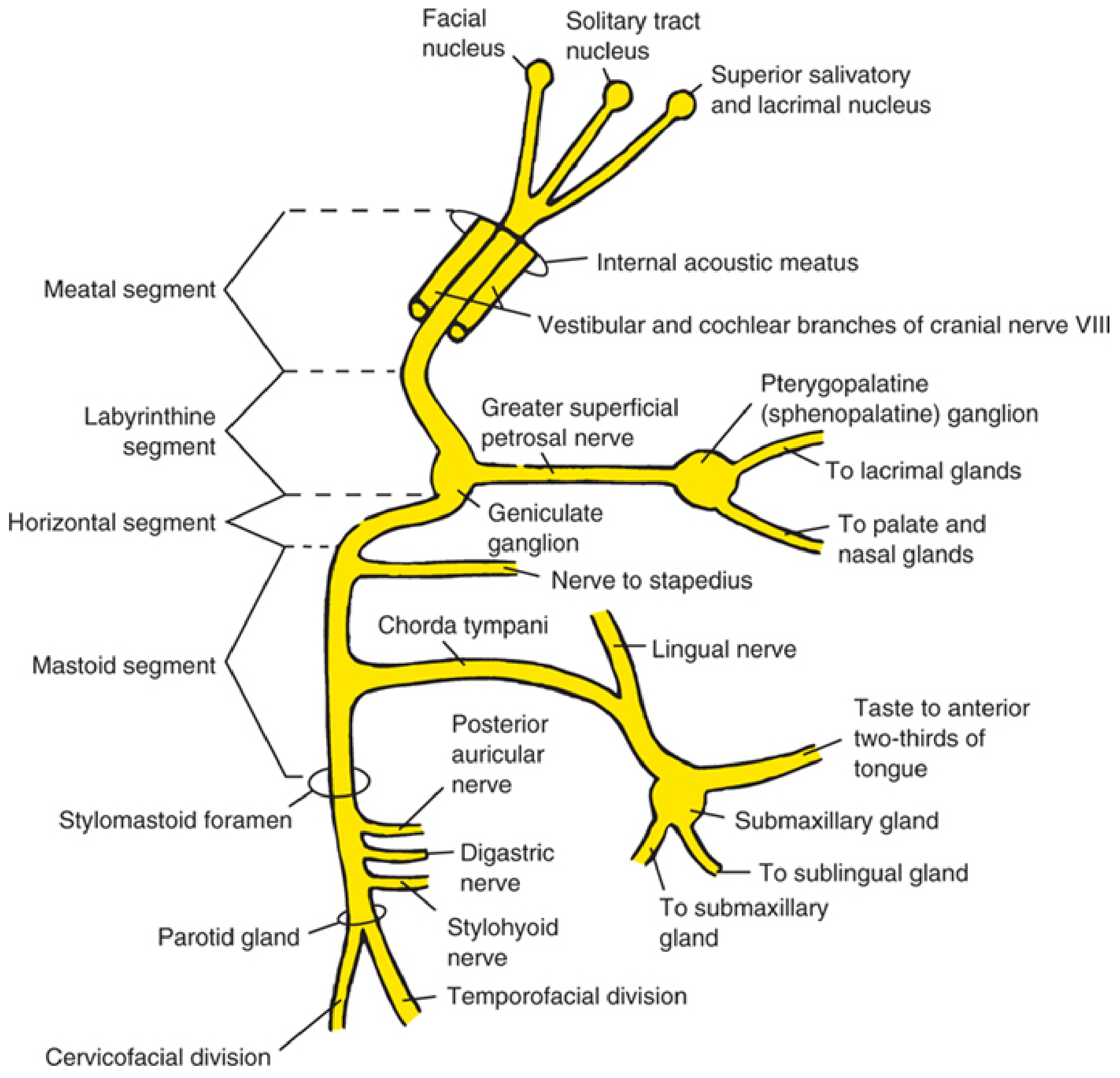
Complete anatomy of CN VII - Localization in Clinical Neurology, 8e
Functions of CN VII (memorize these)
| Function | Which fibers | What it does |
|---|---|---|
| Motor | General somatic efferent | Moves all muscles of facial expression |
| Motor | General somatic efferent | Nerve to stapedius (dampens loud sounds) |
| Motor | General somatic efferent | Stylohyoid + posterior belly of digastric |
| Taste | Special visceral afferent | Taste from anterior 2/3 of tongue (via chorda tympani) |
| Parasympathetic | General visceral efferent | Tears (lacrimal gland, via greater petrosal nerve) |
| Parasympathetic | General visceral efferent | Saliva (submandibular + sublingual glands, via chorda tympani) |
| Sensation | General somatic afferent | Skin of external ear (small area) |
Mnemonic for CN VII branches at the parotid (terminal motor branches): "Two Zebras Bit My Cat" → Temporal, Zygomatic, Buccal, Marginal mandibular, Cervical
Course of the Facial Nerve (the "Long Journey")
The facial nerve takes a very long, winding path through the skull - this is why it is vulnerable.
BRAINSTEM (Pons)
     ↓
Facial nucleus + Superior salivatory nucleus
     ↓
Internal Acoustic Meatus (with CN VIII - the hearing nerve)
     ↓
INTRATEMPORAL (inside the temporal bone = "fallopian canal")
     ├─ Labyrinthine segment → GENICULATE GANGLION
     │        └── Greater Petrosal Nerve → lacrimal gland (tears)
     ├─ Tympanic (horizontal) segment
     │        └── Nerve to Stapedius
     └─ Mastoid (vertical) segment
              └── Chorda tympani → taste + sublingual/submandibular saliva
     ↓
STYLOMASTOID FORAMEN (exits the skull here)
     ↓
PAROTID GLAND (divides into 5 terminal branches)
     ↓
5 branches to muscles of facial expression
Key exam point: The facial nerve exits the skull at the stylomastoid foramen. In Bell's palsy, the nerve gets swollen and trapped inside the bony canal (specifically at the narrowest part - the labyrinthine segment).
Part 2: What is Bell's Palsy?
Bell's palsy = sudden, one-sided, complete weakness of all the muscles of facial expression due to inflammation of CN VII, with no identifiable cause (idiopathic).
Named after Sir Charles Bell who described the anatomy of CN VII in 1821.
What causes it?
Most likely: reactivation of Herpes Simplex Virus type 1 (HSV-1) - the same virus that causes cold sores - from the geniculate ganglion inside the temporal bone.
Think of it like this: the virus was sleeping quietly in the nerve. Something wakes it up (stress, illness, pregnancy). It causes inflammation. The nerve swells inside its tight bony tunnel. Swollen nerve + tight tunnel = compression = paralysis.
Part 3: The #1 Anatomy Lesson in Bell's Palsy
Upper Motor Neuron (UMN) vs. Lower Motor Neuron (LMN) facial palsy
This is the most important concept for a 1st year student and appears in almost every anatomy exam.
The upper part of the face (forehead) gets nerve supply from BOTH sides of the brain.
The lower part of the face gets nerve supply from only the OPPOSITE side of the brain.
Why? Because the corticobulbar fibers that go to the upper face (frontalis muscle) come from both the left and right cortex. So even if one side of the brain is damaged, the other side still works for the forehead.
| Bell's Palsy (LMN / peripheral) | Stroke (UMN / central) | |
|---|---|---|
| Level of lesion | CN VII nerve itself (outside brainstem) | Motor cortex or internal capsule |
| Forehead | AFFECTED - cannot wrinkle | SPARED - can wrinkle |
| Lower face | Weak | Weak |
| All facial muscles | YES - all affected on that side | NO - only lower face mostly |
Exam shortcut: "Forehead spared = Central lesion." "Forehead affected = Peripheral (Bell's palsy)."
Part 4: Clinical Features (What the Patient Shows)
Because CN VII has SO many branches, the features depend on where exactly the nerve is damaged.
In Bell's palsy, the damage is HIGH UP - near the geniculate ganglion - so nearly ALL functions are affected:
Motor symptoms (you can SEE these):
- Cannot wrinkle the forehead (frontalis muscle paralyzed)
- Cannot close the eye (orbicularis oculi paralyzed) → the eye stays open, even at night → lagophthalmos
- Bell's phenomenon - when the patient tries to close the eye, the eyeball rolls UPWARD (you can see the white of the eye). This is a normal protective reflex that only becomes visible because the lid won't close.
- Drooping of the mouth on the affected side
- Cannot smile symmetrically - mouth pulled to the normal side
- Cannot puff cheeks / whistle - air leaks from the weak side
- Food falls out of the corner of the mouth
- Flat nasolabial fold on the affected side
Non-motor symptoms (from branch involvement):
| Symptom | Which branch affected | What it means |
|---|---|---|
| Loss of taste (anterior 2/3 tongue) | Chorda tympani | Proximal lesion |
| Dry eye / Decreased tears | Greater petrosal nerve | High lesion |
| Hyperacusis (sounds are painfully loud) | Nerve to stapedius paralyzed | Can't dampen sound |
| Pain behind the ear | Post-auricular nerve irritation | Often first symptom, day before palsy |
Simple rule: The more symptoms the patient has (taste + dry eye + hyperacusis), the higher the lesion and the worse the prognosis.
Part 5: Why the Eye is the Emergency
When the orbicularis oculi is paralyzed, the patient cannot close their eye. This means:
- The cornea (transparent front of the eye) is exposed day and night
- It dries out
- It can get infected or develop a corneal ulcer
- This can lead to permanent vision loss
This is why even before any medicines, the doctor protects the eye with artificial tears during the day and taping the eyelid shut at night.
Part 6: Remembering All the Features - A Mnemonic
"BELL'S FACE"
| Letter | Feature |
|---|---|
| B | Bell's phenomenon (eye rolls up) |
| E | Eye cannot close (lagophthalmos) |
| L | Loss of taste (anterior 2/3 tongue) |
| L | Loss of nasolabial fold |
| S | Sound hypersensitivity (hyperacusis) |
| F | Forehead cannot wrinkle (key differentiator) |
| A | Ache behind the ear (post-auricular pain) |
| C | Cannot smile / whistle / puff cheeks |
| E | Eye - dry eye / decreased lacrimation |
Part 7: How the Diagnosis is Made
Bell's palsy is a clinical diagnosis. No special tests are needed in a typical case.
The doctor looks for:
- Sudden onset (within hours to a few days)
- All facial muscles on one side are weak (including forehead - this rules out a stroke)
- No other neurological signs
- No ear disease, no tumor
A simple bedside test: Ask the patient to raise their eyebrows. In Bell's palsy, the eyebrow on the affected side does NOT move. In a stroke, it does.
Part 8: Treatment (Simple Version)
| Treatment | Purpose |
|---|---|
| Eye care (artificial tears + tape lid at night) | Prevent corneal damage - MOST URGENT |
| Corticosteroids (Prednisone) | Reduce nerve swelling; start within 72 hours |
| Antiviral drugs (Acyclovir/Valacyclovir) | May be added, especially if vesicles seen |
| Physiotherapy (facial exercises) | During recovery, to prevent muscle stiffness |
Part 9: Prognosis (Outcome)
The good news for patients:
- 80-90% of patients recover completely, usually within 2-3 months
- The most favorable sign: if some facial movement returns within the first 5-7 days
- If taste comes back in the first week - also a good sign
- A small group (~10-15%) have incomplete recovery with some residual weakness or synkinesis (abnormal linked movements, e.g., eye closes when smiling)
Bad prognostic factors: complete paralysis, age >60, diabetes, hypertension.
Part 10: Comparison Table - Bell's Palsy vs. Stroke (UMN palsy)
This is THE most tested comparison in anatomy/neuroscience exams:
| Feature | Bell's Palsy (LMN) | Stroke (UMN) |
|---|---|---|
| Cause | CN VII nerve inflammation | Cortex/internal capsule lesion |
| Onset | Hours to 1-2 days | Sudden (seconds to minutes) |
| Forehead | PARALYZED | SPARED |
| Lower face | Paralyzed | Paralyzed |
| Eye closure | Cannot close | Usually can close |
| Taste | Lost | Normal |
| Lacrimation | May be reduced | Normal |
| Other limbs | Normal | Arm/leg weakness on same side |
| Reflexes | Normal | Exaggerated (UMN signs) |
Part 11: Quick Exam Revision Summary
What is Bell's palsy? Acute idiopathic unilateral LMN facial nerve (CN VII) palsy due to HSV-1 reactivation causing inflammation and entrapment of the nerve in the fallopian canal.
What is the key clinical sign? Unilateral facial weakness involving the FOREHEAD (distinguishes it from central/UMN palsy).
What is Bell's phenomenon? Upward rolling of the eyeball when attempting eye closure (normal reflex unmasked by orbicularis oculi paralysis).
What is the most urgent complication? Corneal exposure keratopathy due to lagophthalmos → treat with artificial tears + eyelid taping.
What is the treatment? Eye protection (immediate) + Oral corticosteroids (within 72 hours of onset).
What is the prognosis? 80-90% complete recovery.
Lesion site? Labyrinthine segment of CN VII inside the fallopian canal (temporal bone), near the geniculate ganglion - the narrowest and most vulnerable segment.
Anatomy Diagram Summary
Here is the CN VII diagram again for reference - trace the nerve from the facial nucleus in the pons, through the internal acoustic meatus, through the fallopian canal, out the stylomastoid foramen, and into the parotid gland where it splits into its 5 terminal branches:
Study tip: Use this diagram as a checklist. For each segment, ask yourself: "If the nerve is damaged HERE, what functions would be lost?" That is the logic behind all facial nerve clinical questions.
Sources: Localization in Clinical Neurology 8e | Harrison's Principles of Internal Medicine 22e | Adams & Victor's Principles of Neurology 12e | Cummings Otolaryngology | Scott-Brown's Otorhinolaryngology Vol 2
This is a shared conversation. Sign in to Orris to start your own chat.