
Segonds fracture
The Segond fracture is a small avulsion fracture of the lateral tibial plateau, just below the joint line, caused by internal rotation and varus stress on the knee. It is pathognomonic for an ACL tear - present in roughly 75-100% of cases when seen on plain radiographs. The fragment is pulled off by the lateral capsular ligament (and/or the anterolateral ligament).
Searching sources...
Reading File
Searching the Web
Segond fracture lateral tibial plateau avulsion ACL anatomy mechanism
Finding Sources
Searching PubMed
Segond fracture anterolateral ligament ACL
Now let me read the most relevant textbook sections in parallel:
Reading File
Reading File
Reading File
Now let me fetch the X-ray image and get the Campbell's / Rockwood sections on clinical management:
Loading Image
Reading File
Reading File
I now have comprehensive information from multiple authoritative sources. Here is the full answer:
Segond Fracture
Definition
A Segond fracture is a small curvilinear or elliptic avulsion fracture of the lateral tibial plateau, occurring just below the lateral joint line. It is produced by excessive tension at the attachment of the lateral capsular ligament (and/or anterolateral ligament, ALL) to the proximal tibia.
- First described by French surgeon Paul Ferdinand Segond in 1879 from cadaveric experiments
- Also called the "lateral capsular sign" on plain radiographs
Mechanism of Injury
The injury occurs with:
- Knee flexion combined with excessive internal rotation and varus stress
- This combination tightens the lateral capsular structures, avulsing a bone fragment from their tibial insertion
Segond himself described a "pearly, resistant fibrous band" that became taut with internal rotation - this corresponds to the modern concept of the anterolateral ligament (ALL) / lateral capsular complex.
- Rosen's Emergency Medicine, p. 718
Anatomy of the Avulsing Structure
This remains a matter of debate in current literature. Three structures have been implicated:
| Structure | Supporting Evidence |
|---|---|
| Lateral capsular ligament (middle third) | Traditional description - original Rosen's/Grainger textbook teaching |
| Anterolateral ligament (ALL) | Claes et al. - ALL inserts at the exact avulsion site |
| Meniscotibial (coronary) ligament | Rockwood & Green 2025 - meniscotibial component responsible for "at least a portion" |
| Posterior fibers of ITB + lateral capsule | Albers case series - both linked to the bone fragment surgically |
Most current evidence supports the ALL as the primary avulsing structure, with contributions from the lateral capsular complex.
- Rockwood & Green's Fractures in Adults, 10th ed. 2025, p. 3053
Radiographic Features
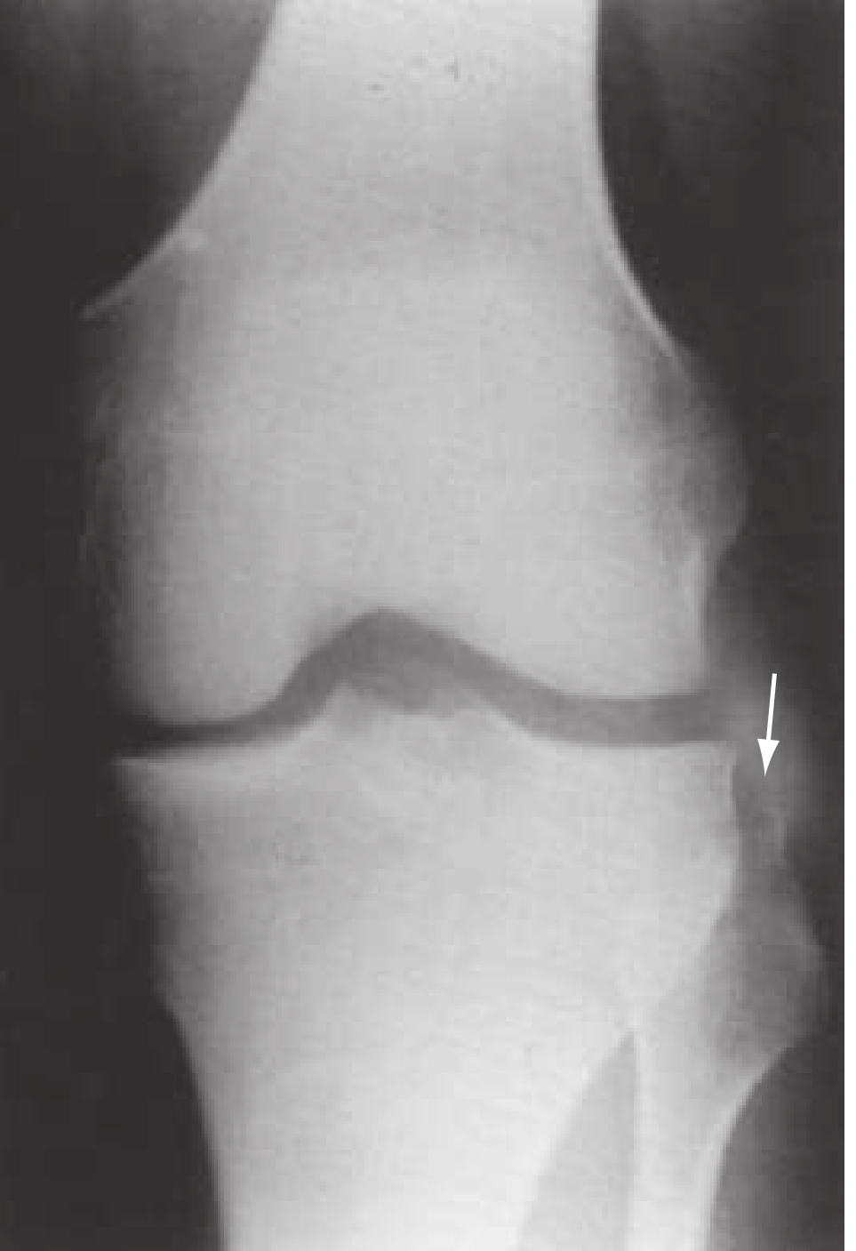
AP view of left knee showing the classic lateral curvilinear avulsion fragment (arrow) with joint effusion - from Rosen's Emergency Medicine
Plain radiograph (AP view):
- Small elliptic/curvilinear bone fragment parallel to the lateral tibial plateau
- Just below the joint line, medial to the fibular head
- Best seen on the anteroposterior (AP) view
- Joint effusion/hemarthrosis is usually present
MRI: Essential in all cases to assess the full extent of associated injury.
- Grainger & Allison's Diagnostic Radiology
Clinical Significance and Associated Injuries
The Segond fracture is pathognomonic for significant internal derangement of the knee. Though the fracture itself is tiny, it signals major ligamentous injury:
| Associated Injury | Frequency |
|---|---|
| ACL tear | 75-100% of cases |
| Medial or lateral meniscal tear | 66-75% (posterior horn most common) |
| Biceps femoris long head avulsion | Less common |
| Fibular collateral ligament avulsion | Less common |
"Its importance is that the mechanism of a forced varus injury that produces this finding results in a very high incidence of rupture of the ACL and tears of the medial meniscus, both said to be as high as 75% to 100% of cases." - Grainger & Allison's Diagnostic Radiology
One important caveat: a 2010 CT study (AJR) found that 8/20 patients with a Segond fracture combined with tibial plateau fracture had an intact ACL, showing the association is not 100% universal in all fracture contexts.
Differential Diagnosis
- Gerdy's tubercle avulsion (Segond noted it is "never Gerdy's tubercle that gives way, but the portion of bone immediately behind it")
- Normal variant ossicle
- Arcuate sign (fibular head avulsion - associated with PCL injury and posterolateral corner injury)
- Lateral tibial plateau fracture
Management
Acute/Emergency:
- Knee immobilizer until orthopedic follow-up
- MRI mandatory to characterize all soft tissue injuries
- Do NOT underestimate the injury based on the small fracture size
Definitive:
- The fracture itself usually heals spontaneously (can heal after ACL reconstruction)
- Treatment is driven by the associated ACL tear, typically requiring ACL reconstruction
- If a Segond fracture is present alongside ACL reconstruction, Campbell's Operative Orthopaedics (15th ed, 2026) lists it as an indication for combined ACL + anterolateral ligament (ALL) reconstruction, which reduces graft failure rate 2.5-3x vs isolated ACL reconstruction in high-risk patients
- Campbell's Operative Orthopaedics, 15th Ed. 2026
- Roberts & Hedges' Clinical Procedures in Emergency Medicine
Recent Evidence
- Systematic review (AJSM 2023, PMID 34668790) - Clinical outcomes after ACL reconstruction in patients with concomitant Segond fracture show no increased re-tear rate, and concomitant Segond fracture is not associated with worse outcomes after ACL reconstruction when managed appropriately.
- Narrative review (J Clin Orthop Trauma 2023, PMID 36860993) - Comprehensive update on anatomy, biomechanics, and clinical implications.
Key takeaway: A Segond fracture is a "don't miss" finding on plain knee radiographs. Its small, innocuous appearance belies its significance as a marker of high-grade lateral capsular disruption and near-certain ACL tear. Always order MRI and arrange orthopedic follow-up.
This is a shared conversation. Sign in to Orris to start your own chat.