
Total no of skull bones and there names
skull bones anatomy labeled diagram
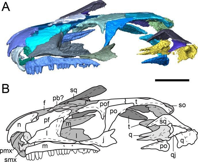
Anatomical diagram and segmented visualization of a skull in left lateral profile, categorized under clinical imaging and skeletal anatomy. Panel A presents a three-dimensional segmented visualization with a color-coded palette to distinguish individual osseous structures. Panel B is a corresponding outline drawing with anatomical labels. Key structures include the nasal (n), frontal (f), and parietal (p) bones of the skull roof; the prefrontal (pf), postfrontal (pof), postorbital (po), and jugal (j) bones surrounding the orbit; and the maxilla (m) and premaxilla (pmx) of the upper jaw containing teeth. Posteriorly, the squamosal (sq), tabular (t), quadrate (q), and quadratojugal (qj) define the temporal region. The diagram highlights a significant temporal emargination. This educational material focuses on cranial osteology, sutural relationships between dermatocranial elements, and the morphological identification of skeletal components for diagnostic or comparative anatomy studies.
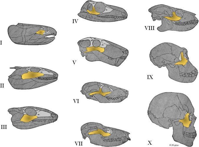
This comparative anatomical diagram illustrates the evolution of the circumorbital bones and skull morphology across ten stages, from lobe-finned fish (I) to modern humans (X). The zygoma (jugal bone) is highlighted in yellow, while the orbital series is depicted in light gray. The series tracks morphological changes through various evolutionary milestones, including primitive amphibians (II), reptiles (III-VI), marsupials (VII), primitive primates (VIII), and anthropoids (IX). Key visual trends include the transition of the zygomatic bone from an elongated, posterior position in fish and early tetrapods to a more robust, compact, and anterior orientation in primates and humans. Concurrently, the skull demonstrates a progression from a flattened, elongated dermatocranium to an expanded, rounded cranial vault with reduced facial prognathism. The diagram highlights the spatial relationship between the zygoma and the orbit, showing how these structures become more integrated and vertically oriented in the facial skeleton over time. This resource serves to teach evolutionary osteology, comparative craniofacial anatomy, and the development of the human skull.
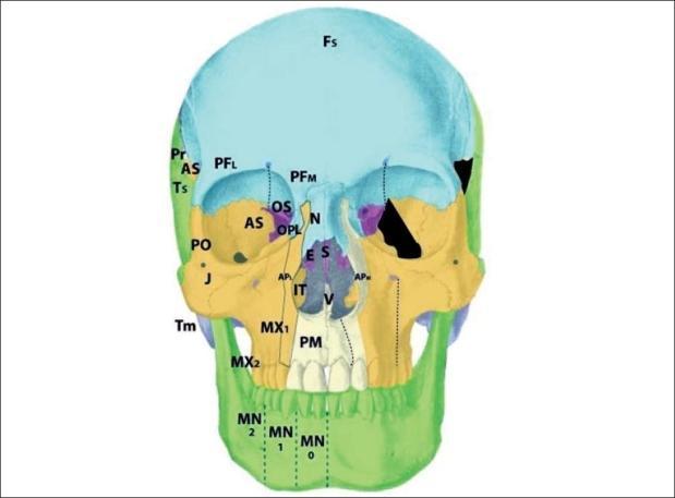
An anatomical diagram of a human skull from an anterior view, color-coded and labeled to demonstrate craniofacial developmental fields and Tessier cleft zones. The diagram highlights the frontal bone (Fs), orbital regions, maxilla (MX), mandible (MN), and nasal structures. Specifically, it illustrates the position of the alisphenoid (AS) or greater wing of the sphenoid in the lateral orbital wall and temporal fossa, bordered by the frontal (Fs), parietal (Pr), squamous temporal (Ts), and zygomatic (J, PO) bones. On the right side of the skull (viewer's right), a black-shaded area indicates a Tessier zone 9 cleft, characterized by a deficiency in the alisphenoid. This defect manifests visually as a significant gap in the lateral orbital wall and sphenofrontal region. Additional labels include the premaxilla (PM), vomer (V), and ethmoid (ES) regions, providing a comprehensive map of the osseous components involved in complex craniofacial clefting patterns.
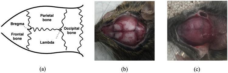
This figure provides a comparative study of mouse cranial anatomy, focusing on critical landmarks for stereotactic neurosurgery. Panel (a) is a schematic diagram illustrating the dorsal view of the skull, identifying the frontal, parietal, and occipital bones separated by sutures. Key reference points are labeled: 'Bregma' (the intersection of coronal and sagittal sutures) and 'Lambda' (the intersection of sagittal and lambdoid sutures). Panel (b) is a clinical photograph of an exposed rodent skull displaying clear visibility of these cranial sutures as dark, irregular lines against the pale bone, with clearly discernible bregma and lambda points. Panel (c) shows a similar surgical exposure where these anatomical landmarks are obscured by soft tissue, vascular markings, or anatomical variation, presenting a challenge for manual stereotactic positioning. The image illustrates the variability in anatomical clarity encountered during neurosurgical procedures in rodent models, emphasizing the educational importance of landmark identification and the potential for computer-guided or deep learning-based localization to improve precision in instrument insertion.
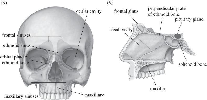
This medical anatomical diagram displays the human skull in two views, focusing on the paranasal sinuses and internal cranial structures. View (a) is an anterior (frontal) perspective of the skull with semi-transparent overlays illustrating the paranasal sinuses. Labeled structures include the frontal sinuses located superior to the orbits, the ethmoid sinus situated medially between the eyes, the orbital plate of the ethmoid bone forming the medial orbital wall, the ocular cavity (orbit), and the large maxillary sinuses within the maxilla. View (b) provides a mid-sagittal section of the facial and mid-cranial region. It highlights the spatial relationship between the nasal cavity and surrounding structures, including the frontal sinus, the perpendicular plate of the ethmoid bone (part of the nasal septum), and the maxilla. Posteriorly, it shows the sphenoid bone containing the sella turcica, where the pituitary gland is seated. This illustration is designed for medical education regarding craniofacial anatomy, specifically for procedures such as Functional Endoscopic Sinus Surgery (FESS) and maxillofacial osteotomies.
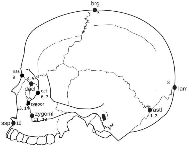
Anatomical diagram of a human skull in a lateral-oblique view, illustrating standard craniofacial landmarks used in forensic anthropology and morphometric analysis. The illustration highlights specific osteological points including: Bregma (brg) at the junction of the coronal and sagittal sutures; Lambda (lam) at the junction of the sagittal and lambdoid sutures; Asterion (astl) where the lambdoid, parietomastoid, and occipitomastoid sutures meet; Nasion (nas) at the intersection of the frontal and nasal bones; and Dacryon (dacl) within the medial orbital wall. Lower facial landmarks include the Ectoconchion (ect) on the lateral orbital margin, Zygoorbitale (zygoor), Zygomaxillare (zygoml) at the zygomatic-maxillary suture, and Subspinale (ssp) at the deepest point of the premaxillary curvature below the anterior nasal spine. Each point is numerically coded for reference to statistical data tables. This content is designed for educational use in anatomy, osteology, and forensic identification.
Total Skull Bones: 22
Part 1 - Cranial Bones (8 bones)
| # | Bone | No. | Notes |
|---|---|---|---|
| 1 | Frontal | 1 | Forehead + roof of orbits |
| 2 | Parietal | 2 | Sides and top of skull (paired) |
| 3 | Temporal | 2 | Sides of skull, houses middle/inner ear (paired) |
| 4 | Occipital | 1 | Back and base of skull; has foramen magnum |
| 5 | Sphenoid | 1 | Central base of skull; "butterfly-shaped" |
| 6 | Ethmoid | 1 | Between nasal cavity and brain; contains cribriform plate |
Part 2 - Facial Bones (14 bones)
| # | Bone | No. | Notes |
|---|---|---|---|
| 1 | Maxilla | 2 | Upper jaw; houses upper teeth (paired) |
| 2 | Zygomatic (Zygoma) | 2 | Cheekbones (paired) |
| 3 | Nasal | 2 | Bridge of the nose (paired) |
| 4 | Lacrimal | 2 | Medial wall of orbit; smallest facial bone (paired) |
| 5 | Palatine | 2 | Form the hard palate and part of nasal cavity floor (paired) |
| 6 | Inferior nasal concha | 2 | Lateral walls of nasal cavity (paired) |
| 7 | Vomer | 1 | Part of the nasal septum |
| 8 | Mandible | 1 | Lower jaw; only movable bone of the skull |
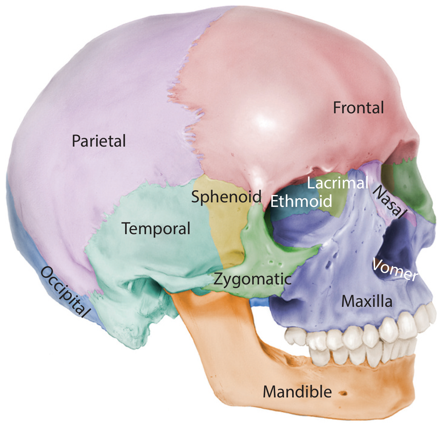
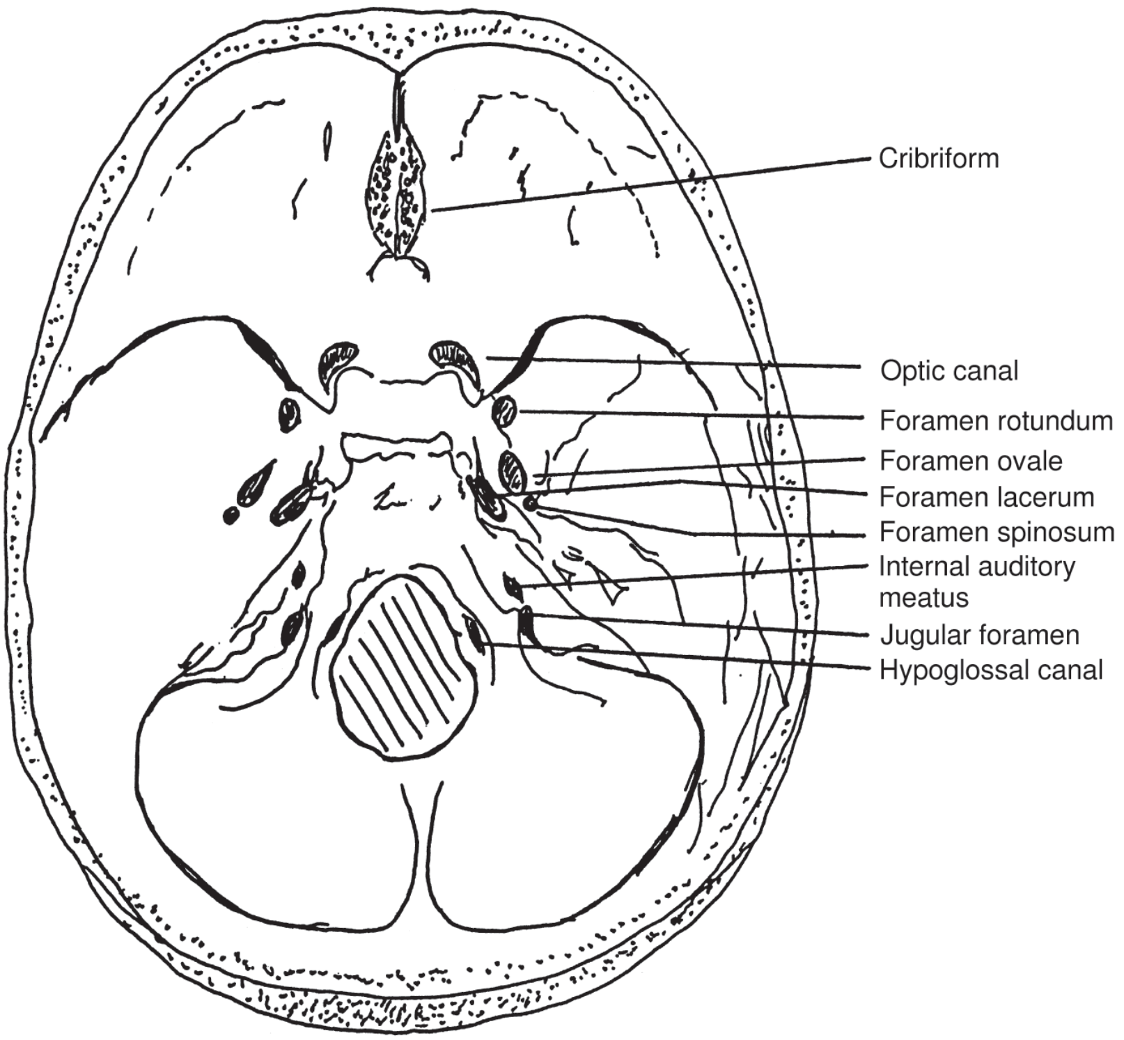
Developmental Classification (from K.J. Lee's Otolaryngology)
- Petrous, Occipital, Ethmoid, Mastoid (part of temporal), Sphenoid
- Frontal, Lacrimal, Nasal, Maxillae, Premaxilla, Palatine, Mandible, Vomer, Sphenoid (partly), Parietal, Zygoma, Tympanic ring, Squamous temporal
Extended Count (28 bones)
- 3 auditory ossicles per ear (malleus, incus, stapes) = 6 total
- Hyoid bone = 1 (though strictly not part of the skull, often grouped here)