
History taking and physical examination: Occupational history techniques. Cultural considerations in patient assessment.
"cultural competency" AND "patient assessment"
CDC taking occupational history work related disease exposure patient assessment cultural humility
https://stacks.cdc.gov/view/cdc/224354
PMID: 41107826
1. Occupational history: techniques in history taking
A. Begin broadly
- “What work do you do now?”
- “Please describe a typical workday.”
- “What jobs have you done in the past?”
- “Has your work changed recently?”
- “Do you think your symptoms relate to, improve away from, or worsen at work?”
B. Take a chronological job history
- Job title and employer/industry
- Dates employed and duration
- Main tasks, materials handled, machinery used
- Work environment, such as indoors/outdoors, confined space, ventilation, heat, noise, dust
- Potential hazards and controls used
- Symptoms or injuries during that period
C. Systematically identify hazards
| Hazard group | Examples | Health concerns |
|---|---|---|
| Dusts and fibres | Silica, asbestos, cotton, flour, wood dust | Asthma, pneumoconiosis, fibrosis |
| Chemicals | Solvents, pesticides, acids, paints, metals | Dermatitis, neurotoxicity, cancer, organ injury |
| Fumes/gases | Welding fumes, smoke, chlorine, carbon monoxide | Airway disease, poisoning |
| Biological agents | Blood, animals, mould, sewage, healthcare waste | Infection, allergy |
| Physical agents | Noise, vibration, radiation, heat/cold | Hearing loss, vibration disease, burns |
| Ergonomic hazards | Repetition, lifting, awkward posture | Back pain, tendinopathy, strain |
| Psychosocial hazards | Long shifts, bullying, violence, high demand | Sleep disturbance, anxiety, burnout |
- “Is there visible dust, smoke, mist, or a chemical smell?”
- “Do you use cleaning agents, paints, solvents, pesticides, welding materials, or metals?”
- “Do you wear gloves, masks, respirators, or other protective equipment?”
- “Is ventilation adequate?”
- “Do other workers have similar symptoms?”
D. Establish the exposure-symptom relationship
- When did symptoms begin in relation to this job or a new task?
- Are symptoms worse during a shift?
- Do they improve on weekends, holidays, or sick leave?
- Did symptoms begin after an accident, spill, fire, renovation, or new product/process?
- Are symptoms related to dose, task, or particular location in the workplace?
E. Include non-paid work and social context
- Domestic work and caregiving
- Informal, temporary, seasonal, migrant, or self-employed work
- Military service
- Hobbies, such as painting, pottery, woodworking, gardening, vehicle repair
- Second jobs and prior jobs with long-latency exposures
- Financial impact of illness, compensation concerns, and return-to-work needs
F. Communication principles
- Explain why you are asking: “Some jobs can affect health, so I ask everyone about their work.”
- Avoid blaming the patient or employer.
- Use simple, nontechnical words.
- Respect confidentiality, especially where employment, immigration status, or compensation is a concern.
- Document concrete facts: job, task, agent, duration, intensity, controls, and symptom timing.
2. Cultural considerations in patient assessment
A. Start with cultural humility, not assumptions
- “What do you think is causing this problem?”
- “What name do you use for this condition?”
- “How serious do you think it is?”
- “What treatment do you think would help?”
- “Are there cultural, spiritual, or family practices that you would like us to consider?”
B. Language and communication
- Determine the patient’s preferred spoken and written language.
- Use a trained professional interpreter when language proficiency is limited.
- Do not use children, relatives, or untrained staff as interpreters for sensitive or complex discussions.
- Speak directly to the patient, not to the interpreter.
- Use short sentences and plain language.
- Confirm understanding with teach-back: “To make sure I explained it clearly, can you tell me how you will take this medicine?”
C. Consent, privacy, and modesty during physical examination
- Explain what will happen and why.
- Obtain consent.
- Offer a chaperone, especially for intimate examination.
- Offer a clinician of the preferred gender when feasible.
- Provide adequate draping and expose only the body part being examined.
- Ask before touching the patient, including hair, head, or religious garments.
D. Family, decision-making, and social roles
- “Who would you like involved in discussions about your care?”
- “Is there anyone you would like us to contact?”
- “Would you prefer time to discuss this with your family?”
E. Illness beliefs and use of traditional healing
- “Are you using any home, herbal, traditional, or spiritual remedies?”
- “What treatments have helped you in the past?”
F. Avoid diagnostic bias
- How pain or distress is expressed
- Whether mental health symptoms are described as physical symptoms
- Health-seeking behavior
- Trust in healthcare systems
- Adherence to treatment
- Interpretation of disability or illness
G. Consider structural and social factors
- Racism, discrimination, stigma, and prior healthcare trauma
- Migration, displacement, legal or immigration concerns
- Health literacy and digital access
- Poverty, housing, food security, transport, and employment conditions
- Access to insurance, medicines, and follow-up care
H. A practical cultural assessment framework
- Cultural identity: language, community, religion, migration background
- Explanatory model: what the illness means to the patient
- Psychosocial environment: family, work, stressors, supports
- Patient-clinician relationship: trust, communication preferences, prior negative experiences
- Treatment preferences and barriers: traditional care, affordability, transport, gender/privacy needs
Key point
Respiratory assessment: Chest X-ray interpretation (pneumoconioses). Spirometry in occupational lung disease.
Chest radiograph silicosis upper lobe small rounded opacities hilar eggshell calcification pneumoconiosis
Chest radiograph asbestosis pleural plaques lower lobe fibrosis pneumoconiosis
"occupational lung disease" AND spirometry
ILO Classification Radiographs of Pneumoconioses spirometry occupational lung disease official guidance
https://www.ilo.org/resource/other/ilo-international-classif…
Respiratory assessment in occupational lung disease
- Exposure and symptom history
- Respiratory examination
- Chest radiograph, interpreted systematically
- Spirometry and, where indicated, lung volumes and DLCO
- HRCT or specialist testing when findings are uncertain or discordant
1. Chest X-ray interpretation in pneumoconioses
A. Basic approach to the chest X-ray
Stepwise reading
-
Check technical quality
- Correct patient and date
- PA versus AP projection
- Adequate inspiration, penetration, rotation, and absence of artifacts
-
Review the lungs
- Are opacities present?
- Are they rounded or irregular?
- What is their size, profusion, and zonal distribution?
- Is there coalescence into large masses?
-
Review pleura
- Pleural plaques
- Diffuse pleural thickening
- Pleural effusion
- Costophrenic-angle blunting
-
Review hila and mediastinum
- Lymph-node enlargement
- Hilar/mediastinal calcification, including eggshell calcification
-
Look for complications
- Progressive massive fibrosis
- Tuberculosis
- Lung cancer
- Emphysema
- Cor pulmonale or pulmonary hypertension
B. ILO classification
Main features coded
- Small opacities
- Shape and size
- Profusion
- Distribution by lung zone
- Large opacities
- Categories A, B, or C
- Pleural abnormalities
- Plaques
- Diffuse pleural thickening
- Costophrenic angle obliteration
- Other features, such as hilar-node calcification and emphysema
Small-opacity types
| Opacity | ILO type | Approximate size |
|---|---|---|
| Rounded | p | Up to 1.5 mm |
| Rounded | q | >1.5 to 3 mm |
| Rounded | r | >3 to 10 mm |
| Irregular | s | Up to 1.5 mm |
| Irregular | t | >1.5 to 3 mm |
| Irregular | u | >3 to 10 mm |
Important limitation: a radiograph may not reflect physiologic impairment precisely. Extensive rounded opacities may coexist with minimal functional impairment, while linear or irregular opacities in asbestosis can underestimate impairment until later disease. Harrison’s Principles of Internal Medicine, 22nd ed., p. 2278.
C. Characteristic X-ray patterns
1. Silicosis
- Bilateral, upper-zone predominant small rounded nodules
- Hilar and mediastinal lymph-node enlargement
- Peripheral lymph-node calcification, called eggshell calcification
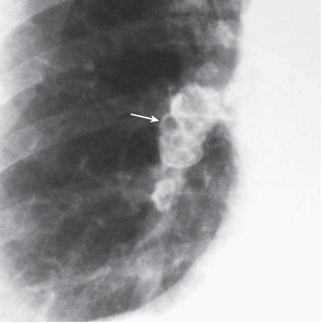
- In advanced disease, coalescent upper-lobe masses due to progressive massive fibrosis
- Progressive massive fibrosis
- Tuberculosis and nontuberculous mycobacterial disease
- COPD
- Lung cancer
- Autoimmune disease, including rheumatoid arthritis
2. Coal workers’ pneumoconiosis
- Small rounded opacities, often upper-zone predominant
- Nodules tend to be smaller than those in silicosis
- May progress to large upper-lobe masses in progressive massive fibrosis
3. Asbestosis
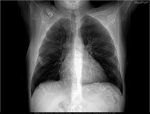
- Bilateral, basal and peripheral linear, reticular, or irregular opacities
- Reduced lung volume in advanced fibrosis
- Pleural plaques, often calcified
- Diffuse pleural thickening
- Costophrenic-angle blunting
4. Beryllium disease
- May resemble sarcoidosis
- Bilateral hilar adenopathy
- Diffuse nodular or reticulonodular infiltrates
- Fibrosis in chronic disease
5. Talcosis, siderosis, and other mineral-dust diseases
| Condition | Typical X-ray pattern |
|---|---|
| Talcosis | Diffuse small nodules, which may coalesce into fibrosis |
| Siderosis | Fine diffuse nodular shadowing, often with relatively minor symptoms |
| Hard-metal disease | Diffuse interstitial or fibrotic pattern |
| Chronic hypersensitivity pneumonitis | Diffuse interstitial changes, often better demonstrated on HRCT |
D. When to request HRCT
- Early interstitial fibrosis
- Small nodules
- Pleural plaques and pleural thickening
- Subpleural reticulation
- Emphysema
- Progressive massive fibrosis
- Alternative diagnoses, such as infection, cancer, or idiopathic pulmonary fibrosis
2. Spirometry in occupational lung disease
A. Purpose
- Obstructive disease: occupational asthma, work-exacerbated asthma, COPD, bronchiolitis
- Possible restrictive disease: pneumoconiosis, asbestosis, other interstitial lung diseases
- Change in lung function during or after workplace exposure
B. Main spirometric variables
| Measure | Meaning |
|---|---|
| FEV1 | Forced expiratory volume exhaled in the first second |
| FVC | Forced vital capacity |
| FEV1/FVC | Main ratio used to identify airflow obstruction |
| Flow-volume loop | Supports recognition of obstruction, restriction, or upper-airway problems |
C. Spirometric patterns
1. Obstructive pattern
- Reduced FEV1
- Reduced FEV1/FVC ratio
- FVC may be normal or reduced due to air trapping
- Occupational asthma
- Work-exacerbated asthma
- Chronic occupational COPD from mineral dust, fumes, vapors, smoke, or irritants
- Obliterative bronchiolitis
2. Restrictive pattern
- Reduced FVC
- Normal or raised FEV1/FVC ratio
- Asbestosis
- Silicosis with significant fibrosis
- Coal workers’ pneumoconiosis with fibrosis
- Chronic hypersensitivity pneumonitis
- Other occupational interstitial lung diseases
- Reduced TLC
- Reduced DLCO, especially in interstitial fibrosis
3. Mixed obstructive-restrictive pattern
- Reduced FEV1
- Reduced FVC
- Reduced FEV1/FVC ratio
- Confirm restriction with TLC
- Pneumoconiosis plus smoking-related COPD
- Advanced coal workers’ pneumoconiosis
- Asbestosis with coexisting COPD
- Dust-related fibrosis plus small-airway disease
D. Bronchodilator reversibility testing
E. Serial testing in suspected occupational asthma
Pre- and post-shift FEV1
- Before the shift
- After the shift
- On several workdays
- Compare with days away from exposure
Serial peak expiratory flow (PEF)
- Several times daily
- During work periods
- During rest days and holidays
- Over several weeks
F. Limitations of spirometry
- Normal spirometry does not exclude early pneumoconiosis.
- Normal spirometry away from work does not exclude occupational asthma.
- Reduced FVC does not confirm restriction without TLC.
- Spirometry does not assess gas transfer, so request DLCO when interstitial disease is suspected.
- Poor technique, smoking, obesity, heart failure, infection, and pre-existing asthma/COPD can confound interpretation.
- A single test should not be used alone for employment, compensation, or causation decisions.
Practical summary
| Clinical suspicion | Chest X-ray pattern | Spirometry/PFT pattern | Useful next step |
|---|---|---|---|
| Silicosis | Upper-lobe rounded nodules, eggshell nodes | Often normal early; restriction in advanced fibrosis | HRCT, TB assessment |
| Coal workers’ pneumoconiosis | Upper-zone small rounded opacities, possible PMF | Normal, obstructive, restrictive, or mixed | HRCT, full PFTs |
| Asbestosis | Basal linear/reticular fibrosis, pleural plaques | Restriction with reduced DLCO | HRCT, full PFTs |
| Occupational asthma | May be normal | Variable obstruction, possible reversibility | Pre/post-shift FEV1, serial PEF |
| Occupational COPD | Often hyperinflation or emphysema | Persistent obstruction | Post-bronchodilator spirometry, exposure assessment |