
Muscle physiology
Reading File
Muscle physiology is a broad topic. Let me clarify what level of detail you need before diving in.
Asking for Preferences
Which aspect of muscle physiology are you interested in? · What level of detail do you need?
Finding Sources
Finding Sources
Reading File
Reading File
Reading File
Reading File
Reading File
Loading Image
Here is a concise overview of muscle physiology covering all three muscle types, drawn from Guyton & Hall, Costanzo, and Miller's Orthopaedics.
Muscle Physiology - Overview
The Three Muscle Types
| Feature | Skeletal | Cardiac | Smooth |
|---|---|---|---|
| Striated? | Yes | Yes | No |
| Voluntary? | Yes | No | No |
| Location | Limbs, trunk, face | Heart walls | Viscera, vessels, airways |
| Control | Motor neuron (ACh) | Intrinsic pacemaker + ANS | ANS, hormones, stretch |
| Speed of contraction | Fast | Intermediate | Slow (1-3 sec total) |
1. Skeletal Muscle
Structure
- Organized into motor units: one alpha-motor neuron + all muscle fibers it innervates
- Small/precise muscles (e.g., extraocular) have <5 fibers/unit; large muscles (e.g., gastrocnemius) have up to 1800 fibers/unit
- Fibers contain actin (thin) and myosin (thick) filaments arranged in sarcomeres
Neuromuscular Junction (NMJ)
- Action potential reaches the motor axon terminal
- Acetylcholine (ACh) is released from presynaptic vesicles into the synaptic cleft (~50 nm wide)
- ACh binds to nicotinic receptors on the sarcolemma → depolarization → muscle action potential
Excitation-Contraction (E-C) Coupling
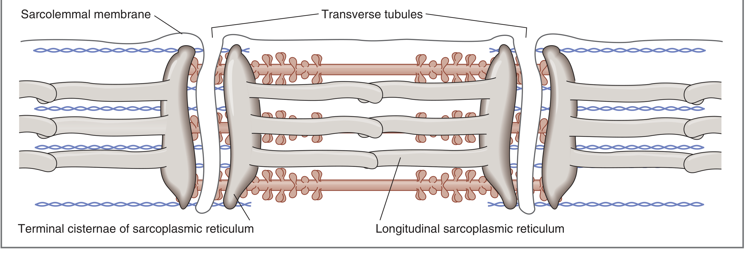
T-tubules (continuous with the sarcolemma) carry depolarization deep into the fiber to reach the SR - Costanzo Physiology, 7th Ed.
Step by step:
- Action potential propagates along the sarcolemma and into T-tubules
- T-tubule depolarization triggers a conformational change in dihydropyridine receptors (DHPR) - voltage sensors
- DHPR mechanically opens ryanodine receptors on the sarcoplasmic reticulum (SR) → Ca²⁺ floods the cytoplasm (from <10⁻⁷ M to 10⁻⁶ M)
- Ca²⁺ binds troponin C on the thin filament (up to 4 ions/molecule, cooperative binding)
- Troponin conformational change moves tropomyosin off the myosin-binding sites on actin
- Myosin heads bind actin → ATP hydrolysis → power stroke (actin slides toward the center of the sarcomere)
- Relaxation: SR Ca²⁺-ATPase pumps Ca²⁺ back into the SR; tropomyosin re-blocks actin
Fiber Types
| Type | Name | Speed | Fatigability | Use |
|---|---|---|---|---|
| Type I | Slow-twitch oxidative | Slow | Fatigue-resistant | Posture, endurance |
| Type IIa | Fast-twitch oxidative | Fast | Moderate | Mixed activities |
| Type IIx/IIb | Fast-twitch glycolytic | Fastest | Fatigues quickly | Short bursts, power |
Types of Contraction
| Type | Muscle length | Tension | Example |
|---|---|---|---|
| Isometric | Unchanged | Generated | Pushing a wall |
| Isotonic concentric | Shortens | Constant | Lifting phase of curl |
| Isotonic eccentric | Lengthens | Generated | Lowering phase of curl |
| Isokinetic | Changes at constant velocity | Maximal throughout ROM | Cybex machine |
Eccentric contractions produce the greatest tension and carry the highest injury risk.
Key Clinical Points
- Myasthenia gravis: IgG antibodies against ACh receptors → fatigable weakness, ptosis, diplopia
- Botulinum toxin: blocks presynaptic ACh release (used for spasticity in cerebral palsy)
- Succinylcholine: depolarizing neuromuscular blocker (short-acting)
- Curare/vecuronium: non-depolarizing blockers, reversed by anticholinesterases (neostigmine)
2. Cardiac Muscle
- Striated, like skeletal muscle, with the same actin/myosin sliding filament mechanism
- Fibers are arranged as a functional syncytium - cells connected by intercalated discs containing gap junctions that allow rapid ion diffusion and electrical coupling
- Three types: atrial, ventricular, and specialized conductive fibers (SA/AV node, Purkinje)
- Left ventricular fibers are arranged in a double helix, producing a twisting (torsion) motion during systole - the apex rotates counterclockwise while the base rotates clockwise
- Key difference from skeletal muscle: Ca²⁺ entry from outside the cell (via L-type channels) is required to trigger SR Ca²⁺ release (calcium-induced calcium release, CICR) - this does NOT occur in skeletal muscle
- Action potential has a prolonged plateau phase (phase 2) due to L-type Ca²⁺ channel inflow, which extends the refractory period and prevents tetany
3. Smooth Muscle
- No sarcomeres; no troponin - instead uses calmodulin to regulate contraction
- Contraction mechanism: Ca²⁺ → binds calmodulin → activates myosin light-chain kinase (MLCK) → phosphorylates myosin → cross-bridge cycling
- Slow cross-bridge cycling: 1/10 to 1/300 the rate of skeletal muscle
- Despite this, maximum force can be 4-6 kg/cm² (greater than skeletal muscle's 3-4 kg/cm²) due to prolonged cross-bridge attachment time
- Energy efficient: requires only 1/10 to 1/300 the energy to maintain the same tension
- Latch mechanism: smooth muscle can maintain sustained tonic contraction for hours at minimal energy cost by keeping cross-bridges "locked" in the attached state
- Total contraction time is ~30x longer than a single skeletal muscle twitch (1-3 seconds vs. ~50-100 ms)
- Triggered by: stretch, hormones (e.g., oxytocin, norepinephrine), autonomic nerves, local metabolites
Quick Comparison Summary
| Property | Skeletal | Cardiac | Smooth |
|---|---|---|---|
| Ca²⁺ sensor | Troponin C | Troponin C | Calmodulin |
| Ca²⁺ source | SR only | SR + extracellular | SR + extracellular |
| Tetany possible? | Yes | No (long refractory period) | No (latch mechanism) |
| Energy use | High | Moderate | Very low |
| Contraction speed | Fast | Moderate | Slow |
Sources: Guyton and Hall Textbook of Medical Physiology; Costanzo Physiology 7th Ed.; Miller's Review of Orthopaedics 9th Ed.
This is a shared conversation. Sign in to Orris to start your own chat.