
Av malformation after dnc
Reading File
An arteriovenous malformation (AVM) after a D&C (dilation and curettage) is known as a uterine AVM - an acquired vascular anomaly where abnormal connections form between uterine arteries and veins. This is a recognized complication of uterine instrumentation and can cause life-threatening hemorrhage.
Searching sources...
Finding Sources
Searching PubMed
uterine arteriovenous malformation dilation curettage
Reading File
Finding Sources
Searching PubMed
uterine arteriovenous malformation diagnosis management embolization
Loading Image
Finding Sources
Reading Article
PMID: 34130801
Here is a thorough summary of uterine arteriovenous malformation (AVM) after D&C:
Uterine AVM After D&C
What It Is
A uterine AVM is an abnormal direct communication between uterine arteries and veins, bypassing the normal capillary bed. It can be congenital (rare) or acquired - the acquired form is far more common and is closely linked to uterine instrumentation, including:
- Dilation and curettage (D&C)
- Suction evacuation
- Caesarean section
- Endometrial biopsy
- Uterine surgery
The D&C-related form is thought to arise from trophoblastic invasion or traumatic disruption of the myometrium, creating pathological arteriovenous channels. It often occurs after evacuation of products of conception, molar pregnancy, or termination of pregnancy.
Presentation
- Recurrent, sudden, heavy vaginal bleeding - the hallmark symptom - often unprovoked and profuse
- Bleeding typically occurs weeks to months after the D&C
- May follow a period of apparently normal recovery
- Can be life-threatening if a large AVM ruptures
- Importantly, if bleeding persists after D&C and beta-hCG has normalized, AVM should be suspected (rather than retained products of conception)
Diagnosis
First-line: Transvaginal Ultrasound with Color Doppler
The classic finding is a hypoechoic or heterogeneous area within the myometrium adjacent to the endometrium, with:
- Turbulent, high-velocity, low-resistance flow on color Doppler
- Aliasing (color mosaic pattern) - a hallmark sign
- Dilated feeding vessels
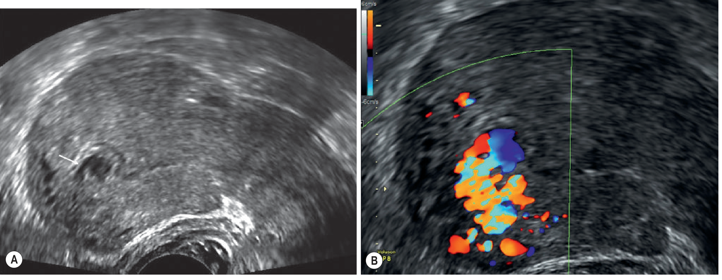
Image: Transvaginal US - (A) focal hypoechoic area; (B) color Doppler aliasing with feeding vessel - classic uterine AVM appearance. Patient had previous surgical removal of retained placenta and presented with recurrent heavy bleeds. (Grainger & Allison's Diagnostic Radiology)
CT Angiography / MRI
- Arterial phase CT shows a hypervascular lesion with early venous opacification
- MRI shows tortuous vessels protruding into the endometrium, with signal voids on T1/T2 weighted sequences and enhancement with contrast
- Selective angiography: shows early venous return (pathognomonic) and can be used therapeutically
Key distinction from retained products of conception (RPOC):
Both can look hypervascular on Doppler, but:
- RPOC typically resolves after b-hCG normalizes
- AVM persists even after b-hCG normalization
- Clinical context (ongoing bleeding, post-D&C) + imaging together guide the diagnosis
(Grainger & Allison's Diagnostic Radiology, p. 917-918)
Management
1. Uterine Artery Embolization (UAE) - First-line for active/severe bleeding
- Selective angiographic embolization is the standard definitive treatment
- The lesion is identified on angiography by early venous return, then embolized with coils or particles
- Preserves fertility in most cases
- High success rate for stopping acute hemorrhage
2. Medical Management - For stable patients or fertility preservation
A 2021 systematic review and meta-analysis (Rosen et al., Fertility and Sterility) found an overall success rate of 88% with medical treatment in 121 women across 32 studies. Agents used:
| Drug | Success Rate | Complication Rate |
|---|---|---|
| Progestins | 82.5% | 10.0% (lowest) |
| GnRH agonists | 89.3% | 10.7% |
| Methotrexate | 90.0% | - |
| Combined hormonal contraception | Included | - |
- No clinical factors reliably predicted success
- 26 subsequent pregnancies reported with no AVM recurrence
- Medical management is reasonable in hemodynamically stable, well-selected patients
3. Surgical Management
- Reserved for cases failing embolization or medical management
- Options: hysteroscopic resection (small AVMs), uterine artery ligation, or as a last resort - hysterectomy
- Blind curettage of a suspected AVM is contraindicated as it can precipitate catastrophic hemorrhage
Key Points Summary
| Feature | Detail |
|---|---|
| Cause after D&C | Traumatic AV fistula formation in myometrium |
| Symptom | Sudden, recurrent, heavy, unprovoked vaginal bleeding |
| Diagnostic clue | Persists after b-hCG normalizes (unlike RPOC) |
| Best first test | Transvaginal USS + color Doppler |
| Gold standard | Selective angiography |
| Treatment of choice | UAE (embolization) |
| Contraindicated | Blind curettage |
| Fertility outcomes | Generally good after UAE or medical management |
Recent evidence note: The Rosen et al. 2021 systematic review (PMID 34130801) supports medical management as a valid option alongside UAE in stable patients. No contradictions to the textbook approach were found in recent literature.
Microadenoma pitutary 3.6mm× 2.8mm
A pituitary microadenoma is defined as a pituitary adenoma less than 10mm in diameter. At 3.6mm x 2.8mm, this is a small microadenoma. The key question is whether it is functioning (hormone-secreting) or non-functioning, as management differs significantly.
Searching sources...
Finding Sources
Searching PubMed
pituitary microadenoma management treatment
Finding Sources
Reading Article
PMID: 37097352
Reading File
Reading File
Finding Sources
Reading File
Reading File
Reading File
Loading Image
Here is a complete clinical overview of a pituitary microadenoma (3.6mm x 2.8mm):
Pituitary Microadenoma - 3.6mm x 2.8mm
Definition & Classification
- A microadenoma is a pituitary adenoma < 10mm in diameter
- Your lesion at 3.6 x 2.8mm is a small microadenoma, well within this category
- Pituitary adenomas make up 10-15% of all intracranial neoplasms
- They are either functioning (hormone-secreting) or non-functioning
Imaging Appearance
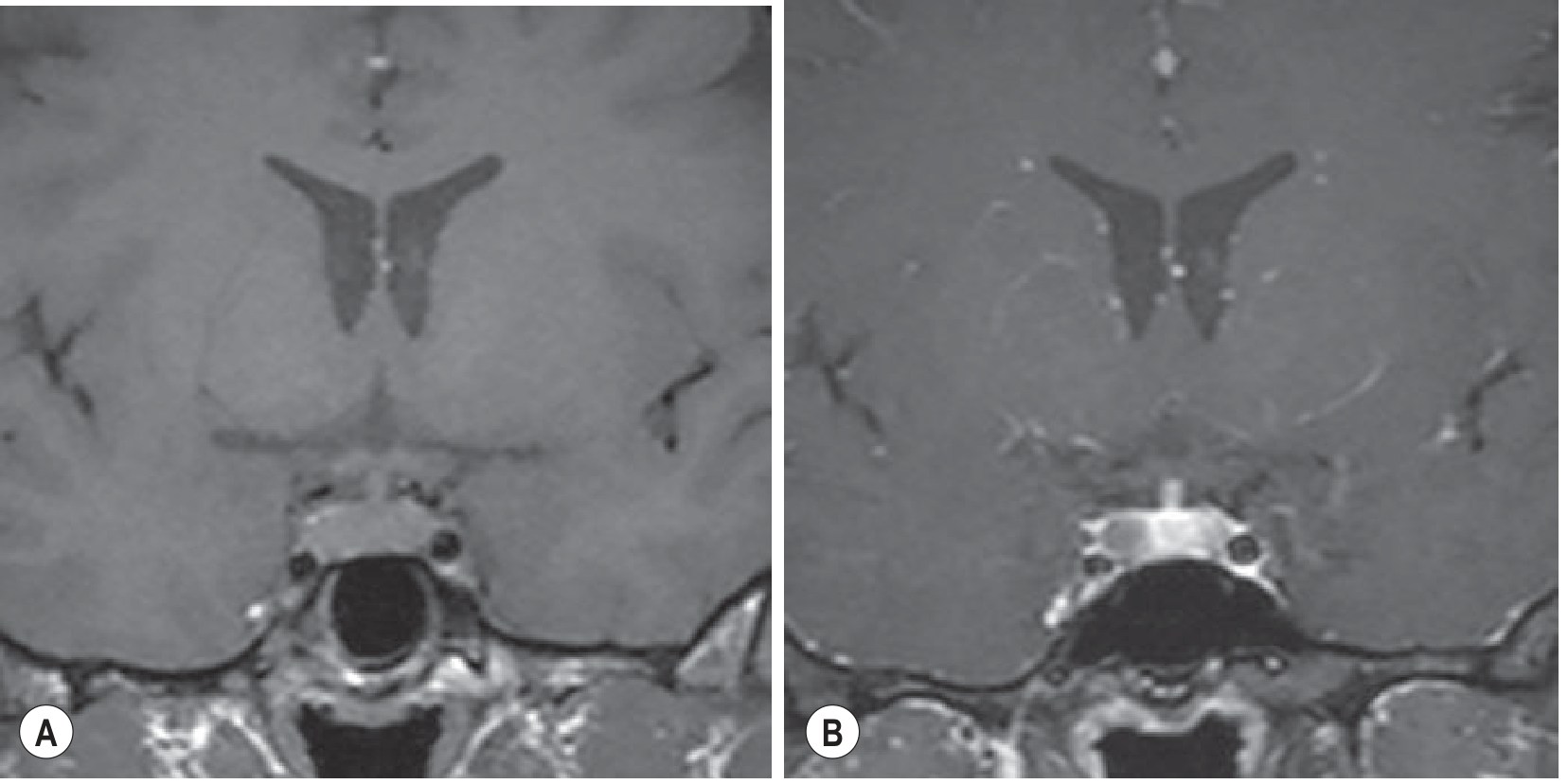
MRI of a pituitary microadenoma. Pre-contrast (A): asymmetric enlargement of anterior pituitary. Post-contrast (B): microadenoma abutting cavernous sinus, enhancing less than normal pituitary tissue - the key radiological sign. (Grainger & Allison's Diagnostic Radiology)
MRI is the investigation of choice. Microadenomas characteristically:
- Enhance later and/or less than normal pituitary tissue on dynamic contrast MRI
- Show as a hypointense/filling defect within the brightly enhancing gland
- Thin 3mm coronal and sagittal slices with gadolinium are standard
- Dynamic pituitary MRI (rapid sequential imaging every 10-15s post-contrast) increases sensitivity
Functional Classification - The Critical Step
All pituitary microadenomas require a full hormonal workup to determine type:
| Type | Hormone | Prevalence | Clinical Features |
|---|---|---|---|
| Prolactinoma | Prolactin (PRL) | ~53% of all adenomas | Amenorrhea, galactorrhea, infertility, loss of libido |
| Corticotropinoma | ACTH | ~4% | Cushing's disease - weight gain, striae, hypertension, buffalo hump |
| Somatotropinoma | GH | ~12% | Acromegaly (adults), gigantism (children) |
| Thyrotropinoma | TSH | Rare | Hyperthyroidism |
| Non-functioning | None | ~30% | Often incidental finding |
Baseline hormone panel to order:
- Serum prolactin (PRL)
- IGF-1 (screens for GH excess)
- ACTH + morning cortisol (± 24h urine cortisol, late night salivary cortisol)
- TSH + free T4
- LH, FSH, testosterone (men) / estradiol (women)
Management by Type
Prolactinoma (most likely for this size)
From Harrison's Principles of Internal Medicine 22E:
"Because microadenomas rarely progress to become macroadenomas, no treatment may be needed if patients are asymptomatic and fertility is not desired; these patients should be monitored by regular serial PRL measurements and MRI scans."
When treatment IS needed:
- First-line: Dopamine agonists - either cabergoline or bromocriptine
- Cabergoline (0.5-1.0 mg twice weekly): achieves normoprolactinemia in ~80% of microadenoma patients; improves/resolves galactorrhea in 90%. Preferred due to once/twice-weekly dosing and fewer side effects
- Bromocriptine: shorter-acting; preferred when pregnancy is desired (more safety data in pregnancy)
- After 2 years of normoprolactinemia, dopamine agonist may be carefully withdrawn with monitoring
- ~5% achieve permanent resolution after long-term treatment
Corticotropinoma (Cushing's disease)
- First-line: Transsphenoidal surgery - success rate >80% for microadenomas
- If microadenoma not found at surgery: hemihypophysectomy
- Requires precise lateralization pre-operatively - inferior petrosal sinus sampling if MRI inconclusive
Somatotropinoma (Acromegaly)
- First-line: Transsphenoidal surgery
- Adjuncts: somatostatin analogues (octreotide, lanreotide), pegvisomant, cabergoline
Non-functioning Microadenoma (Incidentaloma)
- No immediate treatment if asymptomatic and hormones are normal
- Serial MRI: repeat at 6 months, then annually for 3 years, then every 2 years
- Treat only if growth, new symptoms, or visual compromise develops
Prognosis & Natural History
- Only about 5% of microadenomas progress to macroadenomas over the long term
- PRL levels typically remain stable, reflecting slow tumor growth
- A 3.6mm lesion has a very low risk of mass effect on optic chiasm (which requires >10mm suprasellar extension)
- No hypopituitarism expected at this size
- Fertility outcomes are generally good with appropriate treatment
Key Points for This Size (3.6 x 2.8mm)
- No mass effect expected at this size - no visual field testing needed unless symptoms arise
- The most important immediate step is hormonal evaluation to determine if it is functioning
- If prolactinoma with symptoms - start cabergoline
- If asymptomatic and non-functioning - observe with serial MRI
- Surgery (transsphenoidal) is first-line for ACTH/GH-secreting microadenomas, and for prolactinomas resistant to or intolerant of dopamine agonists
(Harrison's Principles of Internal Medicine 22E, p. 3049-3050; Grainger & Allison's Diagnostic Radiology, p. 1424-1425; Tritos & Miller, JAMA 2023)
This is a shared conversation. Sign in to Orris to start your own chat.