
Human body planes
human body anatomical planes sagittal coronal transverse diagram
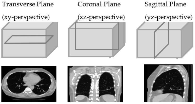
This diagnostic imaging illustration demonstrates the three primary anatomical planes of the human body using Computed Tomography (CT) of the chest. The top row features 3D diagrams of a rectangular volume representing the chest, with an intersecting plane highlighting the orientation for each perspective: Transverse (XY), Coronal (XZ), and Sagittal (YZ). Below these diagrams are corresponding 2D CT slices. The Transverse plane shows a horizontal cross-section of the thorax, displaying the bilateral lungs (dark areas), the central mediastinum including the heart, and the vertebral body. A focal cavitary lesion or nodule is visible in the left lung. The Coronal plane provides a frontal view, illustrating the vertical extent of the lungs, the diaphragm, and the spinal column. The Sagittal plane presents a lateral profile, showing the anterior-posterior depth of the lung, the rib cage, and the curvature of the diaphragm. These views are utilized for volumetric analysis and 3D reconstruction in clinical radiology, particularly for characterizing pulmonary pathology.
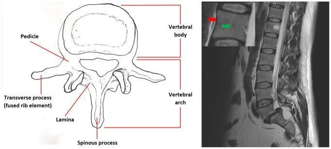
This educational composite compares anatomical structures of the human vertebrae through an anatomical diagram and magnetic resonance imaging. On the left, a coronal anatomical diagram labels the primary components of a single vertebra, including the vertebral body, vertebral arch, pedicle, lamina, spinous process, and transverse process (fused rib element). On the right, a sagittal T2-weighted MRI slice demonstrates the lumbar and lower thoracic spinal column. The MRI shows a sequential stacked configuration of rectangular vertebral bodies separated by intervertebral discs, exhibiting normal spinal curvature. An inset enlargement of one vertebral body uses color-coded arrows to highlight signal intensity variations: a red arrow indicates the cortical bone, which appears as a low-signal (dark) outer boundary, while a green arrow points to the cancellous bone/medullary space inside the vertebral body, characterized by a higher-signal (brighter) grayscale intensity. Posterior to the vertebral bodies, the high-signal cerebrospinal fluid within the spinal canal is clearly visible. This material is designed for medical students to understand spinal anatomy and T2-weighted radiological findings.
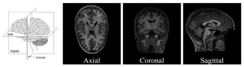
This educational graphic illustrates the primary anatomical planes used in neuroimaging, specifically structural MRI (sMRI). The figure consists of an anatomical diagram followed by three representative diagnostic images. The initial diagram demonstrates the three-dimensional orientation of the axial (transverse), coronal (frontal), and sagittal (longitudinal) planes relative to a human brain model. The subsequent panels present T1-weighted sMRI slices corresponding to these orientations: 1) Axial plane, showing a superior-to-inferior view with visible ventricles and symmetrical cerebral hemispheres; 2) Coronal plane, providing a front-to-back perspective highlighting the brain's vertical structures and cortical thickness; and 3) Sagittal plane, offering a lateral view of the midline structures including the brainstem, cerebellum, and corpus callosum. The educational focus is on standard radiological orientation and spatial classification of neuroanatomical data, essential for diagnostic workflows and computational neuroanalysis, such as Autism Spectrum Disorder (ASD) research.
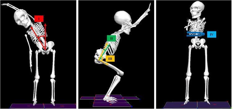
This medical anatomical diagram illustrates the three-dimensional biomechanical analysis of joint angles using a human skeletal model, relevant for clinical motion analysis and physical assessment. The image is divided into three panels representing different anatomical planes. Panel I (left) shows the Coronal Plane, demonstrating the Thorax-with-respect-to-Pelvis (TWRP) bend angle, indicated by red arrows measuring lateral flexion from the pelvic midline. Panel II and III (middle) illustrate the Sagittal Plane during a squatting motion; green arrows define the TWRP flexion/extension angle (II), while yellow arrows define the hip flexion/extension angle (III) relative to the femur and pelvis. Panel IV (right) shows the Transverse Plane, with a blue circular arrow indicating TWRP rotation around the vertical axis. These visual representations are used in biomechanical studies to quantify range of motion (maxROM) and movement symmetry during standardized tests like the Biomechanical Assessment and Symmetry Evaluation (BASE). The diagram serves as an educational tool for understanding segmental orientation and musculoskeletal kinematics in pain management and physical rehabilitation contexts.
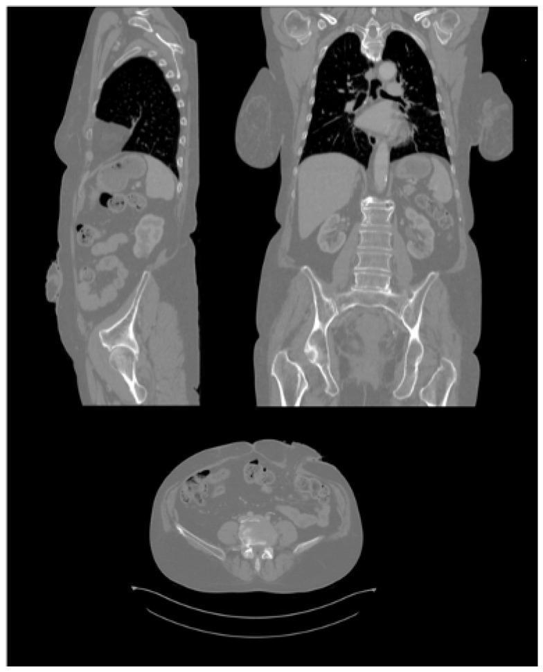
This diagnostic image displays a multiplanar reconstruction of a human abdominal Computed Tomography (CT) scan, presented in sagittal (top left), coronal (top right), and axial (bottom) planes. The sagittal view provides a side perspective of the torso, showing the thoracic cavity with air-filled (hypodense) lungs, the diaphragmatic boundary, abdominal viscera including the liver and intestinal loops, and the spinal column. The coronal view offers a frontal orientation, highlighting the bilateral symmetry of the lungs, the heart in the mediastinum, the liver, kidneys, and the pelvic girdle. The axial view displays a transverse cross-section through the lower abdomen, prominently showing the vertebral body, surrounding musculature, subcutaneous adipose tissue, and intestinal segments. Of particular clinical note is the presence of an ostomy site on the anterior abdominal wall, visible as a protrusion in both the sagittal and axial planes. These images serve as primary anatomical data for surgical planning, specifically for designing personalized ostomy patches adapted to the patient's unique abdominal contours.
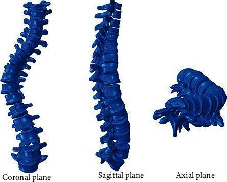
This medical illustration presents a three-dimensional anatomical diagram of the human spine demonstrating scoliosis across three standard orthogonal planes: coronal, sagittal, and axial. In the coronal plane, the spine shows a significant lateral 'S-shaped' curvature, deviating from the normal vertical midline. The sagittal plane view displays the anterior-posterior profile, illustrating alterations in the natural thoracic kyphosis and lumbar lordosis associated with the deformity. The axial plane view highlights the critical rotational component of scoliosis, where vertebral bodies and spinous processes are twisted relative to the longitudinal axis. This visualization serves as an educational tool for orthopedic and radiological study, emphasizing that scoliosis is a complex 3D deformity involving lateral displacement, rotation, and sagittal plane changes. The blue-colored 3D model facilitates the identification of vertebral alignment and the morphological changes used to calculate the Cobb angle for determining clinical severity.
Human Body Planes
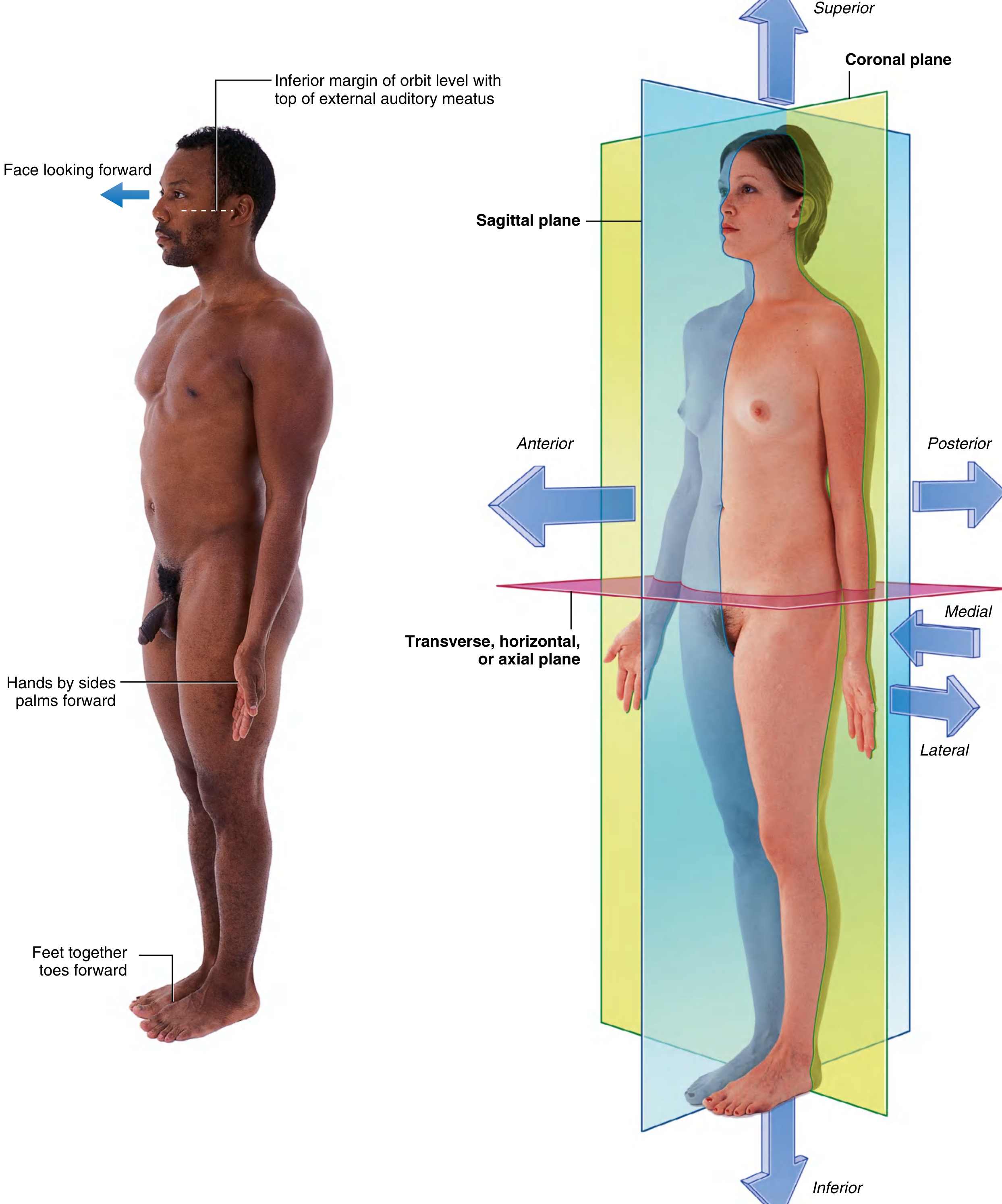
1. Sagittal Plane (Median Plane)
- Orientation: Vertical, running front to back (anterior to posterior)
- Divides the body into: Right and left parts
- Median sagittal plane (midsagittal): The specific sagittal plane that passes exactly through the center of the body, producing equal right and left halves
- Parasagittal planes: Any sagittal plane parallel to but offset from the median plane
- Named after: The sagittal suture of the skull, which runs in this direction
"Any vertical plane passing through the body that is parallel to the median plane."
- The Developing Human: Clinically Oriented Embryology
2. Coronal Plane (Frontal Plane)
- Orientation: Vertical, running side to side (left to right)
- Divides the body into: Anterior (front) and posterior (back) parts
- Named after: The coronal suture of the skull, which runs parallel to this plane; also named after the corona (crown) - approximating the plane of a tiara
- Clinical use: Coronal MRI cuts are used to visualize the brain, spine, and joints from a front-to-back perspective
"Any plane that is parallel to the forehead or to the coronal suture of the skull. In the standing position, it passes vertically through the body from side to side."
- THIEME Atlas of Anatomy
3. Transverse Plane (Axial Plane / Horizontal Plane)
- Orientation: Horizontal, at right angles to the body's long (vertical) axis
- Divides the body into: Superior (upper) and inferior (lower) parts
- Also called: Axial plane, cross-sectional plane
- Clinical use: CT and MRI scans are most commonly displayed in the transverse/axial plane
"Any horizontal, cross-sectional plane that divides the body into upper and lower portions. It is perpendicular to the longitudinal body axis."
- THIEME Atlas of Anatomy
The Three Cardinal Body Axes
| Axis | Runs | Lies at intersection of |
|---|---|---|
| Vertical (longitudinal) | Head to toe (craniocaudal) | Coronal + sagittal planes |
| Sagittal (anteroposterior) | Front to back | Sagittal + transverse planes |
| Transverse (horizontal) | Side to side | Coronal + transverse planes |
Quick Comparison Table
| Plane | Also called | Divides body into | Axis of movement |
|---|---|---|---|
| Sagittal | Median, paramedian | Right / Left | Anteroposterior axis |
| Coronal | Frontal | Anterior / Posterior | Transverse axis |
| Transverse | Axial, horizontal | Superior / Inferior | Vertical axis |
Clinical Relevance
- Axial (transverse) cuts - the default CT view, showing cross-sections of the body
- Coronal cuts - used in brain MRI, abdominal imaging, and chest CT for a front-on perspective
- Sagittal cuts - used for spinal MRI and midline brain structures
- Gray's Anatomy for Students - Chapter 1: Anatomical planes, pp. 18-19
- THIEME Atlas of General Anatomy and Musculoskeletal System - Section 3.1: The cardinal body planes