
Tracheoesophageal anomalies Vacterl
esophageal atresia tracheoesophageal fistula types classification diagram
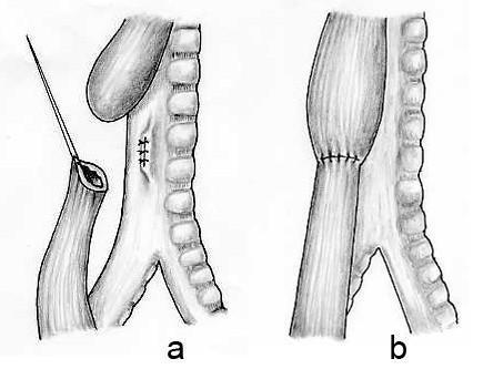
Anatomical illustration depicting the surgical repair of esophageal atresia (EA) with distal tracheoesophageal fistula (TEF). The diagram consists of two panels: (a) Pre-anastomosis state showing a dilated proximal esophageal pouch and a distal esophageal segment originating from the posterior wall of the trachea. Visible surgical interventions include the ligation and division of the fistula, evidenced by suture marks on the tracheal wall. A surgical needle and suture are shown preparing the distal esophagus for mobilization. (b) Post-operative state demonstrating a completed primary end-to-end esophageal anastomosis. The proximal and distal esophageal segments are joined with circumferential sutures, creating a continuous esophageal lumen parallel to the trachea. The trachea is shown with distinct cartilaginous rings and is now anatomically separated from the digestive tract. This illustration serves as an educational tool for pediatric surgery, highlighting the management of Type C esophageal atresia and the restoration of normal foregut anatomy.
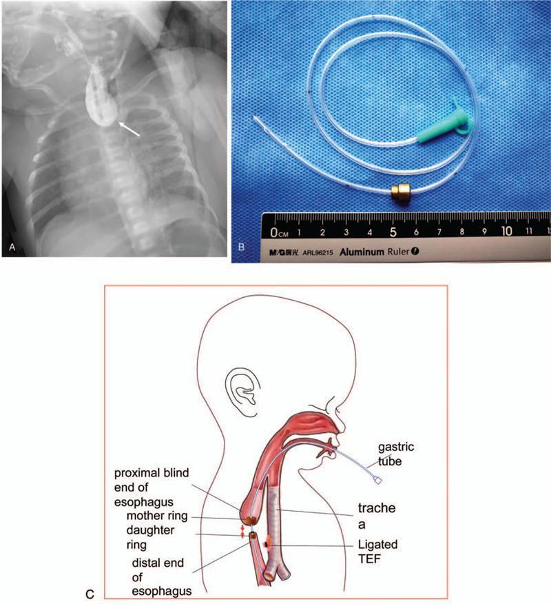
This composite figure illustrates the diagnosis and innovative surgical management of long-gap esophageal atresia (LGEA) with tracheoesophageal fistula (TEF). Panel A is an anteroposterior chest radiograph of a neonate featuring a contrast-filled, dilated proximal esophageal blind pouch terminating at the T2 vertebral level (indicated by a white arrow), a classic radiographic finding for esophageal atresia. Panel B is a clinical photograph showing the customized magnetic compression anastomosis (MCA) device, consisting of a larger 'mother ring' (7mm) and a smaller 'daughter ring' (5mm) threaded onto a gastric tube. Panel C provides a schematic diagram of the surgical approach: the mother ring is positioned in the proximal blind pouch and the daughter ring in the distal esophageal segment. The diagram highlights the use of a 6F gastric tube passing through the central holes of both rings to facilitate alignment and magnetic attraction. Anatomical labels identify the trachea and a ligated TEF. This visual set demonstrates the application of magnetic force to approximate esophageal ends for non-manual anastomosis in complex congenital anomalies.
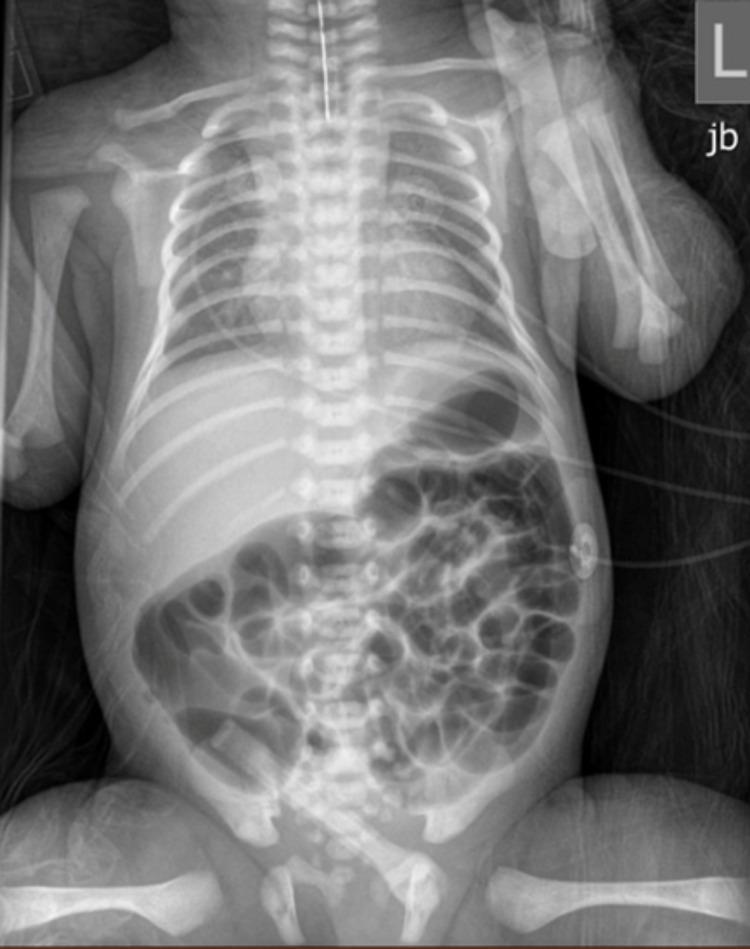
Anteroposterior (AP) neonatal chest and abdominal radiograph demonstrating diagnostic findings for esophageal atresia (EA) with an associated tracheoesophageal fistula (TEF). The primary finding is a radiopaque orogastric feeding tube that terminates abruptly in the proximal esophagus, visualized as a blind pouch in the upper mediastinum, indicating atresia. Despite the esophageal obstruction, there is significant bowel gas distributed throughout the stomach and small intestine in the abdomen. This presence of distal air confirms the existence of a distal tracheoesophageal fistula, allowing air to pass from the trachea into the lower gastrointestinal tract. The skeletal structures show normal neonatal development, and the abdominal gas pattern suggests patency distal to the stomach. This combination of a coiled or superiorly terminating feeding tube and subdiaphragmatic air is a classic radiological sign used in pediatric surgery to classify Type C esophageal atresia. The image is a critical educational example for neonatal intensive care and pediatric radiology training.
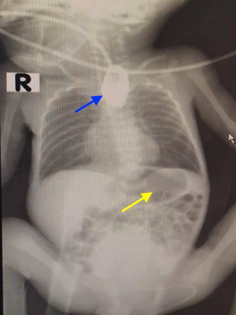
This diagnostic radiograph of a neonate illustrates the classic findings of esophageal atresia (EA) with a distal tracheoesophageal fistula (TEF). The image displays a concentrated, radiopaque contrast-filled blind pouch in the superior mediastinum (indicated by the blue arrow), which demonstrates the termination of the proximal esophagus and confirms esophageal atresia. Simultaneously, the abdominal region shows significant gaseous distension within the stomach and intestinal loops (indicated by the yellow arrow). The presence of subdiaphragmatic gas in the setting of proximal esophageal atresia is pathognomonic for a distal tracheoesophageal fistula, which allows air to pass from the trachea into the distal digestive tract. This preoperative x-ray is essential for classifying the anatomical subtype of the congenital anomaly and planning surgical intervention. The skeletal structures, including the rib cage and vertebral column, serve as landmarks for the localization of the atretic segment relative to the thoracic inlet.
Tracheoesophageal Anomalies & VACTERL
Embryology and Pathogenesis
-
Incidence: 1 in 3,000-4,500 live births; slight male predilection
-
~60-70% have associated anomalies
-
Sleisenger and Fordtran's GI and Liver Disease, p. 708
-
The Developing Human - Clinically Oriented Embryology, p. 576
Classification of EA/TEF (Gross Classification)
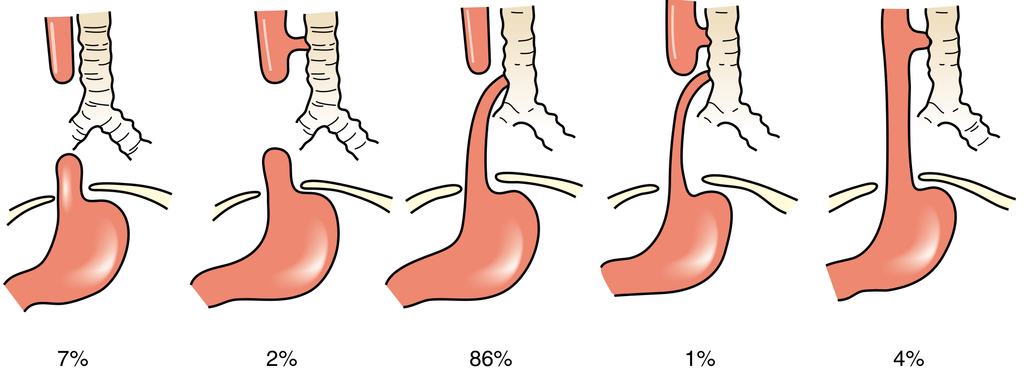
| Type | Description | Incidence |
|---|---|---|
| A | Pure EA - isolated blind proximal pouch, no fistula | 8-10% |
| B | EA + proximal TEF (proximal pouch communicates with trachea) | 1% |
| C | EA + distal TEF (proximal blind pouch + distal esophagus connects to trachea) | 85-86% (most common) |
| D | EA + double TEF (both proximal and distal segments communicate with trachea) | 2% |
| E | H-type TEF - TEF without EA (esophageal continuity intact) | 4-8% |
- Schwartz's Principles of Surgery, p. 1745
- Sabiston Textbook of Surgery, p. 2679
Clinical Presentation
EA (Types A, B, C, D)
- Excessive drooling / pooling of secretions from birth
- Choking, coughing, cyanosis at first feeding
- Regurgitation immediately after feeding
- Abdominal distension (air entering GI tract via distal TEF in type C/D)
- Scaphoid / gasless abdomen = strongly suggests isolated EA (type A) - no fistula pathway for air to enter bowel
- Maternal polyhydramnios (most pronounced in type A - 86%) - fluid cannot pass to stomach for absorption
- Prenatal US: absent stomach bubble + polyhydramnios
H-Type TEF (Type E) - unique presentation
-
No atresia, so diagnosis often delayed to childhood or adulthood
-
Recurrent aspiration pneumonia
-
Coughing/choking with feeds
-
Diagnosis often requires esophagram; can be missed due to small fistula size
-
Sleisenger and Fordtran's GI and Liver Disease, p. 710
Diagnosis
| Finding | Significance |
|---|---|
| NG/OG tube coiled at thoracic inlet (T2 level) on CXR | Confirms proximal EA |
| Air below diaphragm on plain film | Confirms distal TEF |
| Absent bowel gas + failed tube insertion | Suggests pure EA (type A) |
| Contrast esophagram (1 mL water-soluble) | Confirms anatomy if doubt; oral contrast CONTRAINDICATED due to aspiration risk |
| Bronchoscopy | Evaluates tracheomalacia and TEF location; assesses for proximal/secondary fistula |
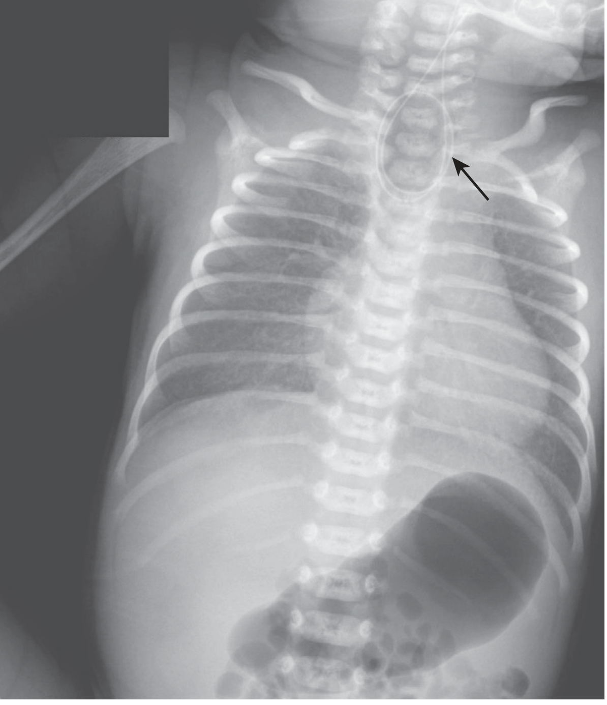
- Sabiston Textbook of Surgery, p. 2679
VACTERL Association
| Letter | Component | Frequency in EA |
|---|---|---|
| V | Vertebral anomalies (hemivertebrae, absent vertebrae) | 6-20% |
| A | Anorectal malformations (imperforate anus) | 10-15% |
| C | Cardiac defects (VSD, ASD, tetralogy of Fallot) | 15-35% |
| TE | Tracheoesophageal fistula + Esophageal atresia | - |
| R | Renal anomalies (agenesis, horseshoe kidney) | 5-15% |
| L | Limb defects (radial ray - absent radius, thumb hypoplasia) | 5-20% |
- Mulholland and Greenfield's Surgery, p. 5402
- Fischer's Mastery of Surgery, p. 7994
- Sleisenger and Fordtran's GI, p. 709 (Table 43.1)
Other Associated Syndromes
| Syndrome | Gene | Key Features |
|---|---|---|
| CHARGE | CHD7 | Coloboma, Heart defects, Atresia choanae, Retardation, Genital/Ear anomalies |
| Feingold | MYCN | EA/duodenal atresias, microcephaly, syndactyly |
| Fanconi Anemia | >20 genes | Bone marrow failure, radial ray defects |
| Anophthalmia-esophageal-genital | SOX2 | Anophthalmia, EA, urogenital anomalies |
| VACTERL-H | FANCB | VACTERL + hydrocephalus |
Preoperative Management
- Stop oral feeds immediately
- Replogle/sump tube into upper esophageal pouch for continuous suction (decompresses pooled secretions)
- Head-up / prone positioning to reduce aspiration and gastric reflux through fistula
- Broad-spectrum IV antibiotics (aspiration pneumonitis risk)
- Avoid intubation if possible - positive pressure ventilation preferentially inflates GI tract via the fistula, worsening gastric distension and compromising ventilation
- If intubation is necessary: advance ETT distal to the TEF OR place an occluding balloon catheter in the fistula
- Emergent gastrostomy (water seal) if ventilation fails - allows air from the fistula to escape rather than inflate the stomach
- Preoperative echocardiogram - determines aortic arch side (right vs. left) which dictates surgical approach
- Sabiston Textbook of Surgery, p. 2679; Mulholland and Greenfield's Surgery, p. 5402
Surgical Management
Standard Type C EA + Distal TEF
- Rigid bronchoscopy (optional for primary cases; evaluates tracheomalacia, confirms TEF location, excludes secondary fistula)
- Extrapleural dissection; division of azygos vein to expose TEF
- Fistula ligation and division - sequentially divide 1-2 mm segments and close tracheal defect immediately with absorbable interrupted sutures (prevents ventilation loss)
- Mobilize upper pouch maximally (well-vascularized); limit mobilization of lower pouch (segmental/precarious blood supply - ischemia risk)
- Primary end-to-end esophageal anastomosis (single or double layer)
- Thoracoscopic approach now increasingly preferred with equivalent outcomes
- Sabiston Textbook of Surgery, pp. 2679-2680
Long-Gap EA
- Placement of gastrostomy for enteral feeding in neonatal period
- Delay primary anastomosis to ~4-6 months of age (allow spontaneous esophageal elongation with growth; mark distal end with hemoclip on prevertebral fascia)
- Confirm adequate elongation radiographically before attempting anastomosis
- Esophageal replacement (stomach, colon, or jejunal conduit) if adequate length cannot be achieved
- Magnetic compression anastomosis (MCA) - emerging technique for LGEA
Postoperative Complications
| Complication | Notes |
|---|---|
| Anastomotic leak | Most common early complication |
| Anastomotic stricture | Requires esophageal dilation |
| Recurrent TEF | Presents with recurrent aspiration; bronchoscopy with Fogarty catheter for diagnosis |
| Tracheomalacia | Very common; "dying spells" (cyanotic episodes) with feeds; most improve with age; severe cases may need aortopexy |
| GERD | Near-universal; requires long-term management |
| Esophageal dysmotility | Affects long-term swallowing |
Other Tracheoesophageal Anomalies
Laryngotracheoesophageal Cleft (LTEC)
- Failure of complete separation of larynx/upper trachea from esophagus
- Persistent connection of variable length between the two structures
- Symptoms: aspiration, similar to TEF
- Distinguishing feature: aphonia (inability to speak/vocalize)
Tracheal Stenosis
- Usually associated with one of the TEF variants
- Results from unequal partitioning of the foregut
- Can be a web (incomplete atresia) or complete atresia (uniformly fatal)
Tracheal Bronchus (Tracheal Diverticulum)
-
Blind bronchus-like projection from trachea
-
May terminate in normal lung tissue (tracheal lobe)
-
Causes recurrent infection and respiratory distress in infants
-
The Developing Human - Clinically Oriented Embryology, pp. 576-577
Waterston / Spitz Risk Classification (Prognosis)
| Group | Criteria | Survival |
|---|---|---|
| I | Birth weight >1.5 kg, no major cardiac defect | ~97% |
| II | Birth weight <1.5 kg OR major cardiac defect | ~59% |
| III | Birth weight <1.5 kg AND major cardiac defect | ~22% |
- EA/TEF incidence ~1:3,000-4,500; type C (EA + distal TEF) = 85% of cases
- Hallmark diagnosis: failure to pass NG/OG tube + gas below diaphragm on plain film
- VACTERL = Vertebral + Anorectal + Cardiac + TEF/EA + Renal + Limb; requires 2+ features; present in 10-21%
- Preop: Replogle tube, head-up, avoid PPV, echo (aortic arch side)
- Surgery: right extrapleural thoracotomy (or thoracoscopy); TEF ligation + primary anastomosis
- H-type TEF (type E) presents late with recurrent aspiration pneumonia, often diagnosed in childhood