
Difference between ranuads disease and burger disease
- Raynaud's disease is a vasospastic disorder where blood vessels (usually in fingers/toes) overreact to cold or stress, causing episodic color changes (white → blue → red). It can be primary (no underlying cause) or secondary (associated with autoimmune diseases).
- Buerger's disease (Thromboangiitis obliterans) is an inflammatory, occlusive disease of small/medium arteries and veins, almost exclusively in young male smokers, leading to progressive ischemia and gangrene.
Raynaud disease color changes fingers
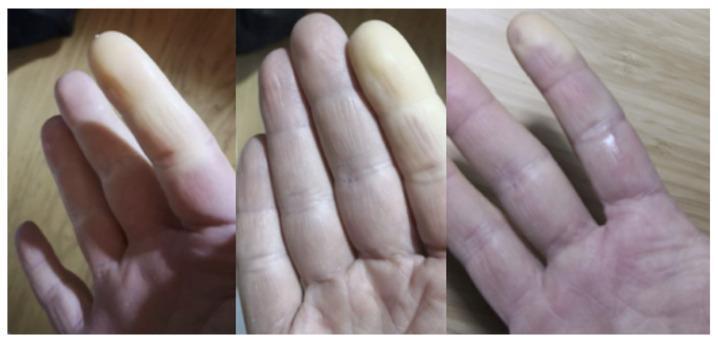
This clinical photograph set comprises three panels illustrating classic Raynaud's phenomenon in the fingers. The images demonstrate significant vascular changes characterized by episodic digital ischemia. Specifically, multiple fingers exhibit distinct, sharply demarcated areas of pallor (whitening), primarily involving the distal phalanges. This chalk-white appearance indicates a complete lack of blood flow due to vasospasm. Adjacent to the ischemic zones, the skin displays a mix of cyanosis (bluish tint) and reactive hyperemia (erythema or redness), showcasing the characteristic triphasic color response. The distribution is asymmetrical across the digits. Skin texture appears slightly taut in the pale areas, with normal dermatoglyphics visible in the unaffected regions. This visual is highly representative of secondary Raynaud's phenomenon, often seen in systemic autoimmune diseases such as systemic sclerosis or idiopathic inflammatory myopathies (IIMs). It serves as a key diagnostic clinical sign for medical students and clinicians assessing peripheral vascular and rheumatological disorders.
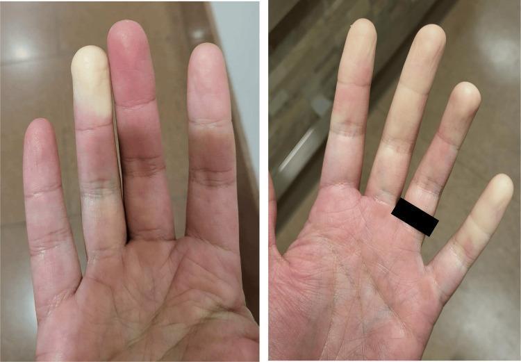
Clinical photograph of a patient's hands demonstrating the characteristic triphasic color changes associated with Raynaud’s phenomenon. The left image shows significant pallor (white phase) in the index finger, representing digital vasospasm and ischemia, contrasted against the erythema (red phase) of the adjacent middle finger, suggesting reperfusion. The right image shows more diffuse, patchy pallor across several digits including the index and ring fingers. These visual findings are hallmark clinical signs of Raynaud’s phenomenon, which may occur as a primary condition or secondary to autoimmune connective tissue diseases. In this clinical context, the findings are associated with a patient positive for anti-centromere antibodies and diagnosed with an autoimmune hepatitis (AIH) and primary biliary cholangitis (PBC) overlap syndrome. The image serves as an educational example of episodic vasospastic disorders and their physical manifestations in the distal extremities.
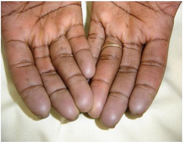
This clinical photograph displays the palmar surface of both hands, illustrating classic features of Raynaud phenomenon, specifically the 'asphyxic' or cyanotic phase. The image demonstrates a clear demarcation of skin color changes: the palms retain a normal, warm-toned pigmentation, while all fingers show a distinct transition to a pale, bluish-gray or cyanotic hue starting near the metacarpophalangeal joints and intensifying towards the distal phalanges. This visual finding is characteristic of tissue hypoxia resulting from vasospasm and sluggish blood flow. In the context of the provided metadata, this manifestation is associated with systemic sclerosis (scleroderma). While the skin overall appears intact, there is evidence of subtle textural changes at the fingertips, which may precede digital pitting or sclerodactyly. The image serves as a clinical reference for the secondary Raynaud phenomenon in autoimmune connective tissue diseases, highlighting the episodic digital ischemia typical of the condition.
Buerger disease thromboangiitis obliterans gangrene
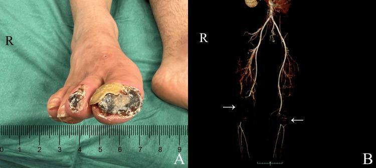
This composite educational image illustrates the clinical and radiological findings of Thromboangiitis Obliterans (Buerger's disease). Panel A is a clinical photograph of a right foot showing advanced ischemic ulcerated lesions and dry gangrene affecting the distal first (hallux) and second toes. The hallux exhibits a large, necrotic black center with an overlying yellowish-tan hyperkeratotic rim, while the second toe shows smaller, deep necrotic lesions. Panel B presents a posterior view of a 3D-reconstructed contrast-enhanced CT arteriogram of the lower extremities. White arrows highlight significant arterial obstructions: occlusion of the right superficial femoral artery extending to the popliteal artery, and occlusion of the left popliteal artery. There is a notable absence of contrast enhancement in the distal runoff vessels, indicating severe limb ischemia. The image demonstrates the correlation between proximal arterial occlusive disease and distal tissue necrosis, emphasizing the diagnostic importance of vascular imaging in peripheral arterial disease management.
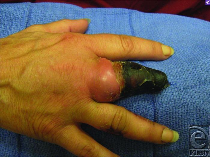
A clinical photograph of a right hand demonstrating advanced digital ischemia and dry gangrene, characteristic of thromboangiitis obliterans (Buerger's disease). The third digit (middle finger) shows a prior partial amputation at the level of the middle phalanx. The residual distal portion of this digit exhibits a dense, black, necrotic eschar consistent with gangrene. There is significant circumferential erythema and swelling proximal to the necrotic tissue, specifically involving the area around the metacarpophalangeal joint, suggesting active inflammation or cellulitis. The surrounding digits show subtle signs of circulatory compromise, including skin pallor and longitudinal nail plate changes. This image serves as an educational example of critical limb-threatening ischemia and the progressive tissue loss associated with small-to-medium vessel vasculitis in tobacco users. The visual findings illustrate the demarcation between necrotic tissue and viable, albeit inflamed, proximal structures.
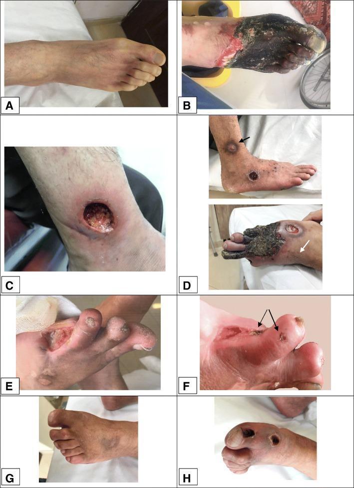
A composite of clinical photographs (panels A-H) demonstrating various stages of dermal gangrene and chronic ulceration in the lower extremities of patients with thromboangiitis obliterans (Buerger’s disease). Panel A shows initial pallor and Raynaud's phenomenon. Panel B illustrates extensive, dark necrotic tissue covering the forefoot and toes with a sharp demarcating border and surrounding erythema. Panel C depicts a deep, circular 'punched-out' ulcer on the lower leg with visible granulation tissue and peripheral edema. Panel D shows multifocal purpuric-like lesions progressing to localized gangrene on the dorsum of the foot and distal digits. Panels E and F provide close-up views of digital ulcers and small gangrenous papules (indicated by arrows) on an amputation stump. Panels G and H show the evolution of localized gangrenous lesions on the toes, transitioning from active necrosis to a dryer, more localized state. This collection highlights the microvascular complications of TAO, including tissue ischemia, necrosis, and subsequent ulceration despite angiogenic medical therapies.
Raynaud's Disease vs. Buerger's Disease
Overview
| Feature | Raynaud's Disease/Phenomenon | Buerger's Disease (Thromboangiitis Obliterans) |
|---|---|---|
| Nature | Functional/vasospastic disorder | Structural/inflammatory-occlusive disease |
| Pathology | Reversible vasospasm - no vessel wall destruction | Segmental thrombosing vasculitis with luminal occlusion |
| Vessels affected | Arteries and arterioles (no structural damage) | Small and medium arteries, veins, and nerves |
1. Raynaud's Disease / Phenomenon
Definition
Types
- Primary (Raynaud's Disease): No associated disorder; affects 3-5% of the general population; predilection for young women; benign course. Ulceration and ischemic gangrene are rare.
- Secondary (Raynaud's Phenomenon): Vascular insufficiency caused by an underlying condition - systemic lupus erythematosus, scleroderma, thromboangiitis obliterans, or atherosclerosis. Every patient should be evaluated for secondary causes.
Mechanism
Classic Triphasic Color Change
Key Clinical Features
- Episodic, triggered by cold or stress
- Bilateral and symmetric
- Resolves with warming
- No permanent ischemia in primary form
- In chronic cases: atrophy of skin, subcutaneous tissue, and muscle may develop
2. Buerger's Disease (Thromboangiitis Obliterans)
Definition
Cause
Pathology
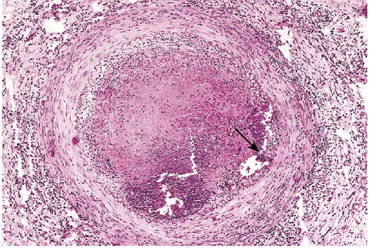
- Acute phase: Segmental thrombosing vasculitis, mixed inflammatory infiltrates, luminal thrombosis, sterile microabscesses (a hallmark), occasionally rimmed by granulomatous inflammation
- Inflammation extends outward into contiguous veins and nerves (rare in other vasculitides)
- Chronic phase: thrombus organizes and recanalizes
- End-stage: artery and adjacent structures encased in fibrous tissue
Key Clinical Features
- Young male smokers, typically onset before age 40
- Foot/hand claudication initially, progressing to rest pain
- Migratory superficial phlebitis (up to 16% of patients)
- Severe pain even at rest (due to nerve involvement)
- Chronic extremity ulcerations progressing to gangrene
- Characteristic angiographic finding: "corkscrew" collaterals, segmental skip lesions, disease confined to distal circulation
Head-to-Head Comparison Table
| Feature | Raynaud's Disease | Buerger's Disease |
|---|---|---|
| Type | Vasospastic (functional) | Inflammatory-occlusive (structural) |
| Age | Young women (primary) | Young men, <40 years |
| Gender | Female predominance | Male predominance |
| Trigger | Cold, stress | Tobacco use |
| Vessel involvement | Arterioles (no wall destruction) | Small/medium arteries, veins, nerves |
| Pathology | Reversible vasospasm | Thrombosis + vasculitis + microabscesses |
| Color changes | Classic white-blue-red triphasic | No triphasic changes (ischemia is persistent) |
| Gangrene | Rare (only secondary form) | Common and progressive |
| Nerve involvement | No | Yes (extends into adjacent nerves) |
| Venous involvement | No | Yes (migratory phlebitis) |
| Symmetry | Bilateral, symmetric | Often asymmetric, multifocal |
| Association | Autoimmune diseases (secondary form) | Heavy tobacco smoking exclusively |
| Angiography | Normal vessel walls | Corkscrew collaterals, segmental occlusions |
| Reversibility | Yes - with warming or treatment | Partial - smoking cessation helps early on, but established lesions do not fully reverse |
| Treatment | Calcium channel blockers, avoid triggers | Strict smoking cessation is the mainstay |
| Outcome | Usually benign | Risk of amputation (31-67% limb loss if smoking continues) |