
Can you search for images which are basically showing anatomical features of supination and pronation of forearm.
supination pronation forearm anatomy bones muscles
radius ulna rotation forearm pronation supination diagram
forearm rotation anatomy
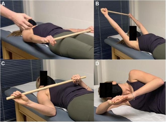
This composite of clinical photographs demonstrates various scapular and shoulder stretching exercises used in the rehabilitation of Pectoralis Minor Syndrome (PMS) and neurogenic thoracic outlet syndrome (NTOS). (A, B) Shoulder flexion stretch: The patient is in a supine position, holding a wooden stick with both hands. This allows the asymptomatic arm to assist the affected arm into maximal overhead flexion, targeting the anterior shoulder and pectoralis minor muscle. (C) External rotation stretch: Also performed supine using a stick, this exercise uses the contralateral arm to facilitate maximal external rotation, stretching the anterior shoulder anatomy. (D) Sleeper stretch: The patient is in a lateral decubitus position (lying on the affected side) with the arm flexed at the elbow. The patient uses the opposite hand to gently push the forearm toward the table, performing passive internal rotation to stretch the posterior shoulder capsule and associated anatomy. These exercises are critical for improving scapular kinematics and relieving neurovascular compression in the retropectoralis minor space.
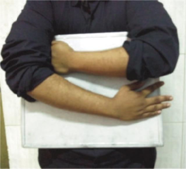
This clinical photograph demonstrates a specific patient positioning technique for simultaneous anteroposterior (AP) radiography of both forearms. The patient is shown holding a white rectangular radiographic cassette against their chest. The positioning involves internal rotation at both shoulders with the forearms crossed horizontally over the cassette to achieve a consistent orientation for imaging. The left forearm is positioned superiorly with the hand clenched, while the right forearm is positioned inferiorly with the hand open and fingers extended against the edge of the cassette. This specialized technique is utilized in orthopedics and radiology to assess bilateral radial bow and forearm anatomy, particularly in patients with limited supination. By ensuring identical positioning, clinicians can more accurately calculate the location of maximum radial bow and compare measurements between the radius and ulna without overlapping shadows, facilitating precise diagnostic evaluation of forearm deformities or injuries.
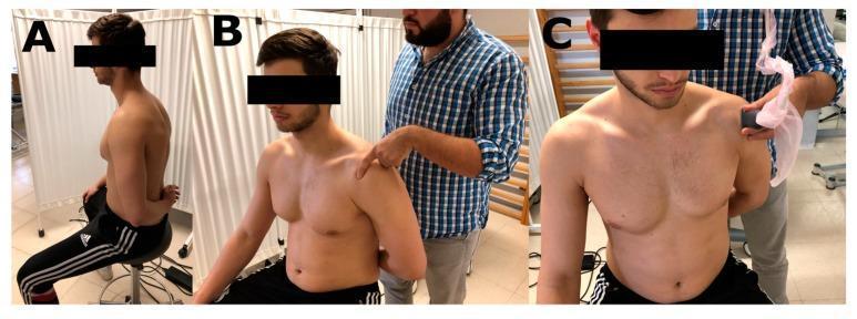
A three-panel clinical photograph series (A, B, C) demonstrating the clinical positioning and physical examination of the supraspinatus muscle tendon (SMT). Panel A shows a male subject seated on a backless stool in the 'Crass position,' with the arm in maximal medial rotation, adduction, and slight hyperextension, placing the forearm behind the lower back. This maneuver exposes the supraspinatus tendon from beneath the acromion for easier accessibility. Panel B illustrates manual surface anatomy palpation, where an examiner uses an index finger to locate the SMT using the acromion and greater humeral tuberosity as bony landmarks. Panel C demonstrates diagnostic corroboration using musculoskeletal ultrasound; an examiner applies a linear ultrasound transducer over the same anatomical region to measure tendon depth and confirm clinical findings. The series illustrates a standardized protocol for identifying the SMT, emphasizing the importance of specific patient positioning in orthopedic physical examination and musculoskeletal imaging.
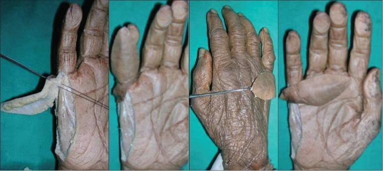
A series of four anatomical photographs from a cadaveric dissection study illustrating the ulnar artery perforator flap on a human hand. The first panel shows a longitudinal flap elevated from the ulnar border of the distal forearm and hypothenar eminence, with metal wires highlighting the underlying vascular perforators and longitudinal arcade arising from the digital artery. The second panel demonstrates the flap partially reflected, showing the subfascial tissue layer and its relationship to the fifth digit. The third panel shows a dorsal view of the hand with a metal probe indicating the arc of rotation, where the skin flap is transferred to the dorsal surface of the proximal phalanx. The fourth panel provides a palmar view of the dissected hand, emphasizing the donor site along the ulnar margin and the reach of the pedicled flap. This sequence illustrates the surgical anatomy, vascular supply, and rotational capabilities of the hypothenar ulnar artery perforator flap for reconstructive surgery of the hand.
pronator teres supinator muscle forearm
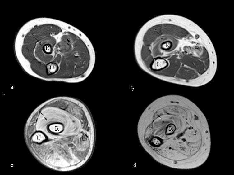
Diagnostic axial T1-weighted MRI images of the proximal forearm across four subjects (labeled a-d), demonstrating various stages of muscle fatty replacement. The cross-sections at the level of the radial tuberosity clearly show the radius (R) and ulna (U). In images 'a' and 'b' (younger ambulant patients), the musculature maintains relatively low signal intensity with preserved architectural definition, indicating mild involvement. In contrast, images 'c' and 'd' (older non-ambulant patients) exhibit progressive hyperintensity within the muscle compartments, characteristic of fatty infiltration. The supinator (indicated by arrows) and the pronator teres (indicated by arrowheads) show the most marked high signal intensity, particularly in image 'd', where normal muscle texture is replaced by bright fatty tissue. This comparison serves as a clinical illustration of disease progression in muscular dystrophies or similar neuromuscular conditions, where increased T1 signal serves as a biomarker for permanent muscle loss and functional decline.
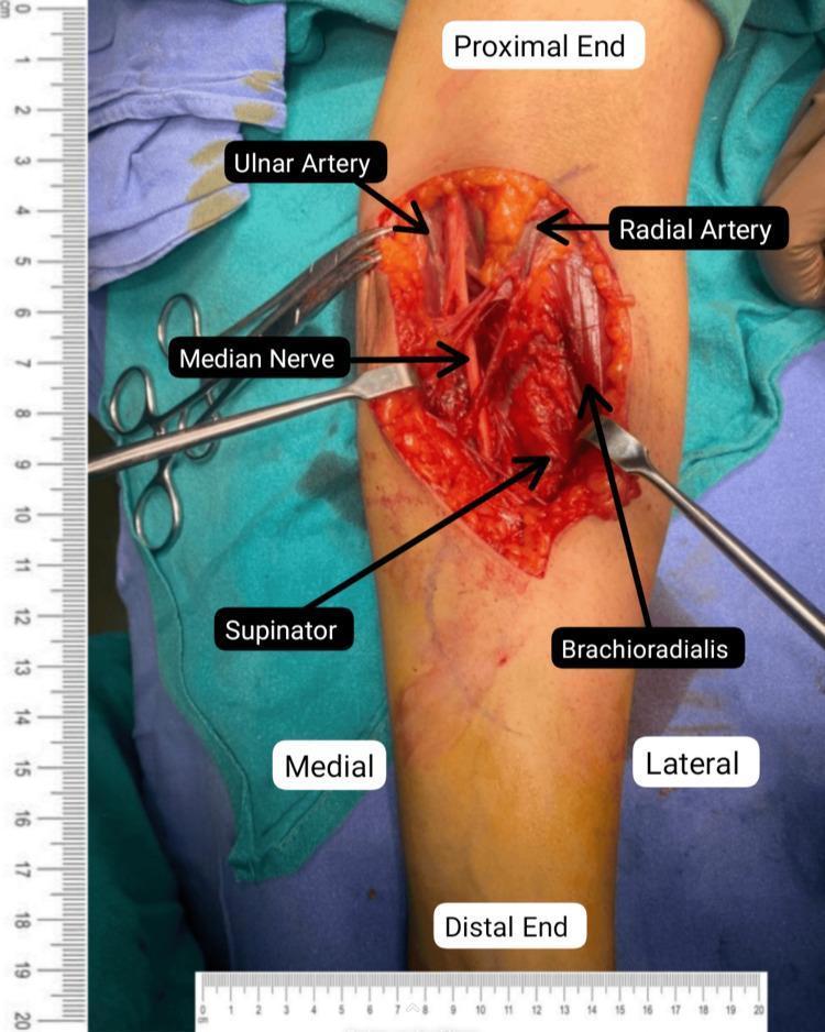
This clinical photograph captures an intraoperative view of a surgical decompression of the left median nerve at the level of the proximal forearm and cubital fossa. The image shows a deep dissection with anatomical structures clearly labeled. Centrally, the decompressed median nerve is visible, positioned between the medially located ulnar artery and the laterally located radial artery. Surrounding musculature is identified, including the brachioradialis muscle on the lateral border and the supinator muscle within the deep field. Surgical instruments, including a metal retractor and forceps, are shown retracting tissues to maintain exposure of the neurovascular bundle. Orientation labels indicate proximal, distal, medial, and lateral directions relative to the patient's limb. The surgical field demonstrates the successful release of potential compression sites, such as the lacertus fibrosus and the pronator teres heads, to treat median nerve entrapment. A centimeter ruler is placed alongside for scale, emphasizing the anatomical precision required for peripheral nerve exploration.
radioulnar joint supination anatomy diagram
Fig. 7.83 - Pronation and Supination (Muscles & Bone Movement)
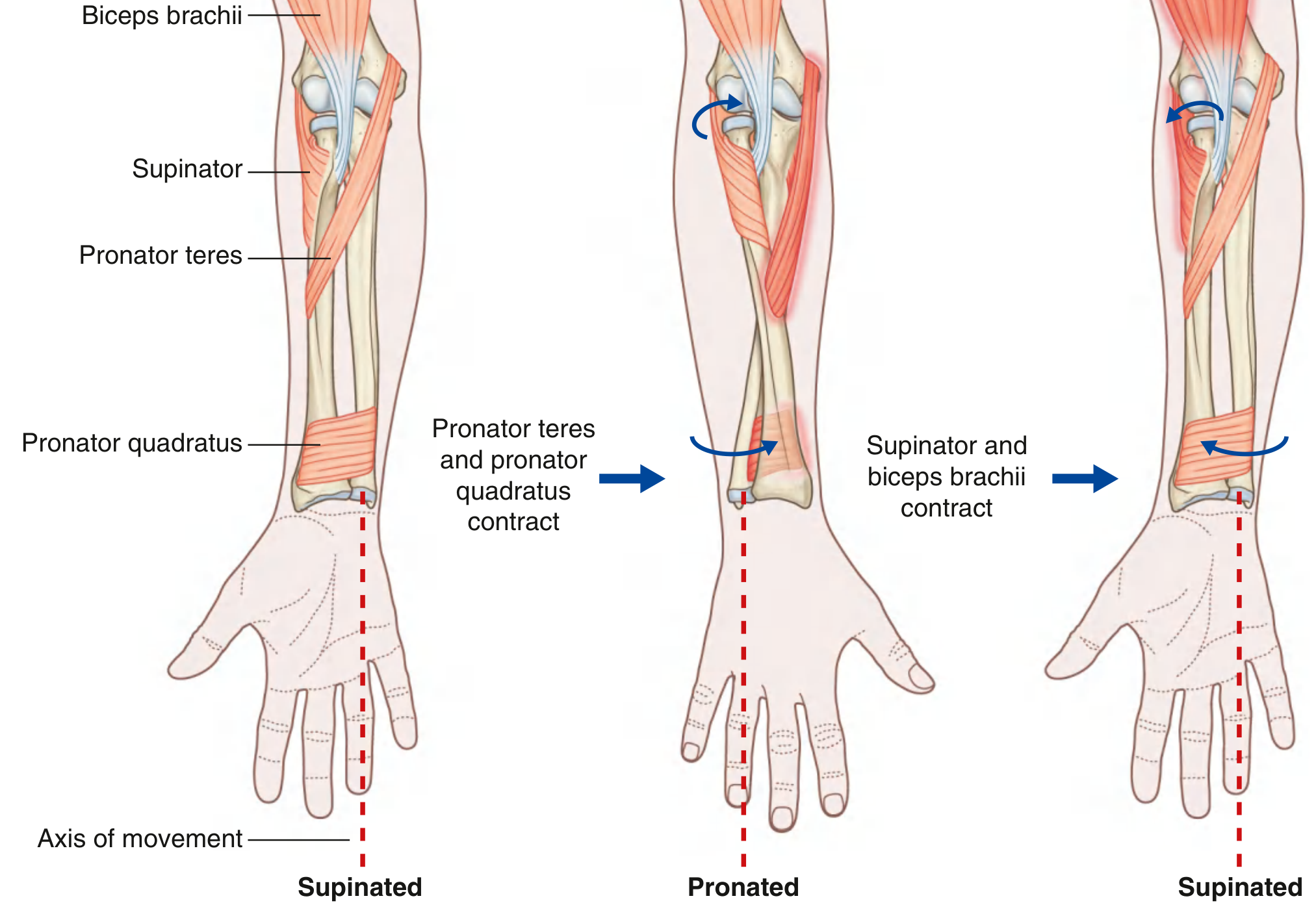
- Left panel (Supinated): The starting neutral/supinated position with the four key muscles labeled - Biceps brachii, Supinator, Pronator teres, and Pronator quadratus. The red dashed line marks the axis of movement.
- Middle panel (Pronated): When Pronator teres and Pronator quadratus contract, the distal radius rotates medially over the ulna, turning the palm posteriorly (pronation). Curved arrows show the direction of radial head rotation at the proximal radioulnar joint.
- Right panel (Supinated again): When Supinator and Biceps brachii contract, they unwrap from the radius and reverse the movement back to the supinated (palm-anterior) position.
Fig. 7.84 - Role of Anconeus During Pronation
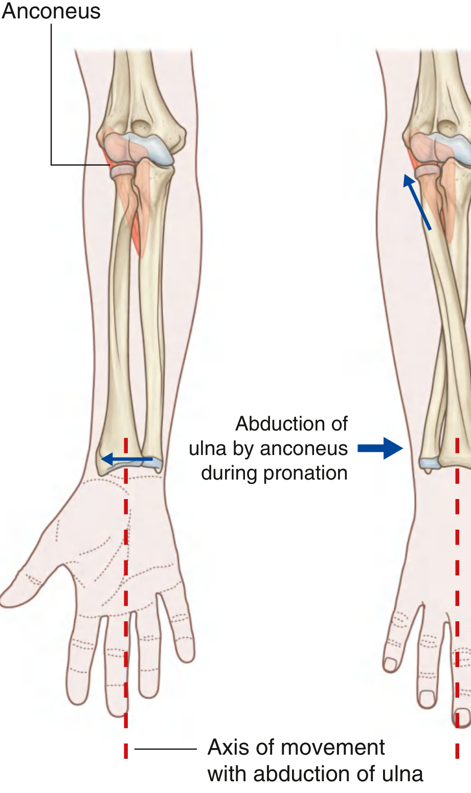
Key Anatomy Summary (from Gray's Anatomy for Students, p. 895-896)
| Movement | Muscles Involved | Joint Action |
|---|---|---|
| Supination | Supinator + Biceps brachii | Radial head spins on capitulum; distal radius slides back laterally |
| Pronation | Pronator teres + Pronator quadratus | Distal radius slides anteriorly/medially over ulnar head |
| Pronation assist | Anconeus | Abducts distal ulna to maintain palm axis |