

98cca4af-c2b5-4568-8411-101f5be260e2.jpeg
Answer these questions with suitable pictures
Reading File
Now let me search the medical library and web in parallel for all the needed content:
Finding Sources
Finding Sources
Searching the Web
superolateral surface of brain cerebrum lobes sulci gyri Brodmann areas anatomy diagram
Finding Sources
Finding Sources
Searching the Web
Brodmann areas brain map motor cortex sensory cortex auditory area Broca Wernicke striate cortex
Loading Image
Loading Image
Loading Image
Searching the Web
blood supply superolateral surface brain middle cerebral artery anatomy diagram
Loading Image
Loading Image
Loading Image
Superolateral Surface of Brain (AN62.2-62.3)
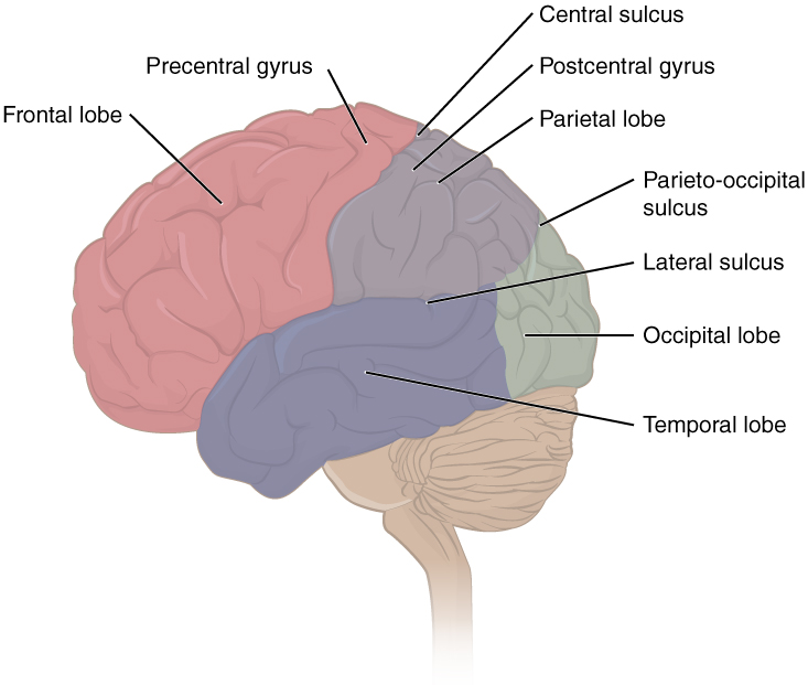
Q1. Borders, Surfaces and Lobes of Cerebrum
Cerebrum - Three Surfaces:
- Superolateral surface - convex, faces the calvaria
- Medial surface - flat, faces the opposite hemisphere (separated by longitudinal fissure)
- Inferior (tentorial) surface - rests on the tentorium cerebelli and floor of anterior/middle cranial fossae
Borders:
- Superomedial border - between superolateral and medial surfaces
- Inferolateral border - between superolateral and inferior surfaces
- Inferomedial (medial occipital) border - between medial and inferior surfaces
Lobes of Cerebrum (4 main):
| Lobe | Boundary |
|---|---|
| Frontal | Anterior to central sulcus; above lateral sulcus |
| Parietal | Posterior to central sulcus; above lateral sulcus |
| Temporal | Below lateral (Sylvian) sulcus |
| Occipital | Posterior to parieto-occipital sulcus |
(Insula = 5th lobe, hidden within lateral sulcus)
Q2. Three Poles of Cerebrum
The cerebrum has three poles:
- Frontal pole - anterior end of the frontal lobe
- Occipital pole - posterior end of the occipital lobe
- Temporal pole - anterior end of the temporal lobe
Q3. How the Cerebral Hemisphere is Divided into Four Lobes
Two key sulci divide the hemisphere into lobes:
- Central sulcus (of Rolando): runs from the superomedial border downward and forward, separating the frontal lobe (anterior) from the parietal lobe (posterior)
- Lateral sulcus (of Sylvius): runs horizontally, separating the temporal lobe (below) from the frontal and parietal lobes (above)
- Parieto-occipital sulcus: on the medial surface, separates the occipital lobe from the parietal lobe (on the superolateral surface, an imaginary line is drawn from this sulcus to the pre-occipital notch)
The temporal lobe is separated from the occipital lobe by an imaginary line drawn from the parieto-occipital sulcus to the pre-occipital notch.
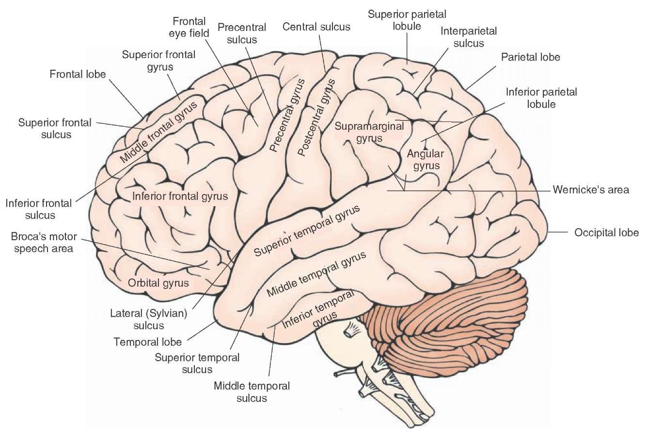
Q4. Sulci, Gyri and Main Functional Areas in Each Lobe
Frontal Lobe
| Sulcus | Gyri |
|---|---|
| Precentral sulcus | Precentral gyrus (motor cortex, BA4) |
| Superior frontal sulcus | Superior frontal gyrus |
| Inferior frontal sulcus | Middle frontal gyrus; Inferior frontal gyrus (pars orbitalis, triangularis, opercularis = Broca's area) |
Key functional areas: Primary motor cortex (precentral gyrus), Broca's speech area (inferior frontal gyrus), Frontal eye field (BA8), Prefrontal cortex
Parietal Lobe
| Sulcus | Gyri |
|---|---|
| Postcentral sulcus | Postcentral gyrus (sensory cortex, BA 1,2,3) |
| Intraparietal sulcus | Superior parietal lobule; Inferior parietal lobule (supramarginal gyrus + angular gyrus) |
Key functional areas: Primary somatosensory cortex, Wernicke's area (angular gyrus / posterior superior temporal), Spatial awareness
Temporal Lobe
| Sulcus | Gyri |
|---|---|
| Superior temporal sulcus | Superior temporal gyrus |
| Middle temporal sulcus | Middle temporal gyrus |
| — | Inferior temporal gyrus |
Key functional areas: Primary auditory cortex (Heschl's gyri in superior temporal gyrus, BA 41/42), Wernicke's area (BA22)
Occipital Lobe
| Sulcus | Gyri |
|---|---|
| Lateral occipital sulcus | Superior/Inferior occipital gyri |
| — | Cuneus, Lingual gyrus (medial surface) |
Key functional areas: Primary visual (striate) cortex (BA17), Visual association cortex (BA18, 19)
Q5. Motor Speech Area (Broca's Area)
| Feature | Details |
|---|---|
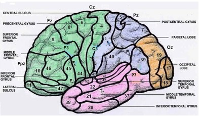
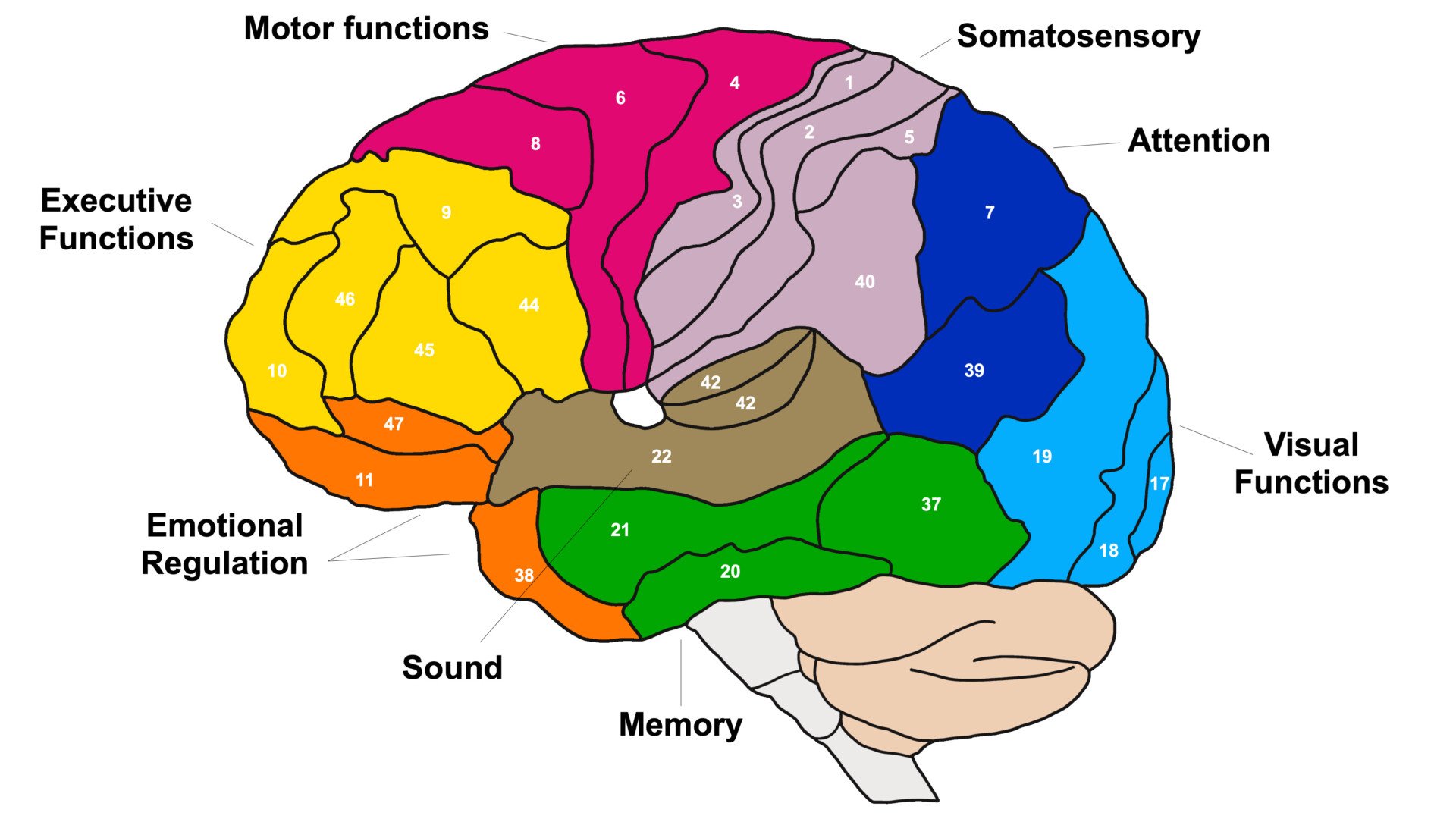
| Brodmann Numbers | Area 44 (pars opercularis) + Area 45 (pars triangularis) |
| Location | Inferior frontal gyrus of the dominant hemisphere (left in 95% of right-handed people), above the anterior part of the lateral sulcus |
| Function | Motor programming of speech - coordinates articulatory movements of lips, tongue, larynx for speech production |
| Applied Anatomy | Damage causes Broca's (expressive/non-fluent) aphasia: patient understands speech but cannot produce fluent speech; they speak in telegraphic, effortful short phrases |
Q6. Wernicke's Area
| Feature | Details |
|---|---|
| Brodmann Number | Area 22 (posterior superior temporal gyrus) - sometimes includes BA 39 & 40 |
| Location | Posterior part of the superior temporal gyrus, in the dominant hemisphere (left), near the junction with the parietal lobe |
| Function | Comprehension of spoken and written language; attaches meaning to words |
| Applied Anatomy | Damage causes Wernicke's (receptive/fluent) aphasia: patient speaks fluently but uses wrong words ("paraphasia"), cannot comprehend speech; "word salad" output. Wernicke's and Broca's areas are connected by the arcuate fasciculus; damage to this causes conduction aphasia |
Q7. Striate Cortex
| Feature | Details |
|---|---|
| Brodmann Number | Area 17 |
| Location | Lips and walls of the calcarine sulcus on the medial surface of the occipital lobe; extends slightly onto superolateral surface at the occipital pole |
| Why "Striate" | Named for the visible white line of Gennari (a myelinated stripe visible to the naked eye in cortical layer IV) |
| Function | Primary visual cortex - receives visual input from the lateral geniculate body (via optic radiation); processes basic visual features (edges, orientation, motion) |
| Applied Anatomy | Unilateral lesion → contralateral homonymous hemianopia. Bilateral lesion → cortical blindness. Macular vision is represented at the posterior pole (macular sparing occurs in posterior cerebral artery strokes because the macular cortex may receive collateral supply from the MCA) |
Q8. Motor Cortex
| Feature | Details |
|---|---|
| Brodmann Number | Area 4 |
| Location | Precentral gyrus, anterior to the central sulcus; also extends onto the anterior wall of the central sulcus |
| Characteristic Cell | Giant Betz cells (largest neurons in the CNS) in layer V |
| Function | Primary motor cortex - initiates voluntary movement; contains the motor homunculus (somatotopic map: lower limb medially on paracentral lobule, face laterally); gives rise to the corticospinal (pyramidal) tract |
| Applied Anatomy | Upper motor neuron (UMN) lesion: contralateral hemiplegia/hemiparesis with spasticity, hyperreflexia, Babinski sign positive. Lesion to the face area → contralateral lower facial palsy (forehead spared). The homunculus shows that the hand and face have disproportionately large representation |
Q9. Sensory Cortex
| Feature | Details |
|---|---|
| Brodmann Numbers | Areas 3, 1, 2 (in anterior-to-posterior order on postcentral gyrus) |
| Location | Postcentral gyrus, posterior to the central sulcus |
| Function | Primary somatosensory cortex - receives pain, temperature, touch, pressure, proprioception from the opposite side of the body via the thalamus (VPL nucleus). Contains the sensory homunculus (somatotopic organization similar to motor homunculus) |
| Applied Anatomy | Lesion → contralateral hemianesthesia (loss of discriminative touch, proprioception, two-point discrimination). Area 3a receives proprioceptive input; area 3b receives cutaneous input; areas 1 and 2 receive both. Parietal lobe lesions also cause astereognosis, sensory neglect, and (dominant hemisphere) Gerstmann's syndrome (dyscalculia, dysgraphia, finger agnosia, right-left disorientation) |
Q10. Auditory Area
| Feature | Details |
|---|---|
| Brodmann Numbers | Area 41 (primary auditory cortex) + Area 42 (secondary auditory cortex) |
| Location | Transverse temporal gyri of Heschl - on the superior surface of the superior temporal gyrus, within the lateral sulcus (Sylvian fissure); you need to "open" the lateral sulcus to see them |
| Function | Area 41 = primary auditory cortex: receives auditory input from the medial geniculate body (via auditory radiation); processes pitch, loudness, and basic sound features. Area 42 = auditory association area: interprets complex sounds, words |
| Applied Anatomy | Each auditory cortex receives bilateral input (from both ears), so unilateral lesions do NOT cause complete deafness but may cause difficulty localizing sounds. Bilateral lesions cause cortical deafness. Lesion of BA41/42 in the dominant hemisphere contributes to Wernicke's aphasia |

Q11. Blood Supply of the Superolateral Surface
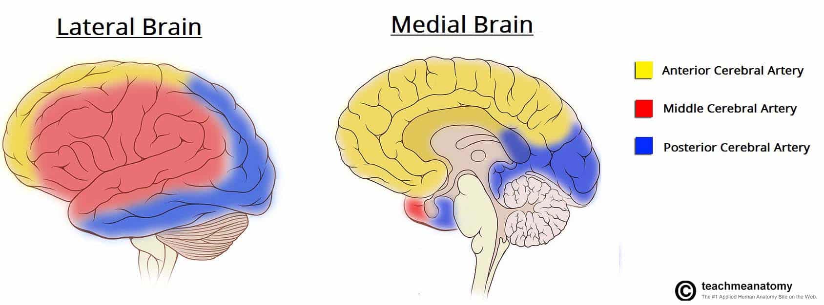
The superolateral surface is mainly supplied by the Middle Cerebral Artery (MCA), with contributions from the other two major cerebral arteries:
| Artery | Territory on Superolateral Surface |
|---|---|
| Middle Cerebral Artery (MCA) - branch of internal carotid artery | Main supply - most of the lateral surface: lateral frontal, parietal, and temporal lobes including motor and sensory cortex (except their upper parts), Broca's and Wernicke's areas, auditory cortex |
| Anterior Cerebral Artery (ACA) - branch of internal carotid artery | Superior strip of the frontal and parietal lobes (upper part of the motor and sensory homunculus - supplies leg/foot areas) |
| Posterior Cerebral Artery (PCA) - from basilar artery | Inferior temporal lobe and most of the occipital lobe including striate cortex |
Venous drainage:
- Superficial cortical veins drain into the superior sagittal sinus (superiorly) and the transverse/sigmoid sinuses (inferiorly)
- Key veins: Vein of Trolard (connects superior sagittal sinus to Sylvian vein), Vein of Labbe (connects Sylvian vein to transverse sinus)
Clinical pearl: MCA stroke (the most common stroke) causes contralateral hemiplegia and hemisensory loss of the face and arm (leg is spared, as it is supplied by ACA), homonymous hemianopia, and aphasia (if dominant hemisphere involved).
Reference: Standring S. Gray's Anatomy; Snell RS. Clinical Neuroanatomy; NCBI/Bookshelf - Cerebral Cortex Anatomy; Kenhub - Brodmann Areas; TeachMeAnatomy - Blood Supply
This is a shared conversation. Sign in to Orris to start your own chat.