

1000157924.jpg
Explain
analyse_media
Reading File
This is a Genetics syllabus table - likely from a biology or medical curriculum. Here is a full explanation of each topic listed:
Genetics Syllabus - Topic Breakdown
The table has 5 numbered topics under "Genetics" (8 lecture hours + 12 practical/self-study hours total).
1. Mendelian Genetics; Monohybrid, Dihybrid, and Trihybrid Cross | Complete, Incomplete, and Codominance
Gregor Mendel established the foundational laws of heredity:
- Monohybrid cross: A cross between two individuals differing in ONE trait (e.g., Tt x Tt). Produces a 3:1 phenotypic ratio in F2.
- Dihybrid cross: A cross involving TWO traits (e.g., TtRr x TtRr). Produces a 9:3:3:1 ratio in F2.
- Trihybrid cross: THREE traits considered simultaneously.
- Complete dominance: One allele fully masks the other (e.g., T over t - tall over short).
- Incomplete dominance: The heterozygote shows an intermediate phenotype (e.g., red x white = pink).
- Codominance: Both alleles are expressed simultaneously (e.g., AB blood type - both A and B antigens present).
2. Nonallelic Gene Interaction and Its Inheritance Characteristics
Nonallelic (epistatic) interactions occur when genes at different loci influence the same trait:
- Epistasis: One gene masks or modifies the expression of another gene at a different locus.
- Types: Dominant epistasis, recessive epistasis, duplicate dominant, complementary, etc.
- Example: Coat color in Labrador dogs involves two genes - the E gene can suppress B gene expression entirely.
- These interactions produce modified Mendelian ratios (e.g., 9:7, 12:3:1, 15:1 instead of 9:3:3:1).
3. Linkage and Crossing Over. Main Genetical Basis of Recombination
- Linkage: Genes located on the same chromosome tend to be inherited together (violating Mendel's law of independent assortment). Named "linked genes."
- Crossing over (recombination): During meiosis I, homologous chromosomes exchange segments at points called chiasmata. This shuffles alleles and produces new combinations (recombinants).
- Genetic basis of recombination: The physical exchange of DNA segments between non-sister chromatids of homologous chromosomes.
- Recombination frequency is used to map genes - genes far apart recombine more frequently than genes close together.
4. Sex Genetics. Sex Linkage. X and Y Chromosome Linkage
- Sex determination: In humans, females are XX and males are XY. The Y chromosome carries the SRY gene which triggers male development.
- Sex-linked inheritance: Genes located on sex chromosomes follow different inheritance patterns.
- X-linked traits: Genes on the X chromosome. Since males have only one X, they express even recessive X-linked traits (e.g., hemophilia, color blindness).
- Y-linked (holandric) traits: Genes on the Y chromosome pass exclusively from father to all sons (e.g., some male fertility genes).
- X-linked dominant vs recessive patterns differ in carrier females vs affected males.
5. Changeability (Variability). Main Types of Variability. Inherited Variability - Mutation
"Changeability" here refers to biological variability/mutability:
- Non-hereditary (phenotypic/modification) variability: Changes due to environment; not passed to offspring.
- Hereditary (genotypic) variability: Changes in genetic material that ARE passed to offspring. Two types:
- Combinative variability: New combinations of existing alleles through sexual reproduction (crossing over, independent assortment, fertilization).
- Mutation: A sudden, heritable change in the DNA sequence.
- Gene (point) mutations: Change in single nucleotide (substitution, insertion, deletion).
- Chromosomal mutations: Structural changes - deletion, duplication, inversion, translocation.
- Genomic mutations: Change in chromosome number - aneuploidy (e.g., trisomy 21 = Down syndrome), polyploidy.
Summary Table:
| # | Topic | Key Concept |
|---|---|---|
| 1 | Mendelian Genetics | Laws of inheritance, dominance patterns |
| 2 | Nonallelic Interactions | Epistasis, modified ratios |
| 3 | Linkage & Crossing Over | Recombination, gene mapping |
| 4 | Sex Genetics | X/Y linkage, sex-linked diseases |
| 5 | Variability & Mutation | Types of mutations, hereditary change |
The numbers (2/1) in the table represent lecture hours per topic, and (3/2/2/2/2) represent practical or seminar hours assigned to each topic.
Heart anatomy
Finding Sources
Finding Sources
Reading File
Finding Sources
Finding Sources
Finding Sources
Reading File
Reading File
Reading File
Loading Image
Loading Image
Reading File
Reading File
Reading File
Here is a comprehensive explanation of heart anatomy, drawn from Gray's Anatomy for Students and Histology: A Text and Atlas.
Heart Anatomy
Overview
The heart is a four-chambered muscular pump located in the middle mediastinum of the thoracic cavity. Its general shape is that of a pyramid resting on one of its sides. It has:
- An apex - formed by the inferolateral part of the left ventricle; lies deep to the left 5th intercostal space, 8-9 cm from the midsternal line
- A base (posterior surface) - directed posteriorly; formed mainly by the left atrium, receiving the pulmonary veins
Layers of the Heart Wall
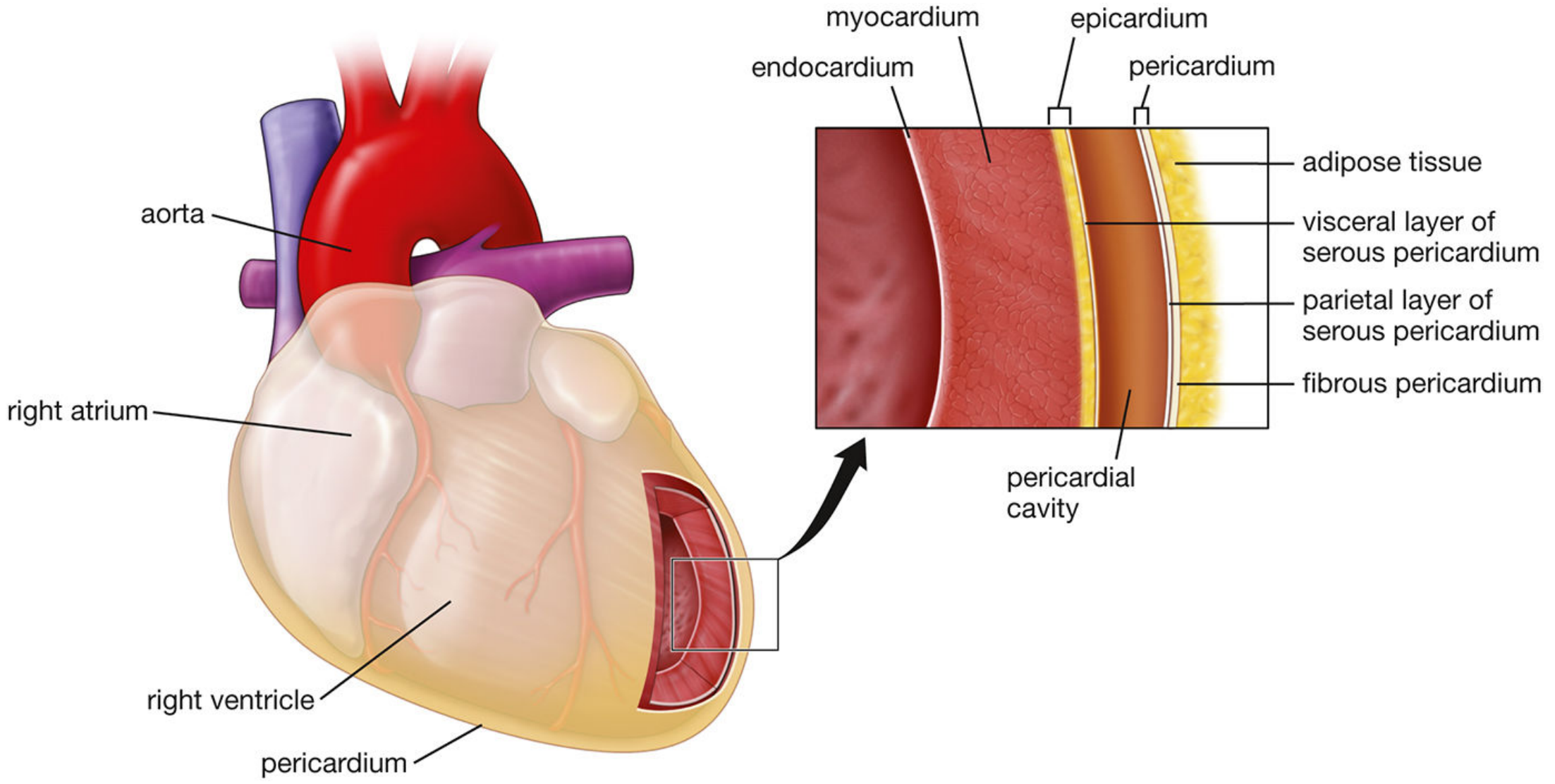
The wall has three layers (outside to inside):
| Layer | Description |
|---|---|
| Epicardium | Outer layer = visceral layer of serous pericardium. Single layer of mesothelial cells + connective/adipose tissue. Contains coronary arteries and cardiac veins. |
| Myocardium | Middle layer = cardiac muscle. The principal pumping component. Thicker in the left ventricle (must pump to the whole body). |
| Endocardium | Inner layer = endothelium + subendothelial connective tissue + subendocardial layer containing cells of the conducting system. |
The Pericardium
The heart sits inside the pericardium, which has two layers:
- Fibrous pericardium - tough outer layer
- Serous pericardium - has two sub-layers:
- Parietal layer - lines the inner surface of the fibrous pericardium
- Visceral layer (epicardium) - adheres to the heart surface
Between the two serous layers is the pericardial cavity, containing 15-50 mL of serous fluid that reduces friction during heartbeats. Excess fluid causes cardiac tamponade.
Four Chambers
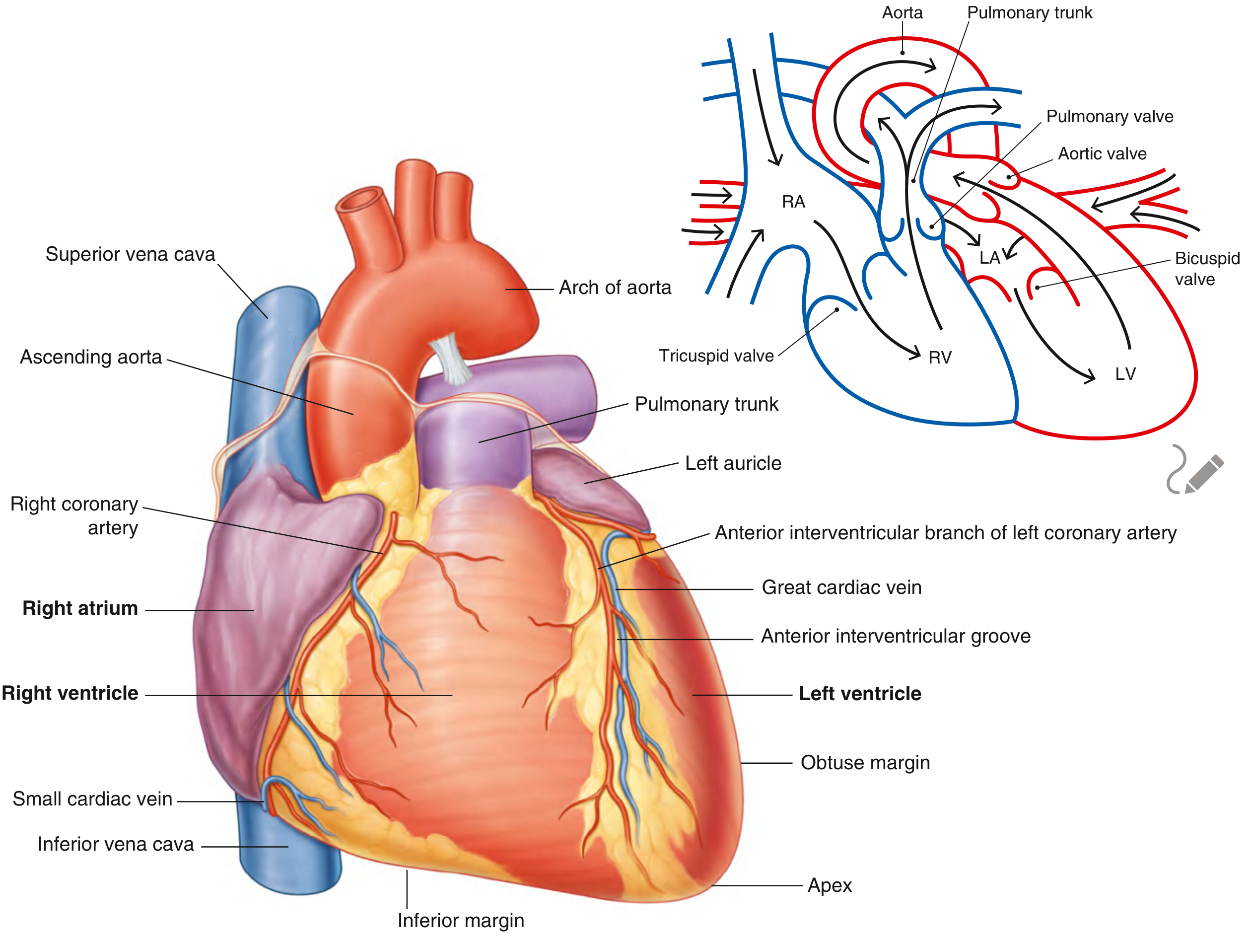
The heart has two atria (thin-walled, receive blood) and two ventricles (thick-walled, pump blood out):
Right Atrium
- Receives deoxygenated blood from the body via the superior vena cava (upper), inferior vena cava (lower), and coronary sinus (cardiac veins)
- Interior divided by the crista terminalis into:
- Sinus of venae cavae (posterior - smooth wall)
- Atrium proper + right auricle (anterior - rough walls with pectinate muscles)
- Blood exits through the tricuspid valve into the right ventricle
Right Ventricle
- Thin-walled (low pressure - pumps to lungs only)
- Receives blood from right atrium through the tricuspid (right AV) valve
- Pumps blood out through the pulmonary valve into the pulmonary trunk → lungs
Left Atrium
- Receives oxygenated blood from lungs via four pulmonary veins
- Smooth interior walls
- Blood exits through the mitral (bicuspid) valve into the left ventricle
Left Ventricle
- Thick, powerful walls (generates high pressure to pump blood to the entire body)
- Separated from right ventricle by the interventricular septum
- Pumps blood out through the aortic valve into the aorta
Valves
| Valve | Location | Function |
|---|---|---|
| Tricuspid (3 cusps) | Right AV orifice | Prevents backflow from RV to RA during systole |
| Mitral/Bicuspid (2 cusps) | Left AV orifice | Prevents backflow from LV to LA during systole |
| Pulmonary | RV - pulmonary trunk junction | Prevents backflow from pulmonary trunk to RV |
| Aortic | LV - aorta junction | Prevents backflow from aorta to LV |
The AV valves (tricuspid and mitral) are anchored by chordae tendineae to papillary muscles projecting from ventricular walls - these prevent the valve leaflets from inverting during contraction.
Each valve leaflet has three histological layers: fibrosa (collagen core), spongiosa (loose CT), and ventricularis (semilunar valves) or atrialis (AV valves).
Conducting System
The heart initiates and propagates its own rhythmic contractions:
- SA node (sinoatrial) - in the right atrium near the SVC; the pacemaker (~60-100 bpm)
- AV node (atrioventricular) - at the junction of atria and ventricles; delays conduction
- Bundle of His - transmits impulse to the ventricles via the interventricular septum
- Left and right bundle branches - run down each side of the septum
- Purkinje fibers - spread impulse through the ventricular myocardium from apex upward
Rate is increased by sympathetic nerves and decreased by parasympathetic (vagal) stimulation.
Coronary Blood Supply
- Left coronary artery (LCA) - gives off the:
- Anterior interventricular (LAD) branch - supplies anterior LV and septum
- Circumflex branch - supplies left atrium and posterior LV
- Right coronary artery (RCA) - runs in the coronary sulcus; supplies the right atrium, RV, and (usually) the SA and AV nodes
Coronary arteries fill primarily during diastole (when the myocardium is relaxed and not compressing the vessels).
External Sulci
- Coronary sulcus - horizontal groove separating atria from ventricles; contains the RCA, circumflex artery, coronary sinus
- Anterior interventricular sulcus - contains the LAD and great cardiac vein
- Posterior interventricular sulcus - contains the posterior interventricular artery and middle cardiac vein
Sources: Gray's Anatomy for Students; Histology: A Text and Atlas (Pawlina); Goldman-Cecil Medicine
Kidney cross section
Finding Sources
Finding Sources
Finding Sources
Finding Sources
Reading File
Reading File
Loading Image
Loading Image
Reading File
Loading Image
Reading File
Loading Image
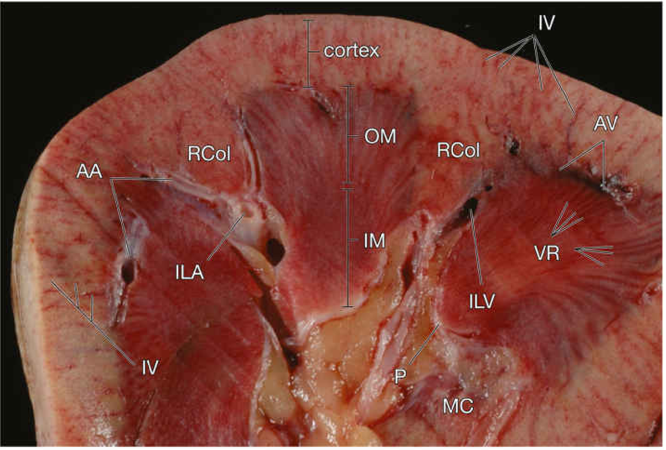
Here is a complete, image-rich explanation of the kidney cross section, drawn from Guyton & Hall, Histology: A Text and Atlas, and Campbell-Walsh Urology.
Kidney Cross Section
Overview Diagram
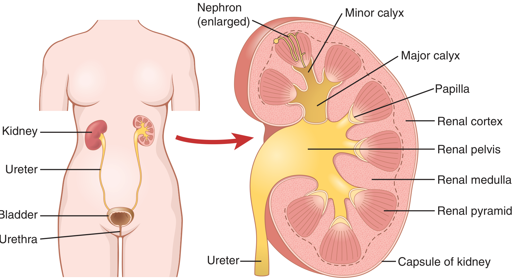
Each kidney weighs ~150 g and lies retroperitoneally on the posterior abdominal wall. Bisecting the kidney from top to bottom reveals two major zones and a central collecting system.
1. Fibrous Capsule
The outermost tough layer that encloses and protects the delicate inner structures of the kidney.
2. Renal Cortex (Outer Region)
- Reddish-brown in the fresh specimen; the outermost parenchymal zone just beneath the capsule
- Contains the main filtration structures:
- Renal corpuscles (glomerulus + Bowman's capsule) - easily identified in histology as spherical structures
- Proximal convoluted tubules (PCT)
- Distal convoluted tubules (DCT)
- Also contains cortical labyrinths (areas between medullary rays with corpuscles and convoluted tubules)
- Medullary rays - cone-shaped extensions of medullary tissue projecting into the cortex, containing straight tubules and collecting ducts
- Renal columns (of Bertin) - extensions of cortical tissue that dip down between the pyramids, separating them
3. Renal Medulla (Inner Region)
- Paler than the cortex; divided into 8-18 renal pyramids (typically 8-12 in humans)
- Each pyramid is a cone-shaped mass with:
- Base - faces the cortex (at the corticomedullary junction)
- Apex (papilla) - points toward the renal sinus and projects into a minor calyx
The medulla has two sub-zones:
- Outer medulla - further divided into outer stripe and inner stripe; contains thick limbs of the loop of Henle and collecting ducts; straight vessels (vasa recta) are prominent here
- Inner medulla - lighter, more homogeneous; contains thin limbs of the loop of Henle and inner medullary collecting ducts
4. Renal Pyramids
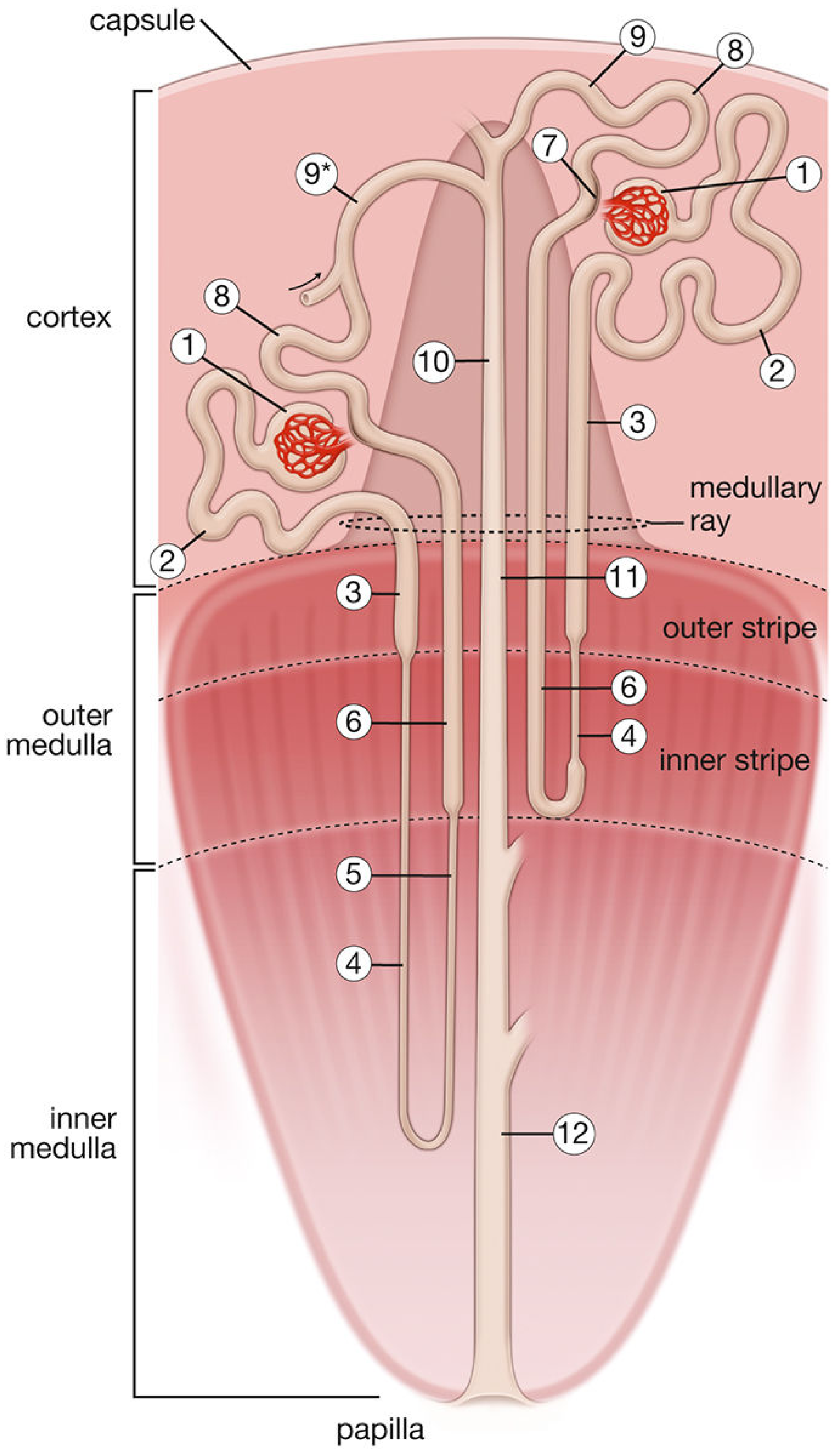
Each pyramid contains parallel arrays of tubules and collecting ducts arranged in a striated pattern. The area cribrosa at the tip of the papilla is perforated by 10-25 openings of the collecting ducts that deliver urine into the minor calyx.
5. Collecting System (Renal Sinus)
The central space (renal sinus) contains fat and the branching urine-collecting passages:
| Structure | Description |
|---|---|
| Minor calyx | Cup-shaped; cups around each papilla; receives urine directly from collecting ducts |
| Major calyx | 2-3 per kidney; formed by fusion of several minor calyces |
| Renal pelvis | Funnel-shaped; formed by fusion of major calyces; narrows at the hilum to become the ureter |
| Ureter | Carries urine to the bladder; walls contain smooth muscle that propels urine by peristalsis |
6. Hilum
The medial indented region through which pass (from front to back):
- Renal vein (most anterior)
- Renal artery
- Ureter (most posterior)
- Lymphatics and autonomic nerves
7. Blood Supply (seen in cross section)
The arterial supply follows this hierarchy:
Renal artery → Interlobar arteries (run between pyramids in renal columns) → Arcuate arteries (arch along the corticomedullary junction) → Interlobular arteries (radiate outward into cortex) → Afferent arterioles → Glomerular capillaries → Efferent arterioles → Peritubular capillaries / Vasa recta
- Vasa recta - long straight capillaries that dip into the medulla alongside the loops of Henle; essential for the countercurrent exchange mechanism that concentrates urine
8. The Nephron - Functional Unit
There are ~2 million nephrons per kidney. Each consists of:
| Segment | Location | Function |
|---|---|---|
| Renal corpuscle (glomerulus + Bowman's capsule) | Cortex | Filtration of blood |
| Proximal convoluted tubule (PCT) | Cortex | Bulk reabsorption (~65% of filtrate) |
| Loop of Henle | Cortex → Medulla → Cortex | Concentration gradient creation |
| Distal convoluted tubule (DCT) | Cortex | Fine-tuned reabsorption/secretion |
| Collecting duct | Cortex → Medulla → Papilla | Final urine concentration; ADH-sensitive |
Two types of nephrons exist:
- Cortical nephrons (85%) - short loops of Henle, stay in outer medulla
- Juxtamedullary nephrons (15%) - long loops reaching deep into the inner medulla; critical for urine concentration
Sources: Guyton and Hall Textbook of Medical Physiology; Histology: A Text and Atlas (Pawlina); Campbell-Walsh-Wein Urology
How does the nephron filter and concentrate urine?
Finding Sources
Finding Sources
Finding Sources
Finding Sources
Reading File
Reading File
Reading File
Finding Sources
Loading Image
Loading Image
Finding Sources
Reading File
Finding Sources
Reading File
Loading Image
Here is a complete, step-by-step explanation of how the nephron filters and concentrates urine, with diagrams from Guyton & Hall and Morgan & Mikhail.
How the Nephron Filters and Concentrates Urine
The nephron uses three fundamental processes to turn 125 mL/min of filtered plasma into just ~1 mL/min of final urine:
Urinary Excretion = Filtration - Reabsorption + Secretion
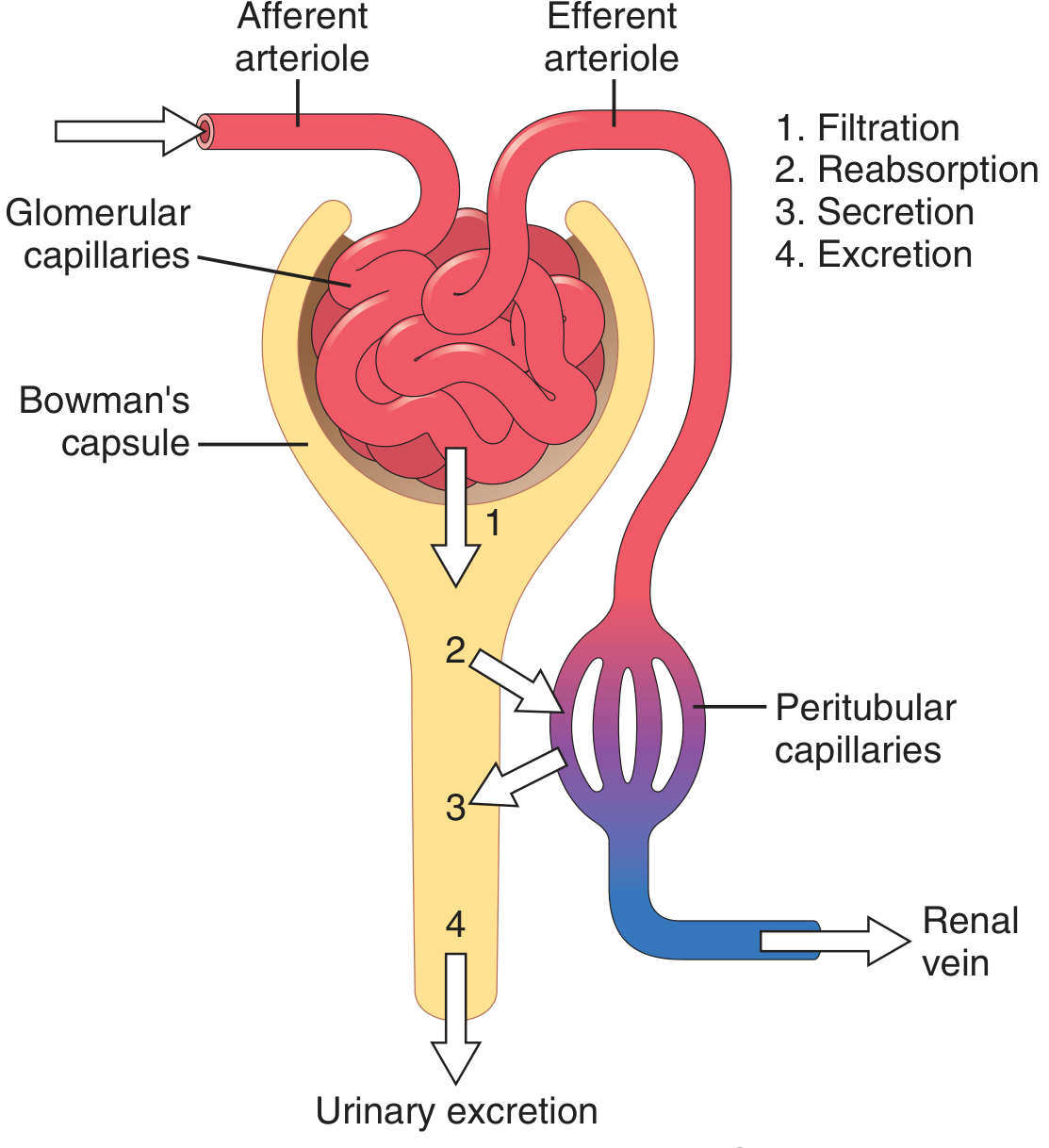
Step 1 - Glomerular Filtration (Renal Corpuscle)
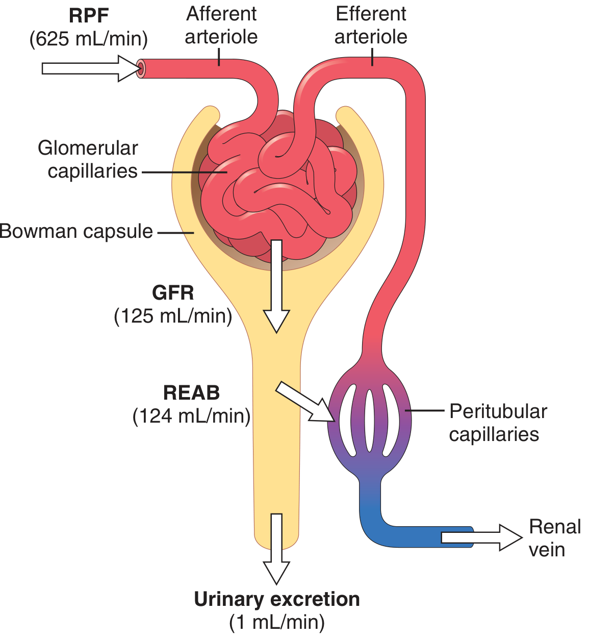
Blood enters the glomerulus via the afferent arteriole under high hydrostatic pressure (~60 mmHg). This pressure forces fluid across the three-layered filtration barrier:
| Layer | Structure | Role |
|---|---|---|
| Fenestrated endothelium | Large pores; negatively charged proteins | Blocks cells; slows proteins |
| Glomerular basement membrane | Collagen + proteoglycan mesh; negative charge | Main barrier to plasma proteins |
| Podocytes (epithelium) | Foot processes (pedicels) with filtration slit diaphragms (nephrin, podocin) | Final filter against protein leak |
What gets filtered: Water, electrolytes (Na+, K+, Cl−), glucose, amino acids, urea, creatinine - all freely filtered. Proteins and cells do NOT pass.
GFR = 125 mL/min (~180 L/day filtered). Urine output is only ~1-2 L/day, meaning >99% of filtered fluid is reabsorbed.
The filtration rate is governed by:
GFR = Kf × [(P_GC - P_BS) - π_GC] (Kf = filtration coefficient; P_GC = glomerular capillary pressure; P_BS = Bowman's space pressure; π_GC = oncotic pressure)
Step 2 - Proximal Convoluted Tubule (PCT) — Bulk Reabsorption
The PCT reabsorbs ~65-67% of the filtered load of Na+, water, and most solutes:
- Na+ is actively pumped out by Na+/K+-ATPase on the basolateral side, creating a gradient that drives Na+ in from the lumen
- Glucose and amino acids: reabsorbed by secondary active transport via SGLT2 (90%) and SGLT1 (10%) co-transporters - virtually 100% of filtered glucose is recovered here (this is the target of SGLT2 inhibitors like dapagliflozin)
- HCO₃⁻: reabsorbed via Na+/H+ exchanger (NHE3)
- Water: follows osmotically through aquaporin-1 (AQP1) channels
- Urea, phosphate, uric acid, low-molecular-weight proteins: also reclaimed here
Fluid leaving the PCT is iso-osmotic (~300 mOsm/L) - large volume removed but osmolality unchanged.
Step 3 - Loop of Henle — Building the Concentration Gradient
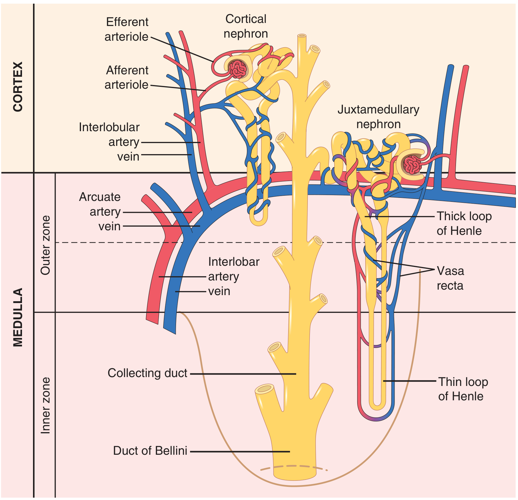
This U-shaped structure is the engine of urine concentration. It works by a countercurrent multiplier mechanism:
Descending Limb (thin)
- Freely permeable to water, relatively impermeable to solutes
- Water is drawn out into the hypertonic medullary interstitium → tubular fluid becomes progressively more concentrated as it descends to the papilla
Ascending Limb (thin then thick)
- Impermeable to water - this is the key
- The thick ascending limb (TAL) actively pumps Na+/K+/2Cl− out via the NKCC2 cotransporter (the target of loop diuretics like furosemide)
- Cl− and Na+ leave into the interstitium, but water cannot follow → tubular fluid becomes hypotonic (~100-200 mOsm/L) as it exits the loop
Net effect: A progressively increasing osmotic gradient builds in the medullary interstitium, from ~300 mOsm/L at the corticomedullary junction to ~1200 mOsm/L at the papilla. This "standing osmotic gradient" is maintained by continuous pumping - the countercurrent multiplier effect.
Urea also contributes significantly to medullary hypertonicity (recycled from the inner medullary collecting duct back into the interstitium).
Step 4 - Distal Convoluted Tubule (DCT)
- Receives hypotonic fluid from the loop of Henle
- Tight junctions - relatively impermeable; maintains the gradient set up by the loop
- Reabsorbs ~5% of filtered Na+ via the NCC cotransporter (target of thiazide diuretics)
- Major site of PTH- and vitamin D-mediated Ca²+ reabsorption
- The macula densa cells (specialized DCT cells at the glomerulus) sense NaCl delivery and regulate GFR via tubuloglomerular feedback
Step 5 - Collecting Duct — Final Concentration (ADH-controlled)
The collecting duct passes down through the progressively hypertonic medullary interstitium. Whether urine is concentrated or dilute depends entirely on ADH (antidiuretic hormone / vasopressin):
With ADH (dehydration, low blood volume):
- ADH released from posterior pituitary
- Binds V2 receptors on collecting duct cells
- Triggers insertion of aquaporin-2 (AQP-2) water channels into the apical membrane
- Water flows out of the tubule down the osmotic gradient into the hypertonic interstitium → small volume of concentrated urine (up to ~1200 mOsm/L)
Without ADH (overhydration):
- AQP-2 channels absent from luminal membrane
- Water cannot leave the tubule
- Large volume of dilute urine produced (~50-100 mOsm/L)
- Absence of ADH response = diabetes insipidus
Aldosterone (from adrenal cortex) also acts on the collecting duct: it upregulates ENaC (epithelial Na+ channels) and the Na+/K+-ATPase, increasing Na+ reabsorption and K+ secretion.
Step 6 - Vasa Recta — Countercurrent Exchange
The vasa recta (straight capillaries alongside the loop of Henle) act as a countercurrent exchanger:
- As blood descends into the hypertonic medulla, NaCl and urea diffuse in and water diffuses out
- As blood ascends, the process reverses
- Net result: solutes are recycled within the medulla and the osmotic gradient is preserved rather than washed away
Summary Table
| Segment | % Na+ Reabsorbed | Key Feature |
|---|---|---|
| PCT | ~65% | Bulk reabsorption; glucose, AA, HCO₃⁻ |
| Thin descending loop | - | Water leaves; fluid concentrates |
| Thick ascending loop | ~25% | Active NaCl pump; water stays → dilutes fluid |
| DCT | ~5% | Fine-tuning; thiazide-sensitive NCC |
| Collecting duct | ~3-5% | ADH-dependent final concentration; aldosterone-sensitive |
| Total reabsorbed | >99% | Only ~1 mL/min excreted |
Sources: Guyton and Hall Textbook of Medical Physiology; Histology: A Text and Atlas (Pawlina); Morgan & Mikhail's Clinical Anesthesiology
This is a shared conversation. Sign in to Orris to start your own chat.