
Tell me everything about Cushing’s syndrome
Cushing's syndrome clinical features buffalo hump moon face
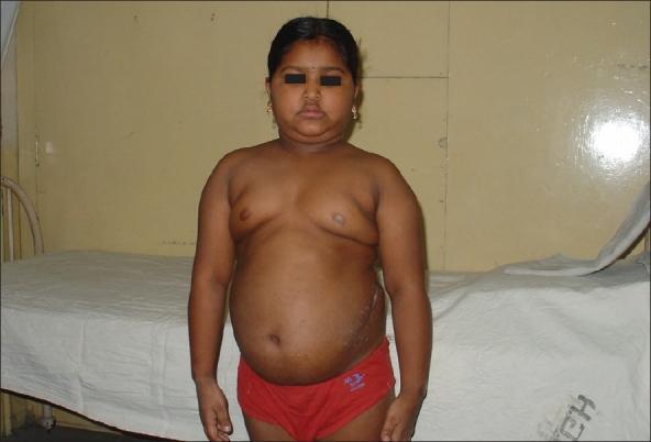
This clinical photograph shows a pediatric patient presenting with characteristic physical features of Cushing syndrome. The face exhibits a rounded, 'moon facies' appearance with associated submental fat deposition (double chin). There is evidence of central adiposity with a protuberant abdomen and fullness in the supraclavicular and upper chest regions, consistent with truncal obesity. A visible dorsocervical fat pad, or 'buffalo hump', is present at the base of the neck. On the patient's left flank, a linear surgical scar is visible, indicating a prior operative intervention. These findings are visually representative of hypercortisolism, which in this case is associated with an ACTH-secreting ganglioneuroma. The image serves as an educational example for identifying the systemic manifestations of glucocorticoid excess in children.
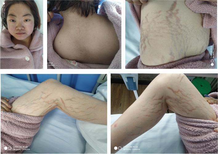
A composite of five clinical photographs illustrating the classic physical manifestations of Cushing's syndrome in a 26-year-old female. The top-left image shows a frontal view of the face, demonstrating a 'moon face' appearance characterized by a rounded facial contour and prominent, plethoric (flushed) cheeks. The top-center photograph displays the posterior cervical and upper thoracic region, showing a significant dorsocervical fat pad, commonly referred to as a 'buffalo hump.' The remaining three images (top-right, bottom-left, and bottom-right) focus on the patient's trunk and limbs, revealing wide, violaceous (purplish) striae distensae across the abdomen and thighs. These striae appear as deep, linear streaks indicating thinning of the dermis. Collectively, these visual findings are hallmark cutaneous and structural signs of chronic hypercortisolism, which in this clinical context was secondary to a left adrenal adenoma. The images serve as educational references for endocrinology and dermatology, highlighting the systemic physical changes associated with cortisol excess.
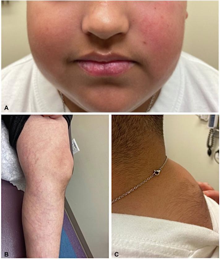
A composite of three clinical photographs demonstrating classic Cushingoid features in a pediatric patient. Panel A: Close-up of the face showing 'moon facies' characterized by rounded cheeks and noticeable facial plethora (erythema). Panel B: View of the left lower extremity showing multiple wide, slightly depressed, erythematous-to-violaceous striae (skin stretch marks) distributed across the proximal thigh and distal leg. Panel C: Lateral view of the upper back and neck region showing a prominent dorsocervical fat pad (buffalo hump) accompanied by significant hypertrichosis (increased hair growth). These visual markers are key clinical indicators of Cushing syndrome, whether endogenous or iatrogenic. The image provides educational value for medical students and clinicians in identifying dermatological and morphological manifestations of hypercortisolism.
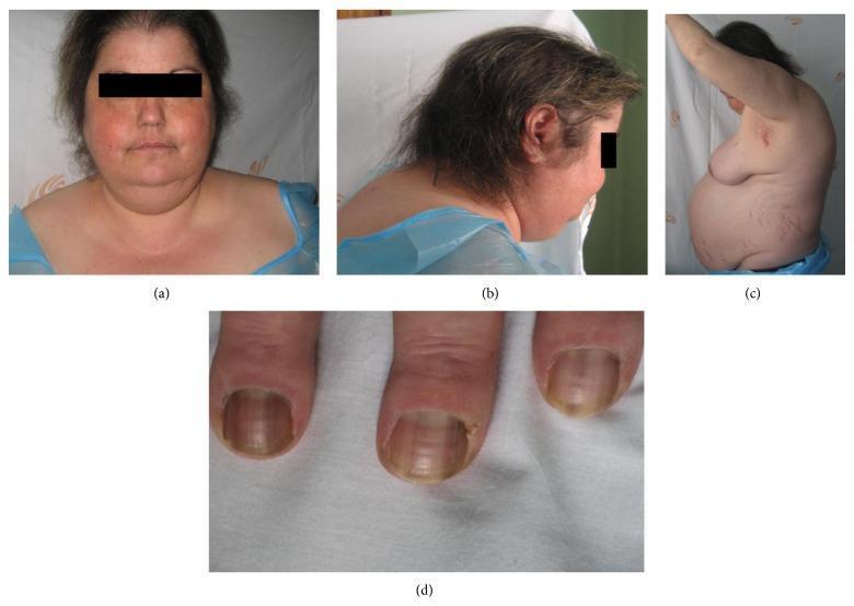
A composite of four clinical photographs demonstrating the classic physical manifestations of Cushing's syndrome. Image (a) is a frontal view of the face showing 'moon facies' characterized by facial rounding and plethora. Image (b) provides a lateral view of the upper back and neck, highlighting a prominent dorsocervical fat pad (buffalo hump). Image (c) shows a profile of the torso, illustrating centripetal obesity and wide, purplish striae distensae along the abdomen and flanks. Image (d) is a close-up of the fingernails, displaying hyperpigmentation and longitudinal ridging, which may suggest associated adrenocorticotropic hormone (ACTH) excess. The collection serves as an educational reference for medical students to identify systemic signs of hypercortisolism, including skin atrophy and telangiectasias.
Cushing syndrome diagnosis treatment
Cushing's Syndrome: A Comprehensive Overview
Definition
"Cushing disease" specifically refers to hypercortisolism caused by an ACTH-secreting pituitary adenoma. "Cushing syndrome" is the broader term encompassing all causes.
Pathophysiology and the HPA Axis
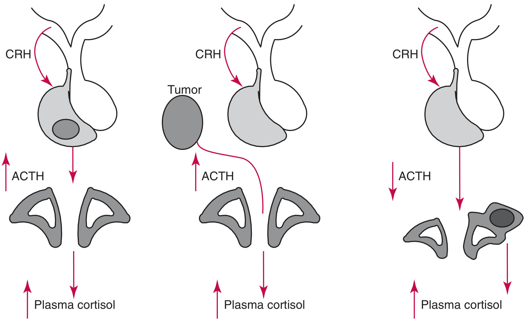
- The hypothalamus releases CRH (corticotropin-releasing hormone) - driven by stress, circadian rhythm, and low serum cortisol.
- CRH stimulates the anterior pituitary's corticotropic cells to secrete ACTH (cleaved from the pro-opiomelanocortin [POMC] precursor).
- ACTH drives the adrenal cortex to produce cortisol.
- Cortisol exerts negative feedback on both the hypothalamus and pituitary, suppressing CRH and ACTH.
Classification / Causes
1. Exogenous (Iatrogenic) - MOST COMMON
- Chronic use of exogenous corticosteroids (systemic, topical, inhaled, nasal, conjunctival)
- Serum cortisol is low (adrenal cortex is suppressed)
- Especially common in children with topical applications
2. ACTH-Dependent (~80% of endogenous cases)
| Subtype | Details | % of Endogenous |
|---|---|---|
| Cushing disease (pituitary adenoma) | Microadenoma secreting ACTH; bilateral adrenal hyperplasia | ~68% |
| Ectopic ACTH syndrome | ACTH secreted by non-pituitary tumors (small cell lung cancer, carcinoid, pheochromocytoma, MEN2A) | ~12% |
| Ectopic CRH secretion | Rare; CRH-producing tumors stimulate pituitary | Rare |
3. ACTH-Independent (~20% of endogenous cases)
| Subtype | Details |
|---|---|
| Adrenal adenoma | Unilateral, benign; autonomous cortisol production; contralateral adrenal atrophies |
| Adrenal carcinoma | Often large, unilateral, aggressive |
| Bilateral adrenal hyperplasia | Includes PPNAD (Primary Pigmented Nodular Adrenocortical Disease) - seen in 30% of Carney complex |
| McCune-Albright syndrome | Rare association |
| MEN type I | Rare feature |
4. Pseudo-Cushing Syndrome
Clinical Features
Central / Fat Redistribution
- Moon facies - rounded face from fat deposition
- Buffalo hump (dorsocervical fat pad) - fat deposition at the back of the neck/upper thoracic region
- Central/truncal obesity - with sparing of the limbs
- Supraclavicular fat pads
- Submental fat deposition ("double chin")
Skin (Dermatologic Signs - highly characteristic)
- Wide, violaceous (purple) striae - abdomen, buttocks, breasts, upper arms, thighs (>1 cm wide, purplish; unlike pale stretch marks of obesity)
- Skin thinning and fragility - "cigarette paper" wrinkling
- Easy bruising / ecchymoses
- Plethora - reddish flush of the cheeks, anterior neck, and V of the chest
- Hypertrichosis - facial lanugo in women
- Acne
- Poor wound healing
- Susceptibility to superficial fungal infections (dermatophyte, Pityrosporum)
- Liddle sign - skin peels off easily when adhesive tape is removed
- Occasionally: livedo reticularis, purpura, poikiloderma-like changes
Musculoskeletal
- Proximal muscle weakness (myopathy) - difficulty rising from a chair
- Osteoporosis - vertebral fractures, kyphosis, back pain
- Loss of height
Cardiovascular / Metabolic
- Hypertension (very common, due to mineralocorticoid effects of cortisol)
- Generalized arteriosclerosis
- Hyperglycemia / diabetes mellitus - in ~20% of patients
- Hypokalemia
- Dyslipidemia
Endocrine / Reproductive
- Hirsutism and oligomenorrhea/amenorrhea in women (androgen co-secretion)
- Loss of libido in men and women
- Hypogonadotropic hypogonadism
Neuropsychiatric
- Depression, anxiety, emotional lability
- Psychosis (in severe cases)
- Cognitive impairment
Other
- Hyperpigmentation - only in ACTH-dependent causes (excess ACTH stimulates melanocortin receptors); especially noticeable on knuckles, oral mucosa, scars
- Polycythemia
- Urolithiasis (nephrolithiasis)
- Immunosuppression
Distinguishing Cushing's Syndrome from Simple Obesity
- Wide purple striae (>1 cm)
- Proximal muscle weakness
- Skin thinning and easy bruising
- Unexplained osteoporosis in a young person
Diagnosis
Phase 1 - Screening (choose one of three)
| Test | Principle | Sensitivity | Notes |
|---|---|---|---|
| 24-hour urinary free cortisol (UFC) | Direct measure of free bioavailable cortisol; integrated over 24h | ~95% | Values >4× upper limit of normal are diagnostic; unreliable in GFR <30 mL/min; may miss subclinical disease |
| Overnight low-dose dexamethasone suppression test (DST) | 1 mg dexamethasone at 11 PM; measure serum cortisol at 8 AM | ~95% | Failure to suppress cortisol to <50 nmol/L (1.8 μg/dL) = positive. Preferred for incidentaloma workup |
| Late-night salivary cortisol (LNSC) | Cortisol nadir is lost in Cushing's; persistent evening elevation | High | Practical outpatient test; reflects free cortisol |
Three laboratory hallmarks: (1) elevated cortisol (serum, UFC, or LNSC), (2) loss of circadian rhythm of ACTH/cortisol, and (3) failure to suppress cortisol with low-dose dexamethasone.
Phase 2 - Confirming Hypercortisolism (if borderline or high suspicion)
- 2-day low-dose DST (48-hour, 0.5 mg q6h dexamethasone)
- Midnight plasma cortisol (in-hospital; >207 nmol/L is diagnostic)
Phase 3 - Localizing the Source
- ACTH suppressed (<5 pg/mL) → ACTH-independent (adrenal source) → CT/MRI of adrenal glands
- ACTH elevated or normal (>15 pg/mL) → ACTH-dependent → need further testing
- Pituitary Cushing disease: cortisol suppresses by >50% (pituitary adenoma retains some sensitivity to very high-dose dexamethasone)
- Ectopic ACTH: cortisol does NOT suppress
- Pituitary MRI with gadolinium for pituitary adenoma (may be small; up to 40% are microadenomas invisible on MRI)
- CT/MRI adrenals for adrenal lesions
- CRH stimulation test and inferior petrosal sinus sampling (IPSS) for equivocal cases - IPSS is the gold standard for confirming pituitary origin and lateralizing the adenoma
Management
Cushing Disease (Pituitary Adenoma)
- First-line: Transsphenoidal surgery (TSS) - resection of the pituitary adenoma; remission rates 65-90%
- If surgery fails or recurs: repeat TSS, pituitary radiotherapy (stereotactic radiosurgery), bilateral adrenalectomy
- Nelson's syndrome: occurs after bilateral adrenalectomy when the pituitary adenoma grows aggressively due to loss of cortisol feedback (treated with radiosurgery - recent systematic review PMID 40884684)
Adrenal Adenoma (ACTH-independent)
- Unilateral adrenalectomy (laparoscopic preferred)
- Preoperatively: control of hypertension, diabetes, electrolytes
- Metyrapone (inhibits glucocorticoid synthesis) if severe symptoms need rapid control
- Critical: postoperative cortisol replacement is mandatory because the contralateral adrenal is suppressed; HPA axis recovery can take 6-24 months
Adrenal Carcinoma
- Open adrenalectomy (for large tumors)
- Mitotane (adrenocortical cytotoxic agent) - adjuvant therapy
- Consent for adjacent organ removal if needed
Ectopic ACTH Syndrome
- Treat the underlying tumor (resect if possible)
- If unresectable: bilateral adrenalectomy or medical adrenalectomy
- Medical therapy options: metyrapone, ketoconazole, mifepristone
Iatrogenic Cushing's
- Gradual tapering of exogenous corticosteroids (never abrupt discontinuation - risk of adrenal crisis)
Medical Therapy (for all types, especially pre-surgery or inoperable cases)
| Drug | Mechanism |
|---|---|
| Metyrapone | Inhibits 11β-hydroxylase → blocks cortisol synthesis |
| Ketoconazole | Inhibits multiple steroidogenic enzymes |
| Mifepristone | Glucocorticoid receptor antagonist |
| Mitotane | Adrenocortical cytotoxin; used for carcinoma |
| Pasireotide | Somatostatin analogue; reduces ACTH from pituitary |
| Cabergoline | Dopamine agonist; reduces ACTH in some pituitary tumors |
Perioperative Considerations
- Stress-dose steroids intraoperatively and for 24h post-op
- VTE prophylaxis (high thrombotic risk due to hypercoagulable state)
- Antibiotics
- Cardiopulmonary workup
- Management of diabetes and hypertension
- Monitoring for adrenal insufficiency postoperatively
Special Associations
| Condition | Notes |
|---|---|
| Carney complex | Primary Pigmented Nodular Adrenocortical Disease (PPNAD) in 30% |
| MEN1 | Rare feature |
| McCune-Albright | Rare feature |
| MEN2A | Rare ectopic Cushing's syndrome via ACTH-secreting medullary thyroid carcinoma or pheochromocytoma |
| Pseudo-Cushing | Alcohol abuse can perfectly mimic Cushing's clinically |
Key Points Summary
- Cushing's syndrome = chronic cortisol excess from any cause
- Most common cause overall = iatrogenic (exogenous steroids)
- Most common endogenous cause = Cushing disease (pituitary ACTH-secreting adenoma, ~68%)
- Hyperpigmentation only occurs in ACTH-dependent types
- Classic discriminatory features: wide purple striae, proximal myopathy, easy bruising, thin skin, unexplained osteoporosis
- Diagnosis requires biochemical confirmation + source localization
- The three screening tests are 24-hr UFC, overnight DST, and late-night salivary cortisol
- Definitive treatment is surgical in most cases
- Adrenal suppression after cure requires steroid replacement during recovery