
Septic arthritis in CKD patient
"septic arthritis" AND "chronic kidney disease"
PMID: 40800124
Septic Arthritis in CKD Patients
Why CKD Changes Everything
- Uremia-induced immune dysfunction - uremia impairs polymorphonuclear (PMN) cell chemotaxis, phagocytosis, and oxidative burst, producing a functional immunocompromised state even without immunosuppressive drugs. Goldman-Cecil explicitly lists uremia among the metabolic conditions contributing to the "net state of immunosuppression."
- Repeated vascular access - patients on hemodialysis have percutaneous vascular access established several times weekly, creating a portal of entry for S. aureus bacteremia and subsequent hematogenous seeding of joints. The frequency of septic arthritis in hemodialysis patients is "much increased compared with the general population." (Rheumatology, 2-Volume Set, Elsevier 2022)
- Comorbidity burden - CKD frequently coexists with diabetes, vascular disease, and immunosuppression, each of which independently elevates risk.
- Atypical presentation - the uremic immunocompromised state blunts the febrile response and inflammatory markers. The synovial WBC count may be below the usual 50,000/µL threshold despite true infection.
Epidemiology & Organisms
| Clinical Setting | Most Likely Organism |
|---|---|
| Native joint (CKD/HD) | S. aureus (incl. MRSA - HD is a major MRSA risk factor) |
| Hemodialysis access | S. aureus, coagulase-negative staphylococci |
| Immunocompromised/CKD | Gram-negative bacilli (Pseudomonas, Enterobacteriaceae) |
| CKD + diabetes | S. aureus, gram-negatives, atypical organisms |
| IV drug use + CKD | Pseudomonas aeruginosa, S. aureus |
Diagnostic Challenges in CKD
Attenuated Inflammatory Response
- CKD patients may lack fever (>80% of immunocompetent adults have fever; this proportion is lower in the immunocompromised/CKD population)
- ESR and CRP are chronically elevated at baseline in CKD, reducing their diagnostic specificity
- A synovial WBC count < 50,000/µL does NOT exclude septic arthritis in immunocompromised patients - this is a critical pitfall
Diagnostic Mimics in CKD (especially relevant)
- β2-microglobulin amyloid arthropathy - occurs with long-term HD; amyloid deposits cause joint swelling, carpal tunnel, destructive arthropathy. Synovial fluid cell count may be inflammatory.
- Crystal arthropathy - CKD predisposes to gout (urate retention) and pseudogout (calcium pyrophosphate). In CKD, however, crystal presence does NOT exclude septic arthritis - both can coexist when synovial WBC > 50,000/µL.
- Calcium hydroxyapatite deposition - especially peri-articular in HD patients
- Calcium oxalate arthropathy - in dialysis patients (excess oxalate from vitamin C metabolism)
- Erosive enthesopathy from secondary hyperparathyroidism
- Reactive arthritis from recurrent UTIs/bacteremic episodes common in CKD
Diagnostic Algorithm (Goldman-Cecil, 2024)
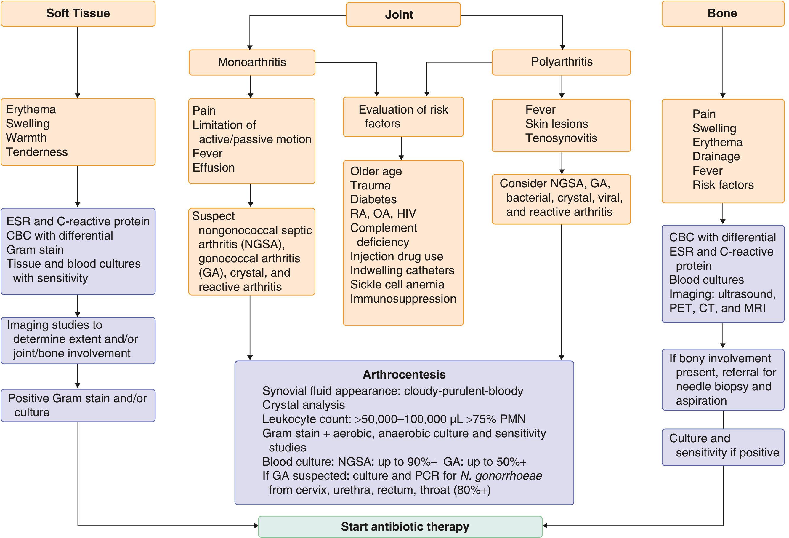
Arthrocentesis is Mandatory
- Synovial fluid: appearance (cloudy/purulent), leukocyte count + differential, crystal analysis, Gram stain, aerobic + anaerobic + fungal + mycobacterial culture
- Blood cultures: positive in up to 90% of nongonococcal septic arthritis
- In CKD with HD and line-associated bacteremia: also culture the access site
- Ultrasound or CT-guided aspiration for deep joints (hip, sacroiliac)
- The Gram stain is positive in fewer than 50% - a negative result does not rule out infection
- PCR of synovial fluid is increasingly useful, especially when prior antibiotics have been given
Treatment
General Principle
Antibiotic Selection
| Gram Stain | Empirical Choice | Rationale in CKD |
|---|---|---|
| Gram-positive cocci | Vancomycin 15 mg/kg IV q12h | MRSA risk is high in HD patients - never use nafcillin/oxacillin empirically |
| Gram-negative bacilli | Cefepime 2 g IV q8-12h, or piperacillin-tazobactam, or meropenem | Pseudomonas coverage needed |
| Negative / unknown | Vancomycin + broad gram-negative cover | Both gram-positives and gram-negatives possible in CKD |
| Organism | First Choice | Alternative |
|---|---|---|
| MSSA | Nafcillin/oxacillin 1.5-2 g IV q4-6h (4-6 wk) or cefazolin 1-2 g IV q8h | Vancomycin 15 mg/kg IV q12h; daptomycin 6-10 mg/kg IV q24h |
| MRSA | Vancomycin 15 mg/kg IV q12h or daptomycin 6-10 mg/kg IV q24h | Linezolid 600 mg PO/IV q12h; dalbavancin |
| Streptococci (pen-sensitive) | Penicillin G 20 MU/24h IV or ceftriaxone 2 g IV q24h | Vancomycin |
| Enterobacteriaceae | Ceftriaxone 1-2 g IV q24h | Ciprofloxacin 500-750 mg PO q12h |
| P. aeruginosa | Cefepime 2 g IV q8-12h | Ceftazidime 2 g IV q8h; imipenem 500 mg IV q6h; meropenem |
CKD-Specific Drug Dose Adjustments
"Doses shown are based on normal renal and hepatic function and may need to be adjusted or serum levels monitored (vancomycin)." - Goldman-Cecil, Table 251-2 footnote
| Drug | CKD/HD Consideration |
|---|---|
| Vancomycin | Requires therapeutic drug monitoring (trough or AUC-guided); dose interval extended in CKD; HD patients need redosing after each session |
| Daptomycin | Dose every 48h (not 24h) when CrCl < 30 mL/min; HD patients dose after dialysis |
| Cefepime | Dose reduce when CrCl < 60 mL/min; neurotoxicity risk in CKD if not adjusted |
| Ciprofloxacin | Reduce dose in severe CKD (CrCl < 30); use with caution - QTc prolongation risk also relevant |
| Imipenem/Meropenem | Dose reduce in CKD; imipenem has a lower seizure threshold at high doses in renal failure |
| Aminoglycosides (gentamicin) | Avoid or use with extreme caution + TDM in CKD due to nephrotoxicity and ototoxicity |
| Linezolid | No renal dose adjustment needed - useful as an oral option in severe CKD |
Duration of Therapy
- Uncomplicated, susceptible organism, non-immunocompromised: 2 weeks (post-drainage)
- CKD/immunocompromised, abnormal joint, MRSA, gram-negative, or complicating osteomyelitis/bacteremia/endocarditis: 4 to 6 weeks (Firestein & Kelley's Textbook of Rheumatology)
- Consider IV-to-oral switch after 2 weeks if clinical improvement + inflammatory markers falling + bioavailable oral agent available
- Immunosuppression and lack of surgical intervention are associated with recurrence
Joint Drainage
- Serial needle aspiration is first-line for most accessible joints
- Surgical drainage is indicated for:
- Hip joint (always)
- Failure to respond to serial aspiration + antibiotics
- Developing life-threatening complications (e.g., necrotizing fasciitis)
- Arthroscopic lavage is an option where available
- Early mobilisation to prevent contractures
Prognosis in CKD
- ~40% of all septic arthritis patients have less than complete recovery
- CKD worsens outcomes through delayed diagnosis, higher MRSA burden, impaired healing
- S. aureus infection alone carries >50% poor joint outcomes and 10-15% mortality - this rate is further elevated in immunocompromised/CKD patients
- Polyarticular sepsis (more common in immunocompromised patients including CKD) carries especially high mortality
Special Considerations: Dialysis-Related Arthropathy
Key Clinical Pearls for CKD
- Don't rely on fever or high WBC to diagnose - both may be blunted in uremia
- Don't rely on a "low" synovial WBC to exclude - counts < 50,000/µL can still represent infection in CKD
- Cover MRSA empirically - hemodialysis is a major risk factor
- Crystal arthritis + infection can coexist - crystals in synovial fluid do not exclude septic arthritis
- Adjust all renally-cleared antibiotics - vancomycin requires TDM; avoid or carefully dose aminoglycosides
- Extend antibiotic duration - 4-6 weeks in immunocompromised/CKD hosts
- Always culture blood + dialysis access site - hematogenous spread from access is a common source
- Screen for endocarditis - S. aureus bacteremia in HD patients has high rates of endocarditis; consider echocardiography
- Goldman-Cecil Medicine, International Edition (PMID-linked textbook, Section 251)
- Harrison's Principles of Internal Medicine, 22nd Ed, 2025 (Chapter on nongonococcal bacterial arthritis)
- Firestein & Kelley's Textbook of Rheumatology (Antibiotic therapy chapter)
- Rheumatology, 2-Volume Set, Elsevier 2022 (Dialysis-related arthropathy section)
- Campbell's Operative Orthopaedics, 15th Ed 2026 (MRSA risk factors)
- Khudair A et al. "The prognostic implications of comorbidities in septic arthritis." Front Med (Lausanne) 2025. PMID: 40800124
Spine TB osteomyelitis in CKD patient on MHD 3/7
"spinal tuberculosis" AND "chronic kidney disease" OR "hemodialysis"
PMID: 32314489
PMID: 40023546
spinal tuberculosis Pott disease MRI vertebral osteomyelitis
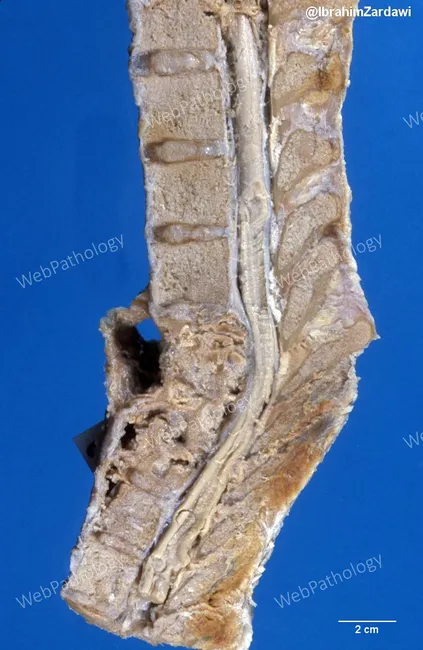
Comprehensive Description: Gross pathology photograph of the lumbar spine showing advanced tuberculous spondylitis (Pott disease). Modality and technique: macroscopic photograph of a sagittal spinal section derived from a lower thoracic–lumbar segment; scale bar included (2 cm). Anatomical context: involves three contiguous lumbar vertebrae with adjacent intervertebral discs, consistent with TB‑related destruction (likely L1–L3). Imaging perspective: sagittal view emphasizing anterior column collapse and disc-space involvement. Visual features: extensive vertebral body destruction with irregular, porous cancellous bone; anterior vertebral column collapse; irregular endplates; disc-space narrowing; preserved posterior elements to varying degrees; surrounding soft-tissue reaction and potential paravertebral changes visible along the spinal canal. Diagnostic interpretation: gross correlate of tuberculous osteomyelitis with granulomatous inflammation; pattern of contiguous vertebral involvement and disc destruction supports tuberculous etiology over pyogenic infection or metastasis. Clinical relevance: highlights risk of spinal canal compromise and paraplegia in Pott disease; informs surgical planning for decompression and stabilization and reinforces need for systemic anti‑tuberculous therapy. Differential considerations: pyogenic spondylitis, brucellar spondylitis, spinal metastasis, fungal vertebral osteomyelitis; clinical correlation and microbiology needed. Educational use: suitable for medical students, residents, and pathology education focusing on spinal tuberculosis gross pathology features. Correlative imaging findings on MRI or CT may reveal vertebral collapse and prevertebral abscess; this specimen-based description complements clinical TB workup.
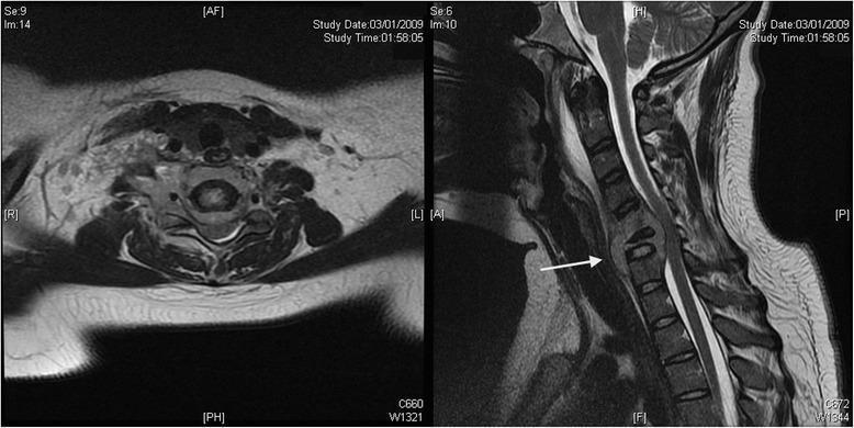
Diagnostic imaging of the cervical spine consisting of axial (left) and sagittal (right) T2-weighted fast spin-echo MRI views. The images demonstrate advanced cervical osteomyelitis and discitis, primarily involving the C6 vertebral level. In the sagittal view, there is significant T2-hyperintensity and destruction of the C6 vertebral body (indicated by a white arrow) with associated collapse. A large prevertebral abscess is visible as a high-signal intensity collection anterior to the vertebral bodies. There is evidence of posterior subligamentous extension of the infectious process into the spinal canal, resulting in focal narrowing of the canal and mild posterior displacement and compression of the cervical spinal cord. The axial view at the C6 level confirms extensive bony destruction and reveals significant circumferential soft tissue edema and inflammatory changes. This presentation is characteristic of severe spinal infection, such as vertebral blastomycosis or tuberculosis (Pott's disease), highlighting the risk of neurological compromise due to epidural extension and mechanical instability.
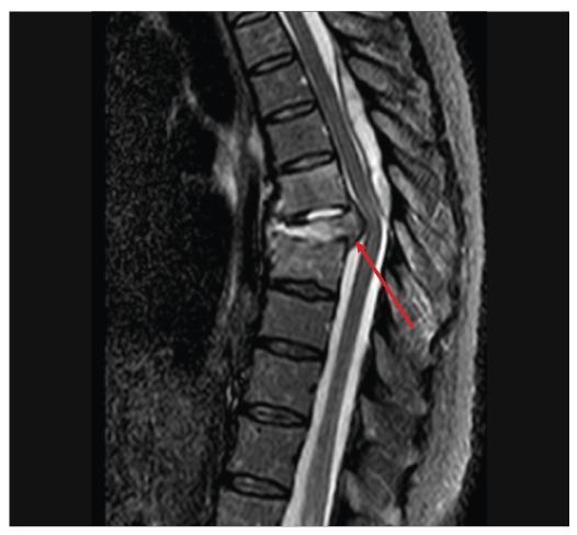
This diagnostic image is a sagittal T2 Short Tau Inversion Recovery (STIR) MRI of the thoracic spine. It demonstrates signs of spondylodiscitis at the T6-T7 level. Key visual features include hyperintensity within the T6-T7 intervertebral disc and the absence of the normal low-signal internuclear cleft, suggesting disc inflammation or edema. The adjacent T6 and T7 vertebral bodies also exhibit significant hyperintensity, indicating bone marrow edema. There is a visible vertebral collapse and a posterior inflammatory/infectious lesion, indicated by a red arrow, that is causing posterior displacement and compression of the dural sac and the spinal cord. This imaging finding is characteristic of infectious discitis-osteomyelitis with secondary neurological compromise, frequently seen in fungal infections like paracoccidioidomycosis or tuberculosis (Pott's disease) in immunosuppressed patients.
Spinal TB Osteomyelitis (Pott's Disease) in CKD on Maintenance Hemodialysis (3×/week)
Why CKD/MHD Dramatically Elevates Risk
- Impaired cell-mediated immunity - uremia suppresses T-lymphocyte function, macrophage activation, and cytokine production. This is the core mechanism allowing TB reactivation or primary progression.
- Malnutrition - common in MHD patients; hypoalbuminemia and low Hb are specific risk factors for TB spondylitis in HD patients (PMID: 32314489 - see below).
- Vitamin D deficiency - nearly universal in CKD; impairs macrophage-mediated mycobacterial killing.
- Zinc deficiency - contributes to immune dysfunction.
- Repeated dialysis access - provides a route for hematogenous dissemination from any bacteremic/mycobacteremic episode.
- Extrapulmonary TB is more common - immunocompromised MHD patients have a higher proportion of extrapulmonary TB, including skeletal/spinal involvement.
TB Spondylitis in Hemodialysis: Clinical Series Data
- Incidence: 1.5/1000 HD patients per year
- Average time from first symptom to diagnosis: 45.4 days (range 11-180 days) - delayed diagnosis is the rule
- Surgery required: 33.3% (4/12 patients)
- Outcome: 91.7% cured or improved with anti-TB therapy ± surgery
- Key risk factors: lower albumin and lower haemoglobin than matched HD controls (p=0.03 and p=0.01 respectively)
Pathology of Spinal TB (Pott's Disease)
- Acid-fast positive, caseating granulomas with or without purulence
- Abscesses expand, containing necrotic debris; skin sinuses may drain spontaneously
- Infection spreads beneath and along the anterior and posterior longitudinal ligaments, sparing the intervertebral discs in adults - this disc-sparing pattern is the hallmark that distinguishes TB from pyogenic spondylodiscitis
- Anterior vertebral body is involved first
- Progressive destruction leads to the characteristic gibbus (kyphotic) deformity
- Epidural spread causes the most severe neurological damage
- Multifocal involvement and skip lesions are well described - always image the entire spine
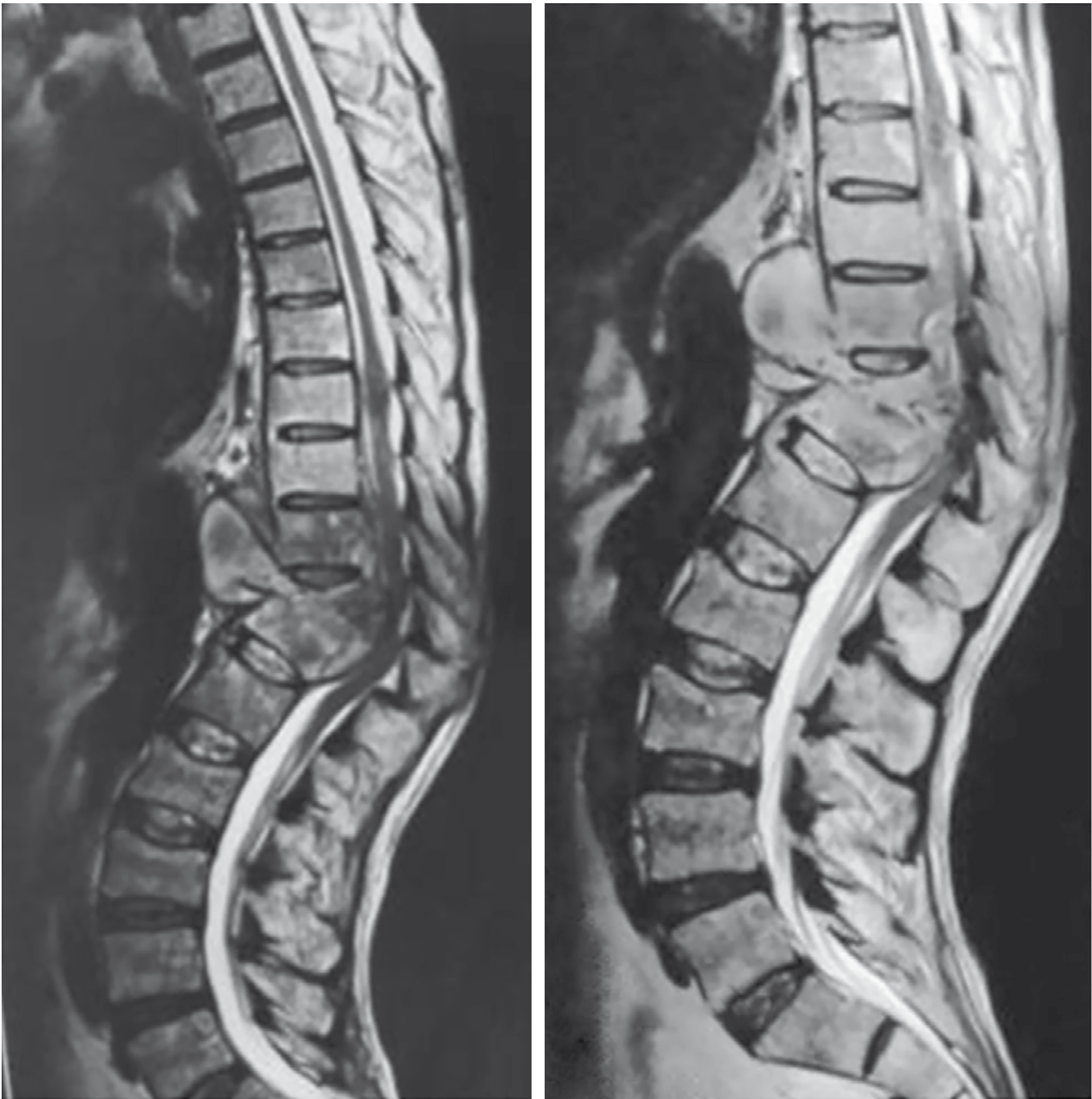
Distribution and Clinical Features
| Feature | Details |
|---|---|
| Most common site | Thoracolumbar junction > lumbar > cervical |
| Onset | Slowly progressive over weeks to months |
| Back pain | Present in 70% at presentation |
| Constitutional symptoms | Weakness, malaise, night sweats, fever, weight loss |
| Neurological deficit | ~30% in resource-limited settings; less in developed countries |
| Presentation delay | Months to years after initial symptoms |
| Cervical disease | Dysphagia, hoarseness (recurrent laryngeal nerve), stridor, risk of sudden death from vascular erosion |
Diagnostic Approach
Laboratory
- Anaemia + hypoproteinemia are common findings suggesting chronic disease (also confounded by CKD baseline)
- ESR - elevated at baseline in CKD, reducing specificity. ESR is normal in >50% of spinal TB patients regardless
- CRP - somewhat more specific but also elevated in CKD
- IGRA (QuantiFERON-TB Gold) is preferred over TST in immunocompromised patients, but both have reduced sensitivity in CKD (TST can be false negative due to anergy; IGRA indeterminate rate is higher in CKD). A negative result does NOT rule out TB.
- TST is contraindicated in patients with suspected reactivation due to risk of skin slough from intense reaction
Imaging
| Modality | Findings |
|---|---|
| Plain X-ray | Subtle disc space narrowing, localized osteopenia (early); vertebral collapse, gibbus (late) |
| CT | Bone destruction detail, paraspinal/psoas abscess delineation |
| MRI with and without contrast | Preferred - demonstrates epidural abscess, cord compression, extent of involvement. T2 hyperintensity in vertebrae and disc space; paravertebral abscess enhancement |
- TB: disc-sparing, multifocal, large paraspinal abscess ("cold abscess"), subligamentous spread, gibbus
- Pyogenic: disc destruction early, single level, fever more prominent, rapid progression
Tissue Diagnosis (Mandatory in MHD Patients)
- CT-guided or fluoroscopy-guided percutaneous biopsy of the lesion - usually adequate
- Specimens for: culture (Lowenstein-Jensen / MGIT), Gram stain, histology (for caseating granulomas), NAAT/PCR (GeneXpert MTB/RIF)
- GeneXpert gives rapid diagnosis AND rifampicin resistance detection within hours - particularly important before committing an MHD patient to a long treatment regimen
- Cultures for mycobacteria may take 4-8 weeks but are essential for drug sensitivity testing
- Blood cultures (mycobacterial) can be positive, especially in immunocompromised patients
Differential Diagnosis in MHD
| Differential | Distinguishing Features |
|---|---|
| Pyogenic spondylodiscitis | Disc destruction early, rapid course, positive blood culture for bacteria |
| Destructive spondyloarthropathy of HD | Involves 3 adjacent vertebrae + 2 discs, subluxation, specific to long-term HD - mimics TB closely on imaging |
| Metastatic malignancy | Usually no disc involvement, multiple vertebrae, known primary |
| Brucella spondylitis | Epidemiological exposure, serology |
| Fungal osteomyelitis (Aspergillus, Candida) | Immunocompromised, blood cultures, biopsy culture |
| Primary bone tumors (myeloma, chondrosarcoma) | Imaging characteristics, serum protein electrophoresis |
Anti-TB Treatment: The MHD Challenge
Standard Regimen Framework
Continuation phase (4-10 months): HR (Isoniazid + Rifampicin)
- For spinal/bone-joint TB: total duration is 9-12 months (not 6 months used for pulmonary TB) - some authorities recommend up to 18-24 months in immunocompromised hosts
- For immunocompromised patients (including MHD): continuation phase extended to total 9+ months
- Patients with normal immune function: may manage with 6 months
Anti-TB Drug Dosing in MHD - Critical Adjustments
Harrison's (22nd Ed, 2025): "Patients with chronic renal failure should never receive aminoglycosides and should receive ethambutol only if serum drug levels can be monitored."
Goodman & Gilman: "Administering drugs after hemodialysis" is the core principle.
| Drug | Standard Dose | CKD/MHD Adjustment | Key Concerns in MHD |
|---|---|---|---|
| Isoniazid (H) | 5 mg/kg/day (max 300 mg) | No dose change in renal failure; usual dose | Peripheral neuropathy risk is HIGHER in CKD - always add pyridoxine (Vitamin B6) 50 mg/day; hepatotoxicity monitoring |
| Rifampicin (R) | 10 mg/kg/day (max 600 mg) | No dose change (primarily hepatic excretion, <10% renal) | Strong CYP inducer - interacts with many drugs used in MHD (immunosuppressants, anticoagulants); hepatotoxicity |
| Pyrazinamide (Z) | 25-35 mg/kg/day | Reduce frequency to 3×/week (25-35 mg/kg TIW) if CrCl is low; redose after each hemodialysis session | Hyperuricemia (worsened by CKD); hepatotoxicity; give TIW (same days as dialysis, AFTER session) |
| Ethambutol (E) | 15-25 mg/kg/day | Switch to 15-25 mg/kg THREE TIMES A WEEK (not daily); redosed after hemodialysis | Ocular toxicity (optic neuritis, loss of red-green discrimination) accumulates with renal excretion impairment - monitor visual acuity + colour vision monthly; hyperuricemia |
| Streptomycin / Aminoglycosides | - | CONTRAINDICATED in CKD | Nephrotoxic + ototoxic; never use |
| Fluoroquinolones (Levo/Moxi) | - | Levofloxacin: reduce dose in CrCl <30; Moxifloxacin: primarily hepatic, less adjustment needed | Useful second-line; QTc monitoring in dialysis patients |
| Linezolid | 600 mg BD | No renal adjustment | Bone marrow suppression, serotonin syndrome; useful in MDR-TB |
| Bedaquiline | Standard | No renal adjustment | QTc prolongation - ECG monitoring |
| Cycloserine | - | Dose adjustment required; avoid if possible | CNS toxicity, seizures - CKD accumulation |
Monitoring in MHD
| Parameter | Frequency | Relevance |
|---|---|---|
| LFTs (AST, ALT, ALP, bilirubin) | Baseline, then monthly | All four first-line drugs are hepatotoxic - risk compounded with malnutrition in MHD |
| Visual acuity + red-green colour discrimination | Baseline, then monthly | Ethambutol optic neuritis accumulates in renal failure |
| Serum uric acid | Monthly | PZA + ethambutol both raise uric acid; CKD baseline is already high |
| Peripheral neuropathy assessment | Monthly | INH neuropathy enhanced by uremia |
| Drug levels (ethambutol) | If possible | Preferred for safe ethambutol use in renal failure (Harrison's) |
| ESR / CRP | Monthly | Trend monitoring for response |
| Neurological exam | At each visit | Early detection of cord compromise |
Surgical Management
Surgical Indications
- Neurological deficit (most urgent)
- Severe kyphosis / spinal instability
- Pain due to instability
- Failure of medical therapy
- Large paraspinal or epidural abscess
- More than 4 vertebral levels involved
- Diagnostic uncertainty requiring open biopsy
Preoperative Preparation in MHD
- Anti-TB drugs 2-4 weeks preoperatively to suppress active infection
- Nutritional optimization (albumin correction) - particularly important in MHD where hypoalbuminaemia is a risk factor
- Manage comorbidities: anaemia correction, fluid balance, blood pressure control
- Full-spine MRI ± CT to identify skip lesions and plan instrumentation
- Dialysis scheduling: aim for dialysis session day before surgery for optimal metabolic state
Surgical Approaches
- Posterior-only approach: fewer perioperative complications, slightly better long-term outcomes in most studies; uses pedicle screw fixation + expandable titanium cages
- Combined anterior-posterior: faster abscess healing and activity recovery; better for large anterior abscesses requiring direct debridement
- Anterior-only: useful for direct neural decompression with anterior compressive lesion
- Neurological improvement is equivalent regardless of approach
- Post-surgical anti-TB therapy remains mandatory regardless of approach
Outcomes
- Fusion of involved levels occurs in 80% with early diagnosis and treatment
- Kyphosis progression: ~20° at thoracic/thoracolumbar levels; ~3°/level in lumbar spine
- Delayed diagnosis (>3 weeks of symptoms before treatment) predicts worse outcome and higher rate of excision arthroplasty
Diagnostic Dilemma: Destructive Spondyloarthropathy of HD vs. TB Spondylitis
| Feature | Destructive Spondyloarthropathy (CKD-HA) | Tuberculous Spondylitis |
|---|---|---|
| Mechanism | Beta-2-microglobulin amyloid + pyrophosphate | Mycobacterial infection |
| Vertebrae involved | Typically 3 adjacent + 2 discs | 2 adjacent vertebrae + disc-sparing (adults) |
| Disc | Destroyed early | Relatively spared |
| Subluxation | Common | Uncommon |
| Paraspinal abscess | Absent | Hallmark (cold abscess) |
| Enhancement on MRI | Minimal | Present (rim enhancement) |
| Systemic features | Absent | May be present (fever, weight loss) |
| ESR/CRP | Normal or mildly elevated | Elevated (though may be suppressed in uremia) |
| Treatment | Supportive | Anti-TB drugs ± surgery |
Gross Pathology Illustration
Summary: Key Clinical Pearls for MHD Patients with Spinal TB
- Risk is 10-25× higher than the general population - maintain a low threshold for TB workup in any MHD patient with back pain
- Hypoalbuminaemia and anaemia are specific risk factors - correct nutritional deficiencies
- Diagnosis is delayed by average 45 days in HD patients - pursue CT-guided biopsy + GeneXpert early
- IGRA preferred over TST but both have reduced sensitivity in CKD - a negative test never rules out TB
- Destructive spondyloarthropathy of HD is the critical mimic - disc-sparing on MRI and paraspinal abscess favour TB
- Never use aminoglycosides (streptomycin, amikacin) - absolutely contraindicated in CKD
- Ethambutol: 3×/week after dialysis - monitor visual acuity monthly; use drug levels if available
- Pyrazinamide: redose after each dialysis session - hyperuricemia will worsen
- Pyridoxine 50 mg/day is mandatory alongside isoniazid in all CKD patients to prevent peripheral neuropathy
- Extended treatment duration: 9-12 months (up to 18-24 months in severe immunocompromise)
- Surgical outcomes are good - 91.7% of HD patients achieve cure/improvement
- Campbell's Operative Orthopaedics, 15th Ed 2026 (Spinal TB section)
- Harrison's Principles of Internal Medicine, 22nd Ed 2025 (Special clinical situations in TB treatment)
- Goodman & Gilman's Pharmacological Basis of Therapeutics (Anti-TB drug pharmacokinetics in renal failure)
- Comprehensive Clinical Nephrology, 7th Ed (TB management in CKD/dialysis)
- Miller's Review of Orthopaedics, 9th Ed (Destructive spondyloarthropathy vs TB)
- Zhang J et al. TB spondylitis in hemodialysis patients. Ther Apher Dial. 2021. PMID: 32314489
- Chandra S & Devassy TV. TB with CKD: epidemiology and management challenges. Indian J Tuberc. 2025. PMID: 40023546