

1000594774.jpg
Explain this
analyse_media
Reading File
Plain Chest X-Ray - PA View: Radiological Explanation
This is a posteroanterior (PA) chest radiograph, one of the most common imaging studies in medicine. Here is a full breakdown:
Technical Assessment
| Parameter | Finding |
|---|---|
| Projection | PA (patient faces the film; X-ray beam enters from behind) |
| Rotation | None - clavicle heads symmetric around the spine |
| Inspiration | Adequate (~9 posterior ribs visible) |
| Penetration | Adequate - spine faintly visible through cardiac shadow |
Normal Structures (Present in This Film)
- Trachea: Midline, no deviation - rules out tension pneumothorax or large mass
- Cardiac silhouette: Normal size; cardiothoracic ratio < 50% (no cardiomegaly)
- Mediastinum: Normal width; aortic knob is normal
- Diaphragm: Both hemidiaphragms dome-shaped and at normal positions; right is slightly higher than left (normal). Gastric air bubble visible under the left hemidiaphragm
- Costophrenic angles: Sharp and clear bilaterally - no pleural effusion
- Bones: No fractures, lytic, or blastic lesions visible in ribs, clavicles, or scapulae
Abnormal Findings
Key finding: Bilateral diffuse reticulonodular opacities
- These are increased interstitial markings seen in both lung fields
- Distribution is predominantly perihilar (around the lung roots) and in the mid-to-lower lung zones
- There is also mild bilateral hilar prominence (slightly enlarged/full hilum on both sides)
Differential Diagnosis
The bilateral reticulonodular (interstitial) pattern with perihilar distribution raises several possibilities:
| Diagnosis | Supporting Feature |
|---|---|
| Sarcoidosis | Perihilar distribution + bilateral hilar prominence is classic (BHL - bilateral hilar lymphadenopathy) |
| Atypical/Viral Pneumonia | Bilateral diffuse interstitial pattern (Mycoplasma, COVID-19, influenza) |
| Miliary Tuberculosis | Fine uniform nodules throughout; consider in endemic areas |
| Pneumoconiosis | Occupational exposure history; upper/mid-zone nodular pattern |
| Interstitial Lung Disease (ILD) | Chronic course; check for fibrotic changes on HRCT |
What This X-Ray Does NOT Show
- No consolidation (no lobar/segmental pneumonia)
- No pneumothorax (no collapsed lung edge)
- No pleural effusion (angles are clear)
- No cardiomegaly
Next Steps
- HRCT chest - gold standard to characterize the interstitial pattern further
- Clinical correlation - symptoms (dry cough, dyspnea, fever?), occupational history, travel history
- ACE levels + LDH if sarcoidosis is suspected
- Sputum/BAL if infection (TB, atypical pneumonia) is high on the differential
Bottom line: This PA chest X-ray shows bilateral interstitial opacities with perihilar predominance. The most classic pattern here fits sarcoidosis with bilateral hilar lymphadenopathy (BHL), but atypical infection (viral/Mycoplasma) and other ILDs must be excluded with clinical context and HRCT.
Create a quick reference PDF on chest X-ray interpretation
Reading File
Running Command
mkdir -p /tmp/workspace/cxr-reference && ls /tmp/workspace/cxr-reference/
Writing File
~/cxr-reference/make_cxr_pdf.py
from reportlab.lib.pagesizes import A4
from reportlab.lib import colors
from reportlab.lib.styles import getSampleStyleSheet, ParagraphStyle
from reportlab.lib.units import mm, cm
from reportlab.platypus import (
SimpleDocTemplate, Paragraph, Spacer, Table, TableStyle,
HRFlowable, KeepTogether
)
from reportlab.lib.enums import TA_LEFT, TA_CENTER, TA_JUSTIFY
from reportlab.platypus import PageBreak
OUTPUT = "/tmp/workspace/cxr-reference/CXR_Quick_Reference.pdf"
# ── Colours ──────────────────────────────────────────────────────────────────
DARK_BLUE = colors.HexColor("#1A3A5C")
MID_BLUE = colors.HexColor("#2E6DA4")
LIGHT_BLUE = colors.HexColor("#D6E8F7")
TEAL = colors.HexColor("#1B7A6E")
LIGHT_TEAL = colors.HexColor("#D0EFEB")
ORANGE = colors.HexColor("#D4500A")
LIGHT_ORANGE= colors.HexColor("#FDE9DC")
YELLOW_BG = colors.HexColor("#FFF8DC")
WHITE = colors.white
GREY_LINE = colors.HexColor("#CCCCCC")
DARK_TEXT = colors.HexColor("#1A1A1A")
LIGHT_GREY = colors.HexColor("#F5F5F5")
RED_ERR = colors.HexColor("#C0392B")
GREEN_OK = colors.HexColor("#1E8449")
# ── Styles ────────────────────────────────────────────────────────────────────
styles = getSampleStyleSheet()
def S(name, **kw):
return ParagraphStyle(name, **kw)
TITLE = S("Title2", fontName="Helvetica-Bold", fontSize=22, textColor=WHITE, alignment=TA_CENTER, spaceAfter=4)
SUBTITLE= S("Sub", fontName="Helvetica", fontSize=11, textColor=LIGHT_BLUE, alignment=TA_CENTER, spaceAfter=2)
H1 = S("H1", fontName="Helvetica-Bold", fontSize=13, textColor=WHITE, alignment=TA_LEFT, spaceAfter=2, spaceBefore=4)
H2 = S("H2", fontName="Helvetica-Bold", fontSize=11, textColor=DARK_BLUE, alignment=TA_LEFT, spaceAfter=3, spaceBefore=6)
H3 = S("H3", fontName="Helvetica-BoldOblique", fontSize=10, textColor=TEAL, alignment=TA_LEFT, spaceAfter=2, spaceBefore=4)
BODY = S("Body2", fontName="Helvetica", fontSize=9, textColor=DARK_TEXT, alignment=TA_JUSTIFY, spaceAfter=2, leading=13)
BULLET = S("Bullet2", fontName="Helvetica", fontSize=9, textColor=DARK_TEXT, alignment=TA_LEFT, spaceAfter=2, leftIndent=12, leading=13)
SMALL = S("Small", fontName="Helvetica", fontSize=8, textColor=colors.grey, alignment=TA_CENTER)
TABLE_H = S("TblH", fontName="Helvetica-Bold", fontSize=9, textColor=WHITE, alignment=TA_CENTER)
TABLE_B = S("TblB", fontName="Helvetica", fontSize=8.5,textColor=DARK_TEXT, alignment=TA_LEFT, leading=12)
TABLE_BC= S("TblBC", fontName="Helvetica", fontSize=8.5,textColor=DARK_TEXT, alignment=TA_CENTER,leading=12)
WARN = S("Warn", fontName="Helvetica-Bold", fontSize=9, textColor=RED_ERR, alignment=TA_LEFT, spaceAfter=2)
NOTE = S("Note", fontName="Helvetica-Oblique",fontSize=8.5,textColor=DARK_BLUE,alignment=TA_LEFT, spaceAfter=2, leading=12)
def section_header(text, colour=DARK_BLUE):
"""Full-width coloured section header bar."""
data = [[Paragraph(text, H1)]]
t = Table(data, colWidths=[175*mm])
t.setStyle(TableStyle([
("BACKGROUND", (0,0), (-1,-1), colour),
("TOPPADDING", (0,0), (-1,-1), 5),
("BOTTOMPADDING", (0,0), (-1,-1), 5),
("LEFTPADDING", (0,0), (-1,-1), 8),
("RIGHTPADDING", (0,0), (-1,-1), 8),
("ROWBACKGROUNDS",(0,0), (-1,-1), [colour]),
]))
return t
def box_table(data_rows, col_widths, header_colour=MID_BLUE, stripe=True):
"""Generic styled table."""
t = Table(data_rows, colWidths=col_widths, repeatRows=1)
style_cmds = [
("BACKGROUND", (0,0), (-1,0), header_colour),
("TEXTCOLOR", (0,0), (-1,0), WHITE),
("FONTNAME", (0,0), (-1,0), "Helvetica-Bold"),
("FONTSIZE", (0,0), (-1,0), 9),
("ALIGN", (0,0), (-1,0), "CENTER"),
("VALIGN", (0,0), (-1,-1), "TOP"),
("GRID", (0,0), (-1,-1), 0.4, GREY_LINE),
("TOPPADDING", (0,0), (-1,-1), 4),
("BOTTOMPADDING", (0,0), (-1,-1), 4),
("LEFTPADDING", (0,0), (-1,-1), 5),
("RIGHTPADDING", (0,0), (-1,-1), 5),
]
if stripe:
for r in range(1, len(data_rows)):
bg = LIGHT_GREY if r % 2 == 1 else WHITE
style_cmds.append(("BACKGROUND", (0,r), (-1,r), bg))
t.setStyle(TableStyle(style_cmds))
return t
# ── Document ──────────────────────────────────────────────────────────────────
doc = SimpleDocTemplate(
OUTPUT, pagesize=A4,
leftMargin=18*mm, rightMargin=18*mm,
topMargin=15*mm, bottomMargin=15*mm,
title="Chest X-Ray Quick Reference",
author="Orris Medical Reference"
)
story = []
W = 174*mm # usable width
# ════════════════════════════════════════════════════════════════════════════
# COVER BANNER
# ════════════════════════════════════════════════════════════════════════════
cover_data = [[
Paragraph("CHEST X-RAY", TITLE),
Paragraph("Quick Reference Guide", SUBTITLE),
Paragraph("Systematic Interpretation · Patterns · Differentials · Key Measurements", SMALL),
]]
cover_t = Table([[
Paragraph("CHEST X-RAY", TITLE),
]], colWidths=[W])
cover_t.setStyle(TableStyle([
("BACKGROUND", (0,0), (-1,-1), DARK_BLUE),
("TOPPADDING", (0,0), (-1,-1), 14),
("BOTTOMPADDING", (0,0), (-1,-1), 2),
("LEFTPADDING", (0,0), (-1,-1), 10),
]))
story.append(cover_t)
sub_t = Table([[Paragraph("Quick Reference Guide for Clinical Practice", SUBTITLE)]], colWidths=[W])
sub_t.setStyle(TableStyle([
("BACKGROUND", (0,0), (-1,-1), DARK_BLUE),
("TOPPADDING", (0,0), (-1,-1), 0),
("BOTTOMPADDING", (0,0), (-1,-1), 14),
("LEFTPADDING", (0,0), (-1,-1), 10),
]))
story.append(sub_t)
story.append(Spacer(1, 6*mm))
# ════════════════════════════════════════════════════════════════════════════
# SECTION 1 — TECHNICAL ADEQUACY (RIPE)
# ════════════════════════════════════════════════════════════════════════════
story.append(section_header("1. TECHNICAL ADEQUACY — The RIPE Checklist"))
story.append(Spacer(1, 3*mm))
ripe_data = [
[Paragraph("Letter", TABLE_H), Paragraph("Parameter", TABLE_H),
Paragraph("How to Assess", TABLE_H), Paragraph("Adequate When…", TABLE_H)],
[Paragraph("<b>R</b>", TABLE_BC), Paragraph("Rotation", TABLE_B),
Paragraph("Medial clavicle ends equidistant from spinous processes", TABLE_B),
Paragraph("Symmetric clavicle-spine gaps", TABLE_B)],
[Paragraph("<b>I</b>", TABLE_BC), Paragraph("Inspiration", TABLE_B),
Paragraph("Count posterior ribs above right hemidiaphragm", TABLE_B),
Paragraph("≥ 6 anterior / ≥ 9–10 posterior ribs visible", TABLE_B)],
[Paragraph("<b>P</b>", TABLE_BC), Paragraph("Projection", TABLE_B),
Paragraph("PA: scapulae lateral to lung fields; AP: scapulae overlap lungs", TABLE_B),
Paragraph("PA preferred — heart appears smaller than AP", TABLE_B)],
[Paragraph("<b>E</b>", TABLE_BC), Paragraph("Exposure / Penetration", TABLE_B),
Paragraph("Look for vertebrae through cardiac shadow", TABLE_B),
Paragraph("Vertebrae just visible; vessels visible in lungs", TABLE_B)],
]
story.append(box_table(ripe_data, [10*mm, 28*mm, 70*mm, 62*mm]))
story.append(Spacer(1, 5*mm))
# ════════════════════════════════════════════════════════════════════════════
# SECTION 2 — SYSTEMATIC APPROACH (ABCDE)
# ════════════════════════════════════════════════════════════════════════════
story.append(section_header("2. SYSTEMATIC APPROACH — ABCDE"))
story.append(Spacer(1, 3*mm))
abcde_data = [
[Paragraph("Letter", TABLE_H), Paragraph("Stands For", TABLE_H),
Paragraph("What to Look For", TABLE_H)],
[Paragraph("<b>A</b>", TABLE_BC), Paragraph("Airway", TABLE_B),
Paragraph("Trachea midline? Any deviation? Carina angle ≤ 70°. Check for foreign bodies, subglottic narrowing.", TABLE_B)],
[Paragraph("<b>B</b>", TABLE_BC), Paragraph("Breathing (Lungs)", TABLE_B),
Paragraph("Compare both lung fields: opacities, consolidation, collapse, pneumothorax, interstitial markings, nodules, masses.", TABLE_B)],
[Paragraph("<b>C</b>", TABLE_BC), Paragraph("Cardiac / Mediastinum", TABLE_B),
Paragraph("Cardiothoracic ratio < 0.5 on PA. Check cardiac borders (silhouette sign), mediastinal width < 8 cm, aortic knob.", TABLE_B)],
[Paragraph("<b>D</b>", TABLE_BC), Paragraph("Diaphragm / Below", TABLE_B),
Paragraph("Right hemidiaphragm higher than left (by 1.5–2.5 cm). Sharp costophrenic angles. Free air under diaphragm?", TABLE_B)],
[Paragraph("<b>E</b>", TABLE_BC), Paragraph("Everything Else", TABLE_B),
Paragraph("Bones (ribs, clavicles, spine): fractures, lytic/blastic lesions. Soft tissues: emphysema, foreign bodies, lines/tubes.", TABLE_B)],
]
story.append(box_table(abcde_data, [10*mm, 35*mm, 125*mm]))
story.append(Spacer(1, 5*mm))
# ════════════════════════════════════════════════════════════════════════════
# SECTION 3 — KEY MEASUREMENTS
# ════════════════════════════════════════════════════════════════════════════
story.append(section_header("3. KEY MEASUREMENTS & NORMAL VALUES", TEAL))
story.append(Spacer(1, 3*mm))
meas_data = [
[Paragraph("Measurement", TABLE_H), Paragraph("Normal Value", TABLE_H),
Paragraph("Significance if Abnormal", TABLE_H)],
[Paragraph("Cardiothoracic (CT) ratio", TABLE_B),
Paragraph("< 0.5 on PA view", TABLE_B),
Paragraph("≥ 0.5 = cardiomegaly (CCF, pericardial effusion, dilated cardiomyopathy)", TABLE_B)],
[Paragraph("Mediastinal width", TABLE_B),
Paragraph("< 8 cm at aortic arch level", TABLE_B),
Paragraph("> 8 cm = consider aortic dissection, lymphoma, thymoma", TABLE_B)],
[Paragraph("Carina angle", TABLE_B),
Paragraph("≤ 70°", TABLE_B),
Paragraph("> 70° = left atrial enlargement (widened carina / 'splayed' carina)", TABLE_B)],
[Paragraph("Tracheal position", TABLE_B),
Paragraph("Midline, slight right deviation at aortic arch is normal", TABLE_B),
Paragraph("Deviation away from opacity = tension pneumothorax or large effusion; toward = collapse", TABLE_B)],
[Paragraph("Diaphragm height", TABLE_B),
Paragraph("Right 1.5–2.5 cm higher than left", TABLE_B),
Paragraph("Elevated = paralysis, subphrenic abscess, hepatomegaly, collapse; Flattened = hyperinflation (COPD)", TABLE_B)],
[Paragraph("Costophrenic angles", TABLE_B),
Paragraph("Acute, sharp", TABLE_B),
Paragraph("Blunting = ≥ 200–300 mL pleural fluid; obliteration = larger effusion", TABLE_B)],
[Paragraph("Hilum position", TABLE_B),
Paragraph("Left hilum 0.5–1.5 cm higher than right", TABLE_B),
Paragraph("Bilateral enlargement (BHL): sarcoidosis, lymphoma, TB; Unilateral: malignancy, infection", TABLE_B)],
]
story.append(box_table(meas_data, [48*mm, 42*mm, 80*mm], header_colour=TEAL))
story.append(Spacer(1, 5*mm))
# ════════════════════════════════════════════════════════════════════════════
# SECTION 4 — LUNG OPACITY PATTERNS
# ════════════════════════════════════════════════════════════════════════════
story.append(section_header("4. LUNG OPACITY PATTERNS & DIFFERENTIALS"))
story.append(Spacer(1, 3*mm))
pattern_data = [
[Paragraph("Pattern", TABLE_H), Paragraph("Appearance on CXR", TABLE_H),
Paragraph("Key Differentials", TABLE_H), Paragraph("Clue", TABLE_H)],
[Paragraph("Consolidation", TABLE_B),
Paragraph("Homogeneous opacity, air bronchogram, no volume loss", TABLE_B),
Paragraph("Pneumonia, pulmonary oedema, infarction, haemorrhage, aspiration", TABLE_B),
Paragraph("Air bronchogram = airspace disease", TABLE_B)],
[Paragraph("Collapse / Atelectasis", TABLE_B),
Paragraph("Opacity with volume loss: shifted fissures, trachea/mediastinum pulled toward lesion", TABLE_B),
Paragraph("Mucus plug, endobronchial tumour, foreign body", TABLE_B),
Paragraph("Volume loss is key distinguishing feature", TABLE_B)],
[Paragraph("Pleural Effusion", TABLE_B),
Paragraph("Homogeneous opacity with meniscus; blunted costophrenic angle; opacifies base", TABLE_B),
Paragraph("CCF, malignancy, infection (empyema), TB, PE, trauma, hypoalbuminaemia", TABLE_B),
Paragraph("Blunts CP angle; > 500 mL shifts mediastinum away", TABLE_B)],
[Paragraph("Pneumothorax", TABLE_B),
Paragraph("Visible lung edge with absent lung markings beyond it", TABLE_B),
Paragraph("Spontaneous (tall thin male), trauma, iatrogenic, COPD, asthma", TABLE_B),
Paragraph("Check for tension: tracheal deviation, collapsed lung", TABLE_B)],
[Paragraph("Interstitial Pattern", TABLE_B),
Paragraph("Reticular, nodular, or reticulonodular; Kerley B lines (septal lines)", TABLE_B),
Paragraph("Pulmonary oedema (Kerley B), ILD, sarcoidosis, viral/atypical pneumonia, lymphangitis", TABLE_B),
Paragraph("Kerley B lines at periphery → pulmonary oedema", TABLE_B)],
[Paragraph("Miliary Pattern", TABLE_B),
Paragraph("Fine 1–3 mm nodules uniformly throughout both lungs", TABLE_B),
Paragraph("Miliary TB, haematogenous metastases, sarcoidosis, fungal infection", TABLE_B),
Paragraph("'Snow storm' appearance", TABLE_B)],
[Paragraph("Cavitation", TABLE_B),
Paragraph("Opacity with central lucency (air-fluid level if infected)", TABLE_B),
Paragraph("TB, lung abscess, Klebsiella, squamous cell carcinoma, Wegener's, fungal", TABLE_B),
Paragraph("Thick wall = malignancy/abscess; thin wall = hydatid/bulla", TABLE_B)],
[Paragraph("Mass / Nodule", TABLE_B),
Paragraph("Rounded opacity; > 3 cm = mass; ≤ 3 cm = nodule; ≤ 1 cm = micronodule", TABLE_B),
Paragraph("Primary lung cancer, metastasis, carcinoid, hamartoma, abscess, granuloma", TABLE_B),
Paragraph("Spiculated margins → malignancy; calcified smooth → benign", TABLE_B)],
[Paragraph("Pulmonary Oedema", TABLE_B),
Paragraph("'Bat-wing' / 'butterfly' perihilar haze, Kerley B lines, cephalization", TABLE_B),
Paragraph("Cardiogenic (CCF, MI), ARDS, fluid overload, neurogenic oedema", TABLE_B),
Paragraph("Cardiomegaly + effusions → cardiogenic", TABLE_B)],
[Paragraph("Hyperinflation", TABLE_B),
Paragraph("Flat diaphragms, barrel chest, > 10 posterior ribs, increased AP diameter", TABLE_B),
Paragraph("COPD (emphysema), severe asthma attack", TABLE_B),
Paragraph("Bullae may be visible in emphysema", TABLE_B)],
]
story.append(box_table(pattern_data, [30*mm, 43*mm, 60*mm, 37*mm]))
story.append(Spacer(1, 5*mm))
# ════════════════════════════════════════════════════════════════════════════
# SECTION 5 — SILHOUETTE SIGN
# ════════════════════════════════════════════════════════════════════════════
story.append(section_header("5. THE SILHOUETTE SIGN", TEAL))
story.append(Spacer(1, 3*mm))
story.append(Paragraph(
"The silhouette sign occurs when an opacity lies in the <b>same plane</b> as a cardiac/mediastinal border, "
"causing that border to disappear. It localises the lesion to a specific lobe or segment.",
BODY))
story.append(Spacer(1, 2*mm))
sil_data = [
[Paragraph("Border Lost", TABLE_H), Paragraph("Structure Touching It", TABLE_H),
Paragraph("Lobe / Segment Involved", TABLE_H), Paragraph("Common Cause", TABLE_H)],
[Paragraph("Right heart border", TABLE_B), Paragraph("Right heart", TABLE_B),
Paragraph("Right middle lobe (RML)", TABLE_B),
Paragraph("RML pneumonia / collapse", TABLE_B)],
[Paragraph("Left heart border", TABLE_B), Paragraph("Left ventricle", TABLE_B),
Paragraph("Lingula (left upper lobe)", TABLE_B),
Paragraph("Lingular pneumonia", TABLE_B)],
[Paragraph("Right hemidiaphragm", TABLE_B), Paragraph("Diaphragm", TABLE_B),
Paragraph("Right lower lobe (RLL)", TABLE_B),
Paragraph("RLL consolidation / effusion", TABLE_B)],
[Paragraph("Left hemidiaphragm", TABLE_B), Paragraph("Diaphragm", TABLE_B),
Paragraph("Left lower lobe (LLL)", TABLE_B),
Paragraph("LLL consolidation", TABLE_B)],
[Paragraph("Aortic knob", TABLE_B), Paragraph("Aorta", TABLE_B),
Paragraph("Apical left upper lobe", TABLE_B),
Paragraph("LUL apical consolidation", TABLE_B)],
]
story.append(box_table(sil_data, [42*mm, 38*mm, 50*mm, 40*mm], header_colour=TEAL))
story.append(Spacer(1, 5*mm))
# ════════════════════════════════════════════════════════════════════════════
# SECTION 6 — LOBAR COLLAPSE PATTERNS
# ════════════════════════════════════════════════════════════════════════════
story.append(section_header("6. LOBAR COLLAPSE PATTERNS"))
story.append(Spacer(1, 3*mm))
collapse_data = [
[Paragraph("Lobe", TABLE_H), Paragraph("Trachea / Mediastinum", TABLE_H),
Paragraph("Fissure Movement", TABLE_H), Paragraph("Opacity Location", TABLE_H)],
[Paragraph("Right Upper Lobe (RUL)", TABLE_B),
Paragraph("Trachea deviated right; elevated right hilum", TABLE_B),
Paragraph("Minor fissure moves up; major fissure moves up", TABLE_B),
Paragraph("Right upper zone — 'S-sign of Golden' if mass", TABLE_B)],
[Paragraph("Right Middle Lobe (RML)", TABLE_B),
Paragraph("Minimal shift", TABLE_B),
Paragraph("Minor fissure moves down", TABLE_B),
Paragraph("Triangular opacity at right heart border; loses right heart border (silhouette sign)", TABLE_B)],
[Paragraph("Right Lower Lobe (RLL)", TABLE_B),
Paragraph("Mediastinum may shift right", TABLE_B),
Paragraph("Major fissure moves posteriorly / inferiorly", TABLE_B),
Paragraph("Triangular opacity behind right heart, obscures right hemidiaphragm", TABLE_B)],
[Paragraph("Left Upper Lobe (LUL)", TABLE_B),
Paragraph("Trachea may shift left", TABLE_B),
Paragraph("Major fissure moves anteriorly", TABLE_B),
Paragraph("Veil-like haze over left hemithorax; 'Luftsichel' sign (air crescent around aortic knob)", TABLE_B)],
[Paragraph("Left Lower Lobe (LLL)", TABLE_B),
Paragraph("Mediastinum may shift left", TABLE_B),
Paragraph("Major fissure moves posteriorly", TABLE_B),
Paragraph("Triangular opacity behind the heart; 'sail sign'; obscures left hemidiaphragm", TABLE_B)],
]
story.append(box_table(collapse_data, [32*mm, 42*mm, 45*mm, 51*mm]))
story.append(Spacer(1, 5*mm))
# PAGE BREAK
story.append(PageBreak())
# ════════════════════════════════════════════════════════════════════════════
# SECTION 7 — LINES & TUBES
# ════════════════════════════════════════════════════════════════════════════
story.append(section_header("7. LINES, TUBES & DEVICES — CORRECT POSITIONS", TEAL))
story.append(Spacer(1, 3*mm))
tubes_data = [
[Paragraph("Device", TABLE_H), Paragraph("Correct Position", TABLE_H),
Paragraph("Malposition Signs", TABLE_H)],
[Paragraph("Endotracheal Tube (ETT)", TABLE_B),
Paragraph("Tip 3–5 cm above carina; at level of T3–T4 with head neutral", TABLE_B),
Paragraph("Too low → right mainstem intubation (right lung hyperinflated, left collapsed). Too high → extubation risk.", TABLE_B)],
[Paragraph("Central Venous Catheter (CVC)", TABLE_B),
Paragraph("Tip at cavoatrial junction or distal SVC (T4–T6 level)", TABLE_B),
Paragraph("Too far → into right ventricle (arrhythmia risk). Pneumothorax on ipsilateral side.", TABLE_B)],
[Paragraph("Chest Drain / Intercostal Drain", TABLE_B),
Paragraph("Apical for pneumothorax; basal for effusion. Tip in pleural space", TABLE_B),
Paragraph("Tip in fissure → poor drainage. Kinked tube. Subcutaneous emphysema.", TABLE_B)],
[Paragraph("Nasogastric Tube (NGT)", TABLE_B),
Paragraph("Tip in stomach, below left hemidiaphragm, on the left side of midline", TABLE_B),
Paragraph("Coiling in oesophagus. Tip in right lung → risk of pulmonary instillation.", TABLE_B)],
[Paragraph("Pacemaker / AICD leads", TABLE_B),
Paragraph("RA lead in right atrial appendage; RV lead at RV apex; LV lead via coronary sinus", TABLE_B),
Paragraph("Lead fracture, displacement, perforation into pericardium.", TABLE_B)],
[Paragraph("PICC Line", TABLE_B),
Paragraph("Tip at cavoatrial junction (same as CVC)", TABLE_B),
Paragraph("Tip in axillary/subclavian vein is malpositioned. Tip too far into RV.", TABLE_B)],
]
story.append(box_table(tubes_data, [38*mm, 64*mm, 68*mm], header_colour=TEAL))
story.append(Spacer(1, 5*mm))
# ════════════════════════════════════════════════════════════════════════════
# SECTION 8 — SPECIFIC DIAGNOSES AT A GLANCE
# ════════════════════════════════════════════════════════════════════════════
story.append(section_header("8. SPECIFIC DIAGNOSES AT A GLANCE"))
story.append(Spacer(1, 3*mm))
diag_data = [
[Paragraph("Diagnosis", TABLE_H), Paragraph("Classic CXR Features", TABLE_H),
Paragraph("Key Differentiating Point", TABLE_H)],
[Paragraph("Community-acquired Pneumonia", TABLE_B),
Paragraph("Lobar / segmental consolidation with air bronchogram; unilateral", TABLE_B),
Paragraph("Silhouette sign localises lobe; Strep. pneumoniae = lobar, Mycoplasma = bilateral interstitial", TABLE_B)],
[Paragraph("Pulmonary Oedema (CCF)", TABLE_B),
Paragraph("Cardiomegaly, perihilar haze, Kerley B lines, pleural effusions (bilateral), upper lobe blood diversion (cephalization)", TABLE_B),
Paragraph("'ABCDE' of CCF: Alveolar oedema, Kerley B lines, Cardiomegaly, Dilated upper lobes, Effusions", TABLE_B)],
[Paragraph("ARDS", TABLE_B),
Paragraph("Bilateral diffuse alveolar infiltrates (ground-glass); no cardiomegaly; no Kerley B lines", TABLE_B),
Paragraph("Unlike CCF: no cardiomegaly, no Kerley B, history of sepsis/trauma", TABLE_B)],
[Paragraph("Pulmonary Embolism (PE)", TABLE_B),
Paragraph("Often normal. Hampton's hump (wedge-shaped pleural opacity). Westermark sign (oligaemia). Palla's sign (dilated PA).", TABLE_B),
Paragraph("CXR cannot exclude PE — CTPA is required", TABLE_B)],
[Paragraph("Sarcoidosis", TABLE_B),
Paragraph("Bilateral hilar lymphadenopathy (BHL) ± parenchymal infiltrates; Stage I = BHL only", TABLE_B),
Paragraph("Symmetrical BHL is almost pathognomonic; ACE level elevated", TABLE_B)],
[Paragraph("Tuberculosis (Primary)", TABLE_B),
Paragraph("Ghon focus (lower/mid zone) + hilar node = Ranke complex; pleural effusion; no cavitation", TABLE_B),
Paragraph("Primary TB: lower zone opacity + unilateral hilar LN", TABLE_B)],
[Paragraph("Tuberculosis (Post-primary)", TABLE_B),
Paragraph("Upper lobe cavitation, fibrosis, patchy consolidation; bilateral; old calcified nodules", TABLE_B),
Paragraph("Upper lobe cavity: always think post-primary TB until proven otherwise", TABLE_B)],
[Paragraph("Tension Pneumothorax", TABLE_B),
Paragraph("Tracheal deviation away from affected side; complete lung collapse; depressed ipsilateral diaphragm", TABLE_B),
Paragraph("Medical emergency — do NOT wait for CT; needle decompression first", TABLE_B)],
[Paragraph("Aortic Dissection", TABLE_B),
Paragraph("Widened mediastinum > 8 cm; indistinct aortic knob; left pleural effusion; tracheal deviation right", TABLE_B),
Paragraph("Any mediastinal widening in chest pain → urgent CT aortogram", TABLE_B)],
[Paragraph("Mesothelioma", TABLE_B),
Paragraph("Unilateral pleural thickening / lobulated pleural mass; pleural effusion; volume loss on affected side", TABLE_B),
Paragraph("Unlike effusion: pleural thickening persists after drainage; asbestos history", TABLE_B)],
[Paragraph("Lung Cancer", TABLE_B),
Paragraph("Hilar mass, peripheral nodule/mass, mediastinal widening, collapse, unilateral effusion, rib destruction", TABLE_B),
Paragraph("Any new mass > 3 cm, cavitating lesion, or Pancoast (apical) tumour → urgent CT + bronchoscopy", TABLE_B)],
]
story.append(box_table(diag_data, [40*mm, 74*mm, 56*mm]))
story.append(Spacer(1, 5*mm))
# ════════════════════════════════════════════════════════════════════════════
# SECTION 9 — QUICK PATTERN MATCHING MNEMONICS
# ════════════════════════════════════════════════════════════════════════════
story.append(section_header("9. QUICK MNEMONICS", TEAL))
story.append(Spacer(1, 3*mm))
mnem_left = [
["RIPE", "Technical Adequacy", "Rotation, Inspiration, Projection, Exposure"],
["ABCDE", "Systematic Review", "Airway, Breathing (Lungs), Cardiac/Mediastinum, Diaphragm, Everything Else"],
["5 T's", "Superior Mediastinal Mass", "Thymoma, Teratoma, Terrible lymphoma, Thyroid, Thoracic aortic aneurysm"],
["CT ratio", "Cardiomegaly", "≥ 0.5 on PA = cardiomegaly"],
["Hampton's Hump", "PE", "Wedge-shaped pleural-based opacity in PE"],
["Luftsichel", "LUL Collapse", "Air crescent around aortic knob in left upper lobe collapse"],
["S-sign of Golden", "RUL Collapse + Mass", "S-shaped minor fissure — hilar mass causing RUL collapse"],
["Westermark sign", "PE", "Focal oligaemia (reduced vascular markings) distal to PE"],
]
mnem_data = [[Paragraph("Mnemonic / Sign", TABLE_H), Paragraph("Used For", TABLE_H), Paragraph("Meaning", TABLE_H)]]
for row in mnem_left:
mnem_data.append([Paragraph(f"<b>{row[0]}</b>", TABLE_B), Paragraph(row[1], TABLE_B), Paragraph(row[2], TABLE_B)])
story.append(box_table(mnem_data, [38*mm, 40*mm, 92*mm], header_colour=TEAL))
story.append(Spacer(1, 5*mm))
# ════════════════════════════════════════════════════════════════════════════
# SECTION 10 — EMERGENCY FINDINGS
# ════════════════════════════════════════════════════════════════════════════
story.append(section_header("10. DO NOT MISS — EMERGENCY CXR FINDINGS", colors.HexColor("#8B0000")))
story.append(Spacer(1, 3*mm))
emerg_data = [
[Paragraph("Finding", TABLE_H), Paragraph("CXR Sign", TABLE_H),
Paragraph("Immediate Action", TABLE_H)],
[Paragraph("Tension Pneumothorax", TABLE_B),
Paragraph("Tracheal deviation away, absent lung markings, depressed diaphragm", TABLE_B),
Paragraph("Needle decompression 2nd ICS MCL immediately", TABLE_B)],
[Paragraph("Massive Haemothorax", TABLE_B),
Paragraph("Unilateral complete opacification, trachea deviates away", TABLE_B),
Paragraph("Large-bore chest drain, cross-match, surgical review", TABLE_B)],
[Paragraph("Aortic Dissection", TABLE_B),
Paragraph("Widened mediastinum, blurred aortic knob, left haemothorax", TABLE_B),
Paragraph("Urgent CT aortogram, cardiothoracic surgery consult", TABLE_B)],
[Paragraph("ETT in Right Mainstem", TABLE_B),
Paragraph("Tip below carina level, left lung collapse", TABLE_B),
Paragraph("Withdraw ETT, recheck position", TABLE_B)],
[Paragraph("Free Air Under Diaphragm", TABLE_B),
Paragraph("Crescentic lucency below right hemidiaphragm (erect film)", TABLE_B),
Paragraph("Perforated viscus — urgent surgical review", TABLE_B)],
[Paragraph("Malpositioned NGT", TABLE_B),
Paragraph("Tip in lung, or coiling in oesophagus", TABLE_B),
Paragraph("Do NOT feed — resite tube", TABLE_B)],
]
t_emerg = Table(emerg_data, colWidths=[40*mm, 70*mm, 60*mm], repeatRows=1)
t_emerg.setStyle(TableStyle([
("BACKGROUND", (0,0), (-1,0), colors.HexColor("#8B0000")),
("TEXTCOLOR", (0,0), (-1,0), WHITE),
("FONTNAME", (0,0), (-1,0), "Helvetica-Bold"),
("FONTSIZE", (0,0), (-1,0), 9),
("ALIGN", (0,0), (-1,0), "CENTER"),
("VALIGN", (0,0), (-1,-1), "TOP"),
("GRID", (0,0), (-1,-1), 0.4, GREY_LINE),
("TOPPADDING", (0,0), (-1,-1), 4),
("BOTTOMPADDING", (0,0), (-1,-1), 4),
("LEFTPADDING", (0,0), (-1,-1), 5),
("RIGHTPADDING", (0,0), (-1,-1), 5),
("BACKGROUND", (0,1), (-1,-1), LIGHT_ORANGE),
("FONTNAME", (0,1), (-1,-1), "Helvetica"),
("FONTSIZE", (0,1), (-1,-1), 8.5),
]))
story.append(t_emerg)
story.append(Spacer(1, 5*mm))
# ════════════════════════════════════════════════════════════════════════════
# FOOTER NOTE
# ════════════════════════════════════════════════════════════════════════════
story.append(HRFlowable(width=W, thickness=0.5, color=GREY_LINE))
story.append(Spacer(1, 2*mm))
story.append(Paragraph(
"This quick reference is intended as a clinical aide-mémoire for healthcare professionals. "
"All radiological findings must be interpreted in clinical context. "
"Always correlate with patient history, examination, and additional investigations. "
"This document does not replace formal radiological reporting.",
SMALL))
# ── Build ─────────────────────────────────────────────────────────────────────
def add_page_number(canvas, doc):
canvas.saveState()
canvas.setFont("Helvetica", 8)
canvas.setFillColor(colors.grey)
page_num = canvas.getPageNumber()
canvas.drawCentredString(A4[0]/2, 8*mm, f"CXR Quick Reference · Page {page_num}")
canvas.restoreState()
doc.build(story, onFirstPage=add_page_number, onLaterPages=add_page_number)
print(f"PDF created: {OUTPUT}")
Running Command
cd /tmp/workspace/cxr-reference && python make_cxr_pdf.py 2>&1
Running Command
ls -lh /tmp/workspace/cxr-reference/CXR_Quick_Reference.pdf
Your PDF is ready! Here's the download link:
CXR Quick Reference
PDF Document · PDF
What's Inside (10 Sections)
| Section | Content |
|---|---|
| 1. RIPE Checklist | Technical adequacy - Rotation, Inspiration, Projection, Exposure |
| 2. ABCDE Approach | Systematic review framework for every CXR |
| 3. Key Measurements | CT ratio, mediastinal width, carina angle, diaphragm heights with normal values |
| 4. Opacity Patterns | Consolidation, collapse, effusion, pneumothorax, interstitial, miliary, cavitation, mass, pulmonary oedema, hyperinflation |
| 5. Silhouette Sign | Which border loss localises which lobe |
| 6. Lobar Collapse | Trachea shift, fissure movement, and opacity location for all 5 lobes |
| 7. Lines & Tubes | Correct positions and malposition signs for ETT, CVC, NGT, chest drain, pacemaker, PICC |
| 8. Specific Diagnoses | Classic features of 11 conditions (pneumonia, CCF, ARDS, PE, sarcoidosis, TB, tension pneumothorax, aortic dissection, mesothelioma, lung cancer) |
| 9. Mnemonics | RIPE, ABCDE, Hampton's Hump, Luftsichel, S-sign of Golden, Westermark, and more |
| 10. Don't Miss | Emergency findings requiring immediate action (highlighted in red) |
Histology slides Of thyroid Duodenum pancreas hyaline
Reading File
This is a visual/histology query. Searching for images and textbook content on all three topics in parallel.
Finding Sources
Finding Sources
Finding Sources
Finding Sources
Finding Sources
Finding Sources
Finding Sources
Good - found histology textbook for pancreas, and Robbins for thyroid and hyaline. Now reading all relevant sections in parallel.
Reading File
Reading File
Reading File
Reading File
Good - I have the textbook content. Now let me get the thyroid histology section properly and the duodenum histology from the histology textbook, plus fetch the pancreas images.
Reading File
Finding Sources
Finding Sources
Excellent - found all sections with images. Now fetching the key histology images and reading the duodenum section.
Reading File
Reading File
Loading Image
That image was a craniopharyngioma (not thyroid). Let me fetch the right images - thyroid follicle, duodenum Brunner glands, pancreas acinus, and hyaline cartilage.
Loading Image
Loading Image
Loading Image
Loading Image
Now let me get the thyroid histology image from the Schwartz textbook and the islets of Langerhans image.
Reading File
Reading File
Now let me fetch the thyroid histology image and the islets of Langerhans image.
Reading File
Loading Image
Now let me get the thyroid follicle histology image from Robbins and the islets of Langerhans image.
Reading File
Reading File
Loading Image
Loading Image
I now have all the images and content needed. Let me also get the thyroid follicle histology image specifically from the Sabiston textbook, and the hyaline cartilage image.
Reading File
I have the thyroid histology image URL. Let me fetch it now.
Loading Image
I now have all the images and content needed. Here is the comprehensive histology explanation:
Histology Slides: Thyroid, Duodenum, Pancreas & Hyaline
1. THYROID GLAND
Histology Slide
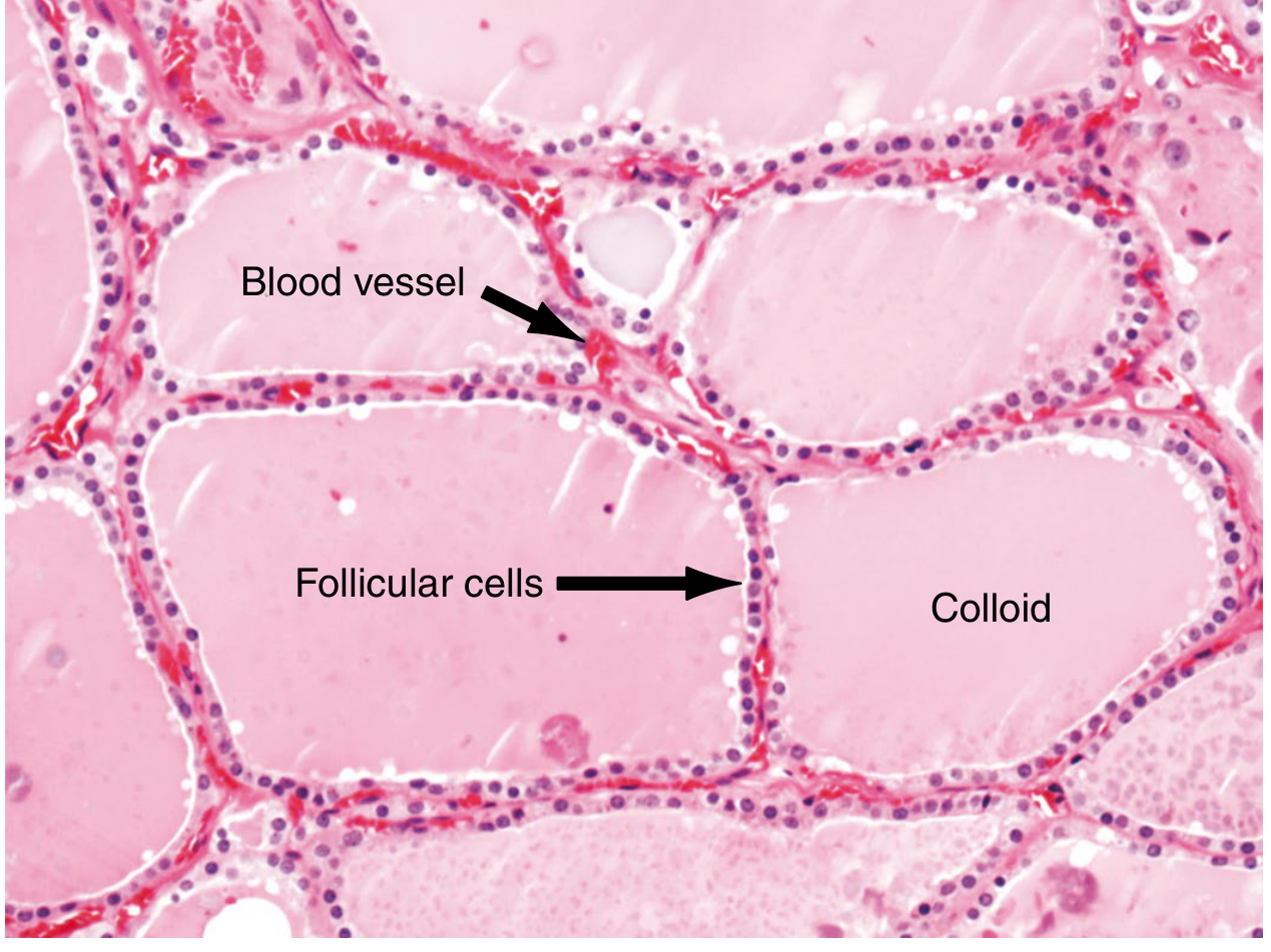
Normal thyroid parenchyma (H&E). Large pale-pink colloid-filled follicles lined by a single layer of cuboidal follicular cells. Blood vessels run in the interfollicular stroma. - Sabiston Textbook of Surgery
What You See on the Slide
| Structure | Appearance | Function |
|---|---|---|
| Follicles | Spherical spaces ~30 µm diameter; ~20-40 per lobule | Structural unit of the thyroid |
| Colloid | Homogeneous, pale eosinophilic (pink) material filling the follicle lumen | Storage form of thyroglobulin (inactive T3/T4) |
| Follicular cells (thyrocytes) | Single layer of cuboidal to low columnar cells lining each follicle | Synthesise T3 and T4 |
| Parafollicular (C) cells | Larger pale cells scattered in interfollicular stroma, especially upper poles | Secrete calcitonin (calcium regulation) |
| Fibrous septa | Thin connective tissue dividing the gland into lobules | Support and vascularity |
Key Functional Points
- Active gland: follicular cells become taller (columnar), colloid appears vacuolated at edges ("resorption vacuoles")
- Inactive/suppressed gland: follicular cells flatten (squamous), colloid is dense and abundant
- Hormone synthesis: TSH → cAMP → follicular cells take up iodide → iodinate thyroglobulin in colloid → resorb it back → release T3/T4 into blood
2. DUODENUM
Histology Slide
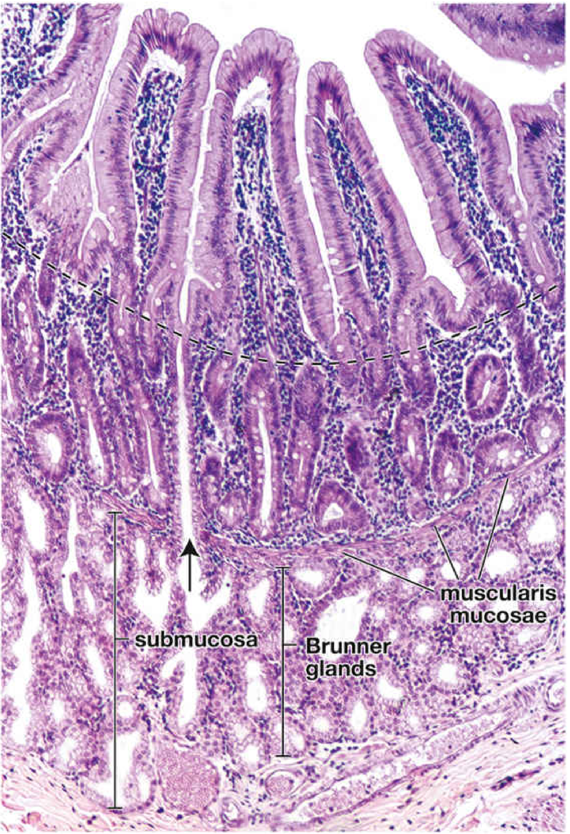
Duodenal wall H&E. Top: tall finger-like villi lined by absorptive epithelium. Middle (dashed line): base of villi / crypts of Lieberkühn. Bottom: submucosa containing the hallmark Brunner glands. - Histology: A Text and Atlas (Pawlina)
What You See on the Slide
| Layer | Structure | Appearance | Significance |
|---|---|---|---|
| Mucosa | Villi | Tall finger-like projections lined by columnar absorptive cells + goblet cells | Massively increases surface area for absorption |
| Mucosa | Crypts of Lieberkühn | Tubular glands at base of villi | Contain stem cells, Paneth cells, enteroendocrine cells |
| Mucosa | Muscularis mucosae | Thin smooth muscle layer | Separates mucosa from submucosa |
| Submucosa | Brunner glands | Branched tubular mucous glands - UNIQUE TO DUODENUM | Secrete alkaline (pH 8.1-9.3) mucus + bicarbonate - neutralise gastric acid chyme |
| Muscularis externa | Inner circular + outer longitudinal smooth muscle | Two muscle layers | Peristalsis + segmentation |
Key Distinguishing Feature
Brunner glands in the submucosa = hallmark of duodenum. No other part of the GI tract has submucosal glands (except the oesophagus). Their alkaline secretion protects the duodenal mucosa from acid and activates pancreatic enzymes.
3. PANCREAS
The pancreas is a dual gland - exocrine and endocrine components side by side.
Exocrine Pancreas: Acinar Cells
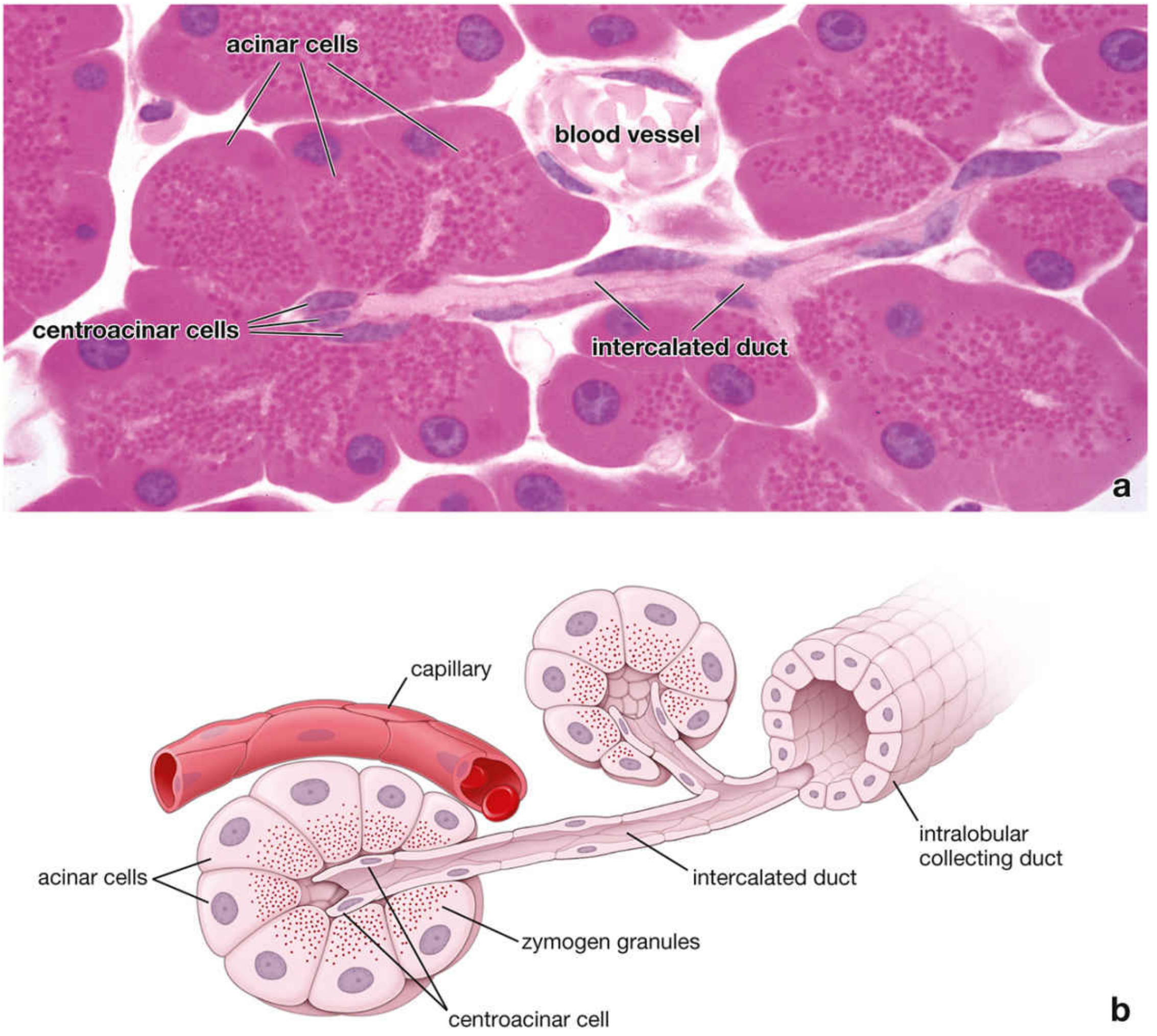
Pancreatic acinus - H&E (top) and schematic (bottom). Pyramidal acinar cells with zymogen granules (red apical dots), centroacinar cells at the duct opening, and the intercalated duct. - Histology: A Text and Atlas (Pawlina)
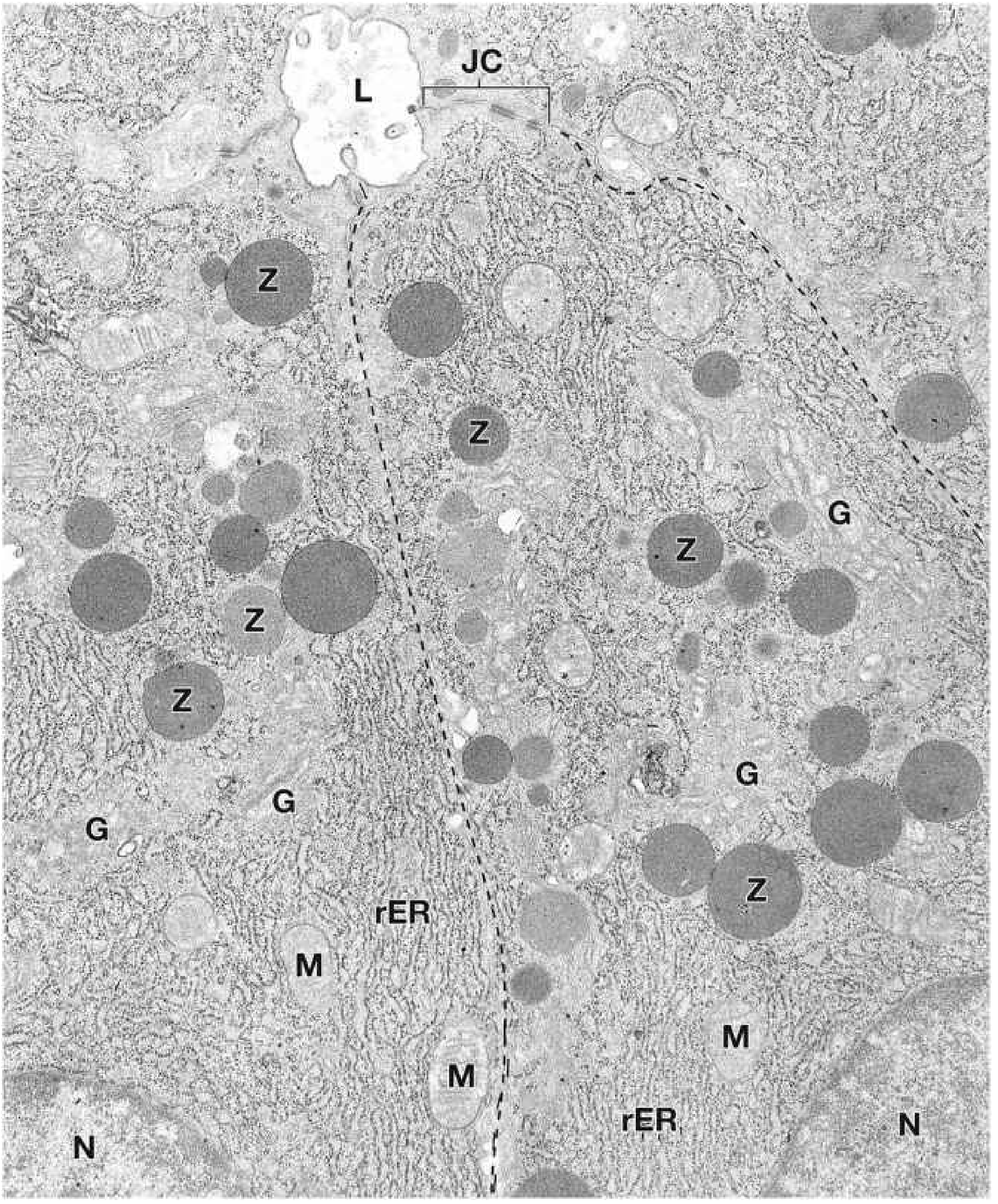
EM of pancreatic acinar cells. Z = large round zymogen granules at apical pole; rER = abundant rough ER at basal pole (basophilia on H&E); G = Golgi; M = mitochondria; L = lumen of acinus; JC = junctional complex. - Histology: A Text and Atlas (Pawlina)
Endocrine Pancreas: Islets of Langerhans
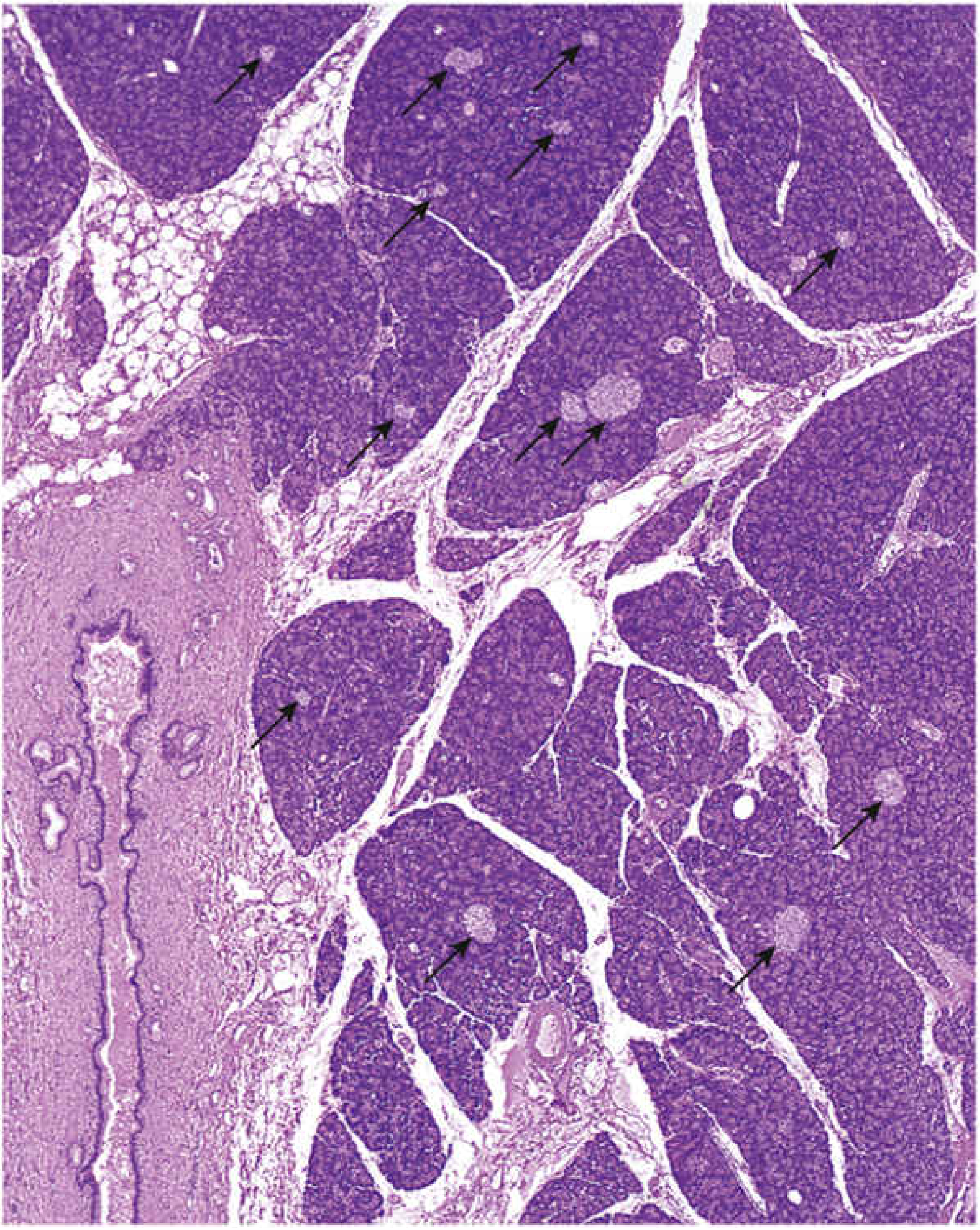
Pancreas low-power H&E. The dark purple background = densely packed exocrine acini. Arrows point to islets of Langerhans - paler, less densely staining rounded clusters of endocrine cells amid the acini. - Histology: A Text and Atlas (Pawlina)
What You See on the Slide
| Component | Appearance | Cell Types | Function |
|---|---|---|---|
| Exocrine acini | Rounded clusters of pyramidal cells; basophilic basal cytoplasm (rER), eosinophilic apical zymogen granules | Acinar cells (serous); centroacinar cells | Digest proteins, fats, carbohydrates, nucleic acids |
| Centroacinar cells | Small pale cells at the centre of acinus where duct begins | Special to pancreas | Line intercalated ducts; secrete HCO₃⁻ |
| Intercalated ducts | Very thin ducts originating inside the acinus - unique to pancreas | Duct epithelium | Drain acini; add bicarbonate |
| Islets of Langerhans | Pale, rounded clusters 1-3 million throughout pancreas (~1-2% volume); well-vascularised by fenestrated capillaries | α cells (glucagon), β cells (insulin), δ cells (somatostatin), PP cells | Blood glucose regulation |
Zymogen Granules (Exam Favourite)
- Located at the apical pole of acinar cells
- Appear bright red/eosinophilic on H&E
- Most numerous in fasting individuals
- Contain inactive proenzymes (trypsinogen, chymotrypsinogen, lipase, amylase) - activated only in duodenum
4. HYALINE (Histological Term)
"Hyaline" (from Greek hyalos = glass) is a descriptive histological term - not one specific substance. It means any tissue that appears homogeneous, glassy, and pink (eosinophilic) on H&E stain.
Hyaline Cartilage (Most Common Type)
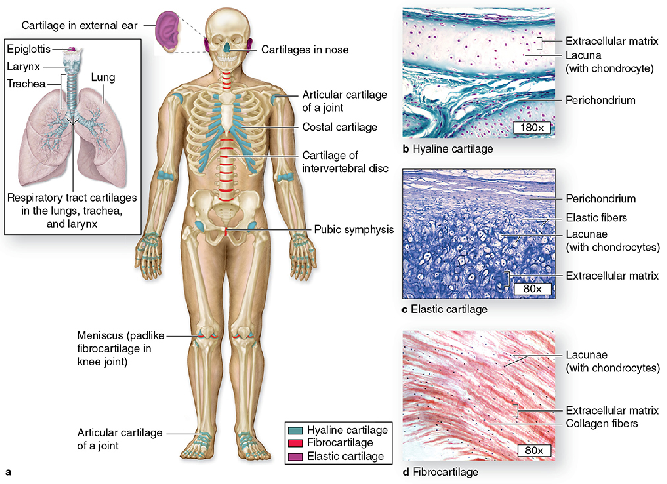
Three types of cartilage: (b) Hyaline cartilage - glassy matrix with chondrocytes in lacunae, dense perichondrium. (c) Elastic cartilage - visible elastic fibres in matrix. (d) Fibrocartilage - parallel collagen bundles, no perichondrium. - Junqueira's Basic Histology
What You See in Hyaline Cartilage
| Structure | Appearance | Significance |
|---|---|---|
| Matrix | Homogeneous, glassy blue-purple (H&E) - appears "hyaline" | Type II collagen + proteoglycans + water - shock absorption |
| Chondrocytes | Round cells in lacunae (spaces); may appear in isogenous groups | Produce and maintain matrix |
| Perichondrium | Dense fibrous layer surrounding cartilage | Appositional growth, blood supply (cartilage is avascular) |
| Lacunae | Empty-appearing spaces around chondrocytes | Shrinkage artifact from fixation |
Other Contexts Where "Hyaline" Appears
| Location | What it Means | Associated Condition |
|---|---|---|
| Arteriole walls | Hyaline arteriolosclerosis | Hypertension, diabetes mellitus |
| Intracellular (hepatocytes) | Alcoholic (Mallory) hyaline | Alcoholic liver disease |
| Renal tubular droplets | Reabsorption droplets | Nephrotic syndrome |
| Russell bodies (plasma cells) | Immunoglobulin accumulation | Chronic inflammation |
| Old scars/tendons | Hyalinised collagen fibers | Post-scar remodelling |
| Amyloid | Congo red + apple-green birefringence | Amyloidosis |
According to Robbins Pathologic Basis of Disease: "The term hyaline usually refers to an alteration within cells or in the extracellular space that gives a homogeneous, glassy, pink appearance in routine H&E sections. It is widely used as a descriptive histologic term rather than a specific marker for cell injury."
This is a shared conversation. Sign in to Orris to start your own chat.