Now I have enough to build a truly comprehensive, structured response. Let me craft this masterpiece module by module.
🏥 B.Sc NURSING - COMPLETE CLINICAL FOUNDATION
Zero to Professional Level | Hinglish Medium | 4th Semester
Aapka Swagat hai! Welcome to your complete nursing foundation. Ham yahan se start karenge - ek ek cheez clearly samjhenge. Jab tak aap confidently hospital mein kaam nahi kar sakte, tab tak yeh series continue rahegi.
📚 MODULE 1: MEDICAL TERMINOLOGY (Part 1)
"Hospital Ki Language - Sabse Pehle Yeh Seekho"
Professor's Note: Hospital mein har jagah abbreviations use hoti hain. Agar inhe nahi jaante, toh doctor ka order bhi nahi samajh payenge. Yeh sabse pehla aur MOST IMPORTANT topic hai.
🔴 PART A: TIMING & FREQUENCY ABBREVIATIONS
(Kitni baar aur kab medicine deni hai)
1. STAT
| Category | Details |
|---|
| Full Form | Statim (Latin) |
| Pronunciation | "STAT" (जैसे "start" में "stat") |
| Hindi Meaning | तुरंत / अभी के अभी / फ़ौरन |
| English Meaning | Immediately, Right Now |
Hospital Use:
- Doctor likhta hai: "Inj. Adrenaline 1mg IV STAT"
- Matlab: Abhi is waqt, bina kisi delay ke do
🏥 Real Hospital Example:
Patient ka BP suddenly drop ho gaya - 70/50 mmHg. Doctor shouts: "Give IV fluids STAT!" - Matlab turant, ek second bhi waste nahi karna.
🧠 Memory Trick:
STAT = Start Treating A.S.A.P. This instant!
Ya yaad karo: "Statue" - jaise statue still rehta hai, waise STAT pe sab ruk jaate hain aur turant kaam karte hain.
⚠️ Common Mistake:
Students sochte hain STAT matlab "jaldi se dena hai" - NAHI! STAT matlab usi waqt, us minute mein dena hai. 15-20 minute delay = dangerous!
2. SOS
| Category | Details |
|---|
| Full Form | Si Opus Sit (Latin) = "If it is necessary" |
| Pronunciation | "S-O-S" |
| Hindi Meaning | जरूरत पड़ने पर / जब लगे तब |
| English Meaning | As needed / If Required |
Hospital Use:
- "Tab. Paracetamol 500mg SOS" = Agar bukhar/dard ho tab do
🏥 Real Hospital Example:
Patient ko occasional headache hai. Doctor writes: "Tab. Paracetamol 1g SOS" - Matlab patient khud keh raha hai tab do, routine nahi hai.
🧠 Memory Trick:
SOS = Ship ka distress signal - jab emergency ho tab signal karo. Medicine bhi tab do jab patient ko "distress" ho (pain/fever).
3. OD (Once Daily)
| Category | Details |
|---|
| Full Form | Omni Die (Latin) = Every Day |
| Pronunciation | "O-D" |
| Hindi Meaning | दिन में एक बार |
| Timing | Usually Morning (unless specified) |
Example: "Tab. Metformin 500mg OD" = Subah ek baar
🧠 Memory Trick: OD = One time a Day. "O" aur "D" dono ek baar likhe = ek baar do!
4. BD (Twice Daily)
| Category | Details |
|---|
| Full Form | Bis Die (Latin) = Twice a Day |
| Pronunciation | "B-D" |
| Hindi Meaning | दिन में दो बार |
| Timing | Morning + Evening (8AM - 8PM) |
Example: "Tab. Amoxicillin 500mg BD" = Subah aur shaam
🧠 Memory Trick: BD = Before Dawn & Dusk = Subah aur sham. Ya: "B" = 2 letters ka word "BD" = 2 times.
5. TDS / TID (Three Times Daily)
| Category | Details |
|---|
| Full Form | Ter Die Sumendus = To be taken 3 times a day |
| Pronunciation | "T-D-S" |
| Hindi Meaning | दिन में तीन बार |
| Timing | 8AM - 2PM - 8PM |
Example: "Tab. Metronidazole 400mg TDS" = Teen baar
🧠 Memory Trick: TDS = Three times Daily Schedule. "T" = Three!
6. QID / QDS (Four Times Daily)
| Category | Details |
|---|
| Full Form | Quater In Die = 4 times a day |
| Pronunciation | "Q-I-D" |
| Hindi Meaning | दिन में चार बार |
| Timing | 6AM - 12PM - 6PM - 12AM |
🧠 Memory Trick: QID = 4 corners of a Square (Q like a circle with 4 sides)
7. HS (At Bedtime)
| Category | Details |
|---|
| Full Form | Hora Somni = At the hour of sleep |
| Pronunciation | "H-S" |
| Hindi Meaning | सोते वक्त / रात को सोने से पहले |
| Timing | 9-10 PM |
Example: "Tab. Digoxin 0.25mg HS" = Raat ko sone se pehle
🧠 Memory Trick: HS = Hit the Sack (English idiom for going to sleep)
8. AC / PC
| Abbreviation | Full Form | Hindi | Timing |
|---|
| AC | Ante Cibum = Before food | खाने से पहले | 30 min before meals |
| PC | Post Cibum = After food | खाने के बाद | 30 min after meals |
🧠 Memory Trick:
- AC = A se pehle C ... wait! Better: Anti = pehle, like Anticipate = pehle se
- PC = Post (baad mein) = PC pe kaam karo khana khane ke baad!
🔵 PART B: ROUTE OF ADMINISTRATION
(Kahan se medicine deni hai)
THE MASTER TABLE OF ROUTES
| Abbreviation | Full Form | Hindi | Where Given | Needle Size |
|---|
| PO | Per Os (by mouth) | मुँह से | Oral | No needle |
| IV | Intravenous | नस में | Into vein | 18-22G |
| IM | Intramuscular | मांसपेशी में | Deltoid/Gluteal | 21-23G |
| SC/SQ | Subcutaneous | चमड़ी के नीचे | Abdomen/arm | 25-27G |
| ID | Intradermal | चमड़ी के अंदर | Forearm inner | 27G |
| PR | Per Rectum | मलद्वार से | Rectum | Suppository |
| SL | Sublingual | जीभ के नीचे | Under tongue | No needle |
| INH | Inhalation | सांस द्वारा | Lungs | Inhaler/Nebulizer |
| TOP | Topical | लगाने के लिए | Skin surface | No needle |
🧠 Memory Trick for Routes:
"PIIISPR" story: "Pissa baar baar Intravenously Inject karo, Intramuscularly Inject karo, SC mein, PR se... sabke alag-alag raste hain!"
IV - Intravenous (Most Important!)
Hindi: नस के अंदर सीधे
Why important: Fastest action - 30 seconds to 1 minute mein drug blood mein pahunch jaata hai.
🏥 Real Example:
Patient ko anaphylaxis (allergic reaction). STAT Adrenaline IV dena hai kyunki IM ya SC se time lagega, IV se turant blood mein jayega.
Sites for IV:
- Dorsum of hand (haath ki peechwali nass) - most common
- Forearm veins
- Antecubital fossa (kehni ke paas)
- Cephalic vein
⚠️ Complications to Watch:
- Phlebitis = nass mein sujan/laalipan (check karo har 4-8 ghante)
- Infiltration = fluid tissue mein jaana (seedha nahin raha cannula)
- Extravasation = toxic drug tissue mein gaya (dangerous!)
IM - Intramuscular
Sites (4 common sites):
| Site | Hindi | Used For |
|---|
| Deltoid | Kandha (shoulder muscle) | Vaccines, small volume (<2ml) |
| Dorsogluteal | Peeche neeche (buttock) | Large volume, oily injections |
| Ventrogluteal | Side buttock | Safest site (less nerve risk) |
| Vastus lateralis | Jaangh (thigh - outer side) | Infants, children |
🧠 Mnemonic for Dorsogluteal site:
"Upper Outer Quadrant" = Cheeks ko 4 parts mein baanto, upar waala aur bahar waala part = safe zone
⚠️ DANGER: Never inject in lower inner gluteal area = Sciatic nerve injury = patient lame ho sakta hai!
SC - Subcutaneous
Hindi: Chamdi ke theek neeche, fat layer mein
Sites: Abdomen (belly), outer arm (upper), outer thigh
Angle: 45° (ya 90° agar patient mota hai)
Examples: Insulin, Heparin, Enoxaparin
🏥 Important: Insulin always SC - kabhi IV nahi (emergency mein alag protocol)
ID - Intradermal
Hindi: Chamdi ki layers ke beech
Angle: 10-15°
Used for: Mantoux test (TB), allergy testing, BCG vaccine
How to confirm correct placement: "Bleb" (chote buble jaisi swelling) bananI chahiye
🟢 PART C: TUBE & SPECIAL ROUTES
NGT - Nasogastric Tube
| Item | Details |
|---|
| Full Form | Nasogastric Tube |
| Hindi | नाक से पेट तक डाली जाने वाली नली |
| Also Called | Ryle's Tube (same thing!) |
| Purpose | Feeding, medicine, gastric decompression, lavage |
🏥 Real Example:
Unconscious patient jis case mein muh se khana nahi kha sakta - NGT dalke liquid diet dete hain.
Sizes:
- Adult: 14-18 Fr
- Child: 8-12 Fr
- Infant: 5-8 Fr
Nursing Check (MOST IMPORTANT - before use):
- Aspirate gastric content (acidic, pH < 5.5)
- Inject air and listen with stethoscope (gurgling sound in stomach)
- Xray = gold standard to confirm
⚠️ DANGER: Never feed through NGT without confirming placement - lungs mein hoga toh patient ko pneumonia/death ho sakti hai!
NPO / NBM
| Abbreviation | Full Form | Hindi |
|---|
| NPO | Nil Per Os | कुछ भी मुँह से नहीं |
| NBM | Nothing By Mouth | कुछ भी मुँह से नहीं |
Both mean exactly the same thing.
When used:
- Pre-surgery: 6-8 hours NPO (no solids), 2 hours (no liquids)
- Unconscious patients
- Post-abdominal surgery
- Before procedures
🏥 Example: "Patient is NPO from midnight for 8 AM surgery" = Raat 12 baje ke baad kuch bhi muh se nahi
KVO
| Item | Details |
|---|
| Full Form | Keep Vein Open |
| Hindi | नस खुली रखो (बहुत धीरे dripchalna) |
| Rate | 10-15 ml/hour |
| Purpose | IV access banaye rakhna bina zyada fluid diye |
🏥 Example: Patient stable hai, IV access chahiye agar emergency ho. Doctor likhta hai: "IV NS @ KVO rate."
🧠 Memory Trick: KVO = Keep the Vein Open = dori mat khaino, dhire dhire chalti rahe
PRN
| Item | Details |
|---|
| Full Form | Pro Re Nata (Latin) |
| Hindi | जरूरत पड़ने पर (same as SOS) |
| Difference from SOS | PRN = nurse assess karke deti hai; SOS = patient maangta hai |
🟡 PART D: MONITORING ABBREVIATIONS
THE VITAL MONITORING TABLE
| Abbreviation | Full Form | Hindi | Normal Value |
|---|
| SpO₂ | Peripheral Oxygen Saturation | खून में ऑक्सीजन का % | 95-100% |
| RR | Respiratory Rate | सांस की गति | 12-20/min (adult) |
| HR | Heart Rate | दिल धड़कने की गति | 60-100 bpm |
| BP | Blood Pressure | रक्तचाप | 120/80 mmHg |
| CRT | Capillary Refill Time | उंगली दबाने पर वापस आने का समय | < 2 seconds |
| GCS | Glasgow Coma Scale | होश का पैमाना | 15/15 = full conscious |
| ABG | Arterial Blood Gas | धमनी से खून का गैस test | pH 7.35-7.45 |
| ECG | Electrocardiogram | दिल की बिजली की लकीरें | Normal sinus rhythm |
🟠 PART E: HOSPITAL AREAS & DEPARTMENTS
| Abbreviation | Full Form | Hindi |
|---|
| ICU | Intensive Care Unit | गहन चिकित्सा इकाई |
| NICU | Neonatal Intensive Care Unit | नवजात गहन चिकित्सा |
| PICU | Pediatric Intensive Care Unit | बच्चों की गहन चिकित्सा |
| CCU | Coronary Care Unit | हृदय रोग गहन इकाई |
| OT | Operation Theatre | शल्य कक्ष |
| ER/ED | Emergency Room / Emergency Department | आपातकालीन विभाग |
| OPD | Outpatient Department | बाह्य रोगी विभाग |
| IPD | Inpatient Department | आंतरिक रोगी विभाग |
| MRD | Medical Records Department | चिकित्सा रिकॉर्ड विभाग |
🧠 Story for Hospital Departments:
"Ek patient OPD se aaya, wahan IPD mein admit hua, fever zyada badhne par ICU mein le jaana pada. Zyada serious hua toh CCU ya PICU mein shift kiya. Operation ki zaroorat thi toh OT gaya. Discharge ke baad papers MRD mein jama ho gaye."
🔴 PART F: LAB TEST ABBREVIATIONS
| Abbreviation | Full Form | Hindi | Normal Range |
|---|
| RBS | Random Blood Sugar | कभी भी blood sugar | < 200 mg/dL |
| FBS | Fasting Blood Sugar | खाली पेट blood sugar | 70-100 mg/dL |
| PPBS | Post Prandial Blood Sugar | खाने के 2 घंटे बाद | < 140 mg/dL |
| CBC | Complete Blood Count | खून की पूरी जांच | (see below) |
| LFT | Liver Function Test | लीवर की जांच | (see below) |
| KFT | Kidney Function Test | गुर्दे की जांच | (see below) |
| PT | Prothrombin Time | खून जमने का समय | 11-13 seconds |
| INR | International Normalized Ratio | खून जमने का अनुपात | 0.8-1.2 (normal) |
| Hb | Hemoglobin | हीमोग्लोबिन | M: 13-17, F: 12-15 g/dL |
| HCT | Hematocrit / PCV | खून में RBC का % | M: 40-52%, F: 36-48% |
| RBC | Red Blood Cells | लाल रक्त कोशिकाएं | 4.5-5.5 million/µL |
| WBC | White Blood Cells | सफेद रक्त कोशिकाएं | 4000-11000/µL |
| CRP | C-Reactive Protein | सूजन/संक्रमण का चिह्न | < 1 mg/dL |
| ESR | Erythrocyte Sedimentation Rate | थोड़े शोध की दर | M: <15, F: <20 mm/hr |
| Troponin | Troponin I or T | दिल की मांसपेशी का protein | < 0.04 ng/mL |
| D-Dimer | D-Dimer | खून के थक्के का marker | < 0.5 µg/mL |
📚 MODULE 2: VITAL SIGNS - "Hospital Ki Nabd"
"Vitals lena = Patient ki kahani sunna"
🏥 INTRODUCTION
Yeh 6 cheezein hain jo nurse SABSE PEHLE check karti hai:
- Blood Pressure (BP)
- Heart Rate / Pulse
- Respiratory Rate (RR)
- Temperature
- SpO₂ (Oxygen Saturation)
- Pain (6th vital sign - modern nursing)
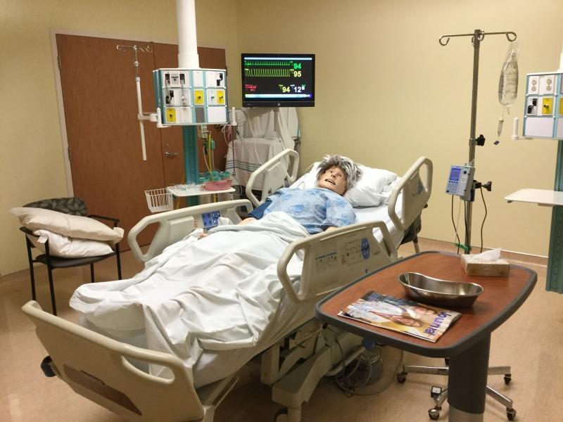
Clinical Monitoring Image:
ICU-style patient monitoring - BP, Pulse, RR, SpO₂ - sabhi ek saath monitor pe dikhte hain
💓 VITAL SIGN 1: BLOOD PRESSURE (BP)
Definition (Simple)
BP = Dil ke dhakkan se arteries (nasson) mein kitna pressure banta hai
Jaise paani ki pipe mein paani ka pressure hota hai, waise hi blood ka pressure hota hai.
Two Numbers in BP:
| Number | Name | Hindi | Meaning |
|---|
| Top number | Systolic | सिस्टोलिक | Dil dhakata hai tab ka pressure |
| Bottom number | Diastolic | डायस्टोलिक | Dil aram mein hai tab ka pressure |
Normal BP = 120/80 mmHg (read as "120 over 80")
BP VALUES TABLE (Must Memorize!)
| Category | Systolic | Diastolic | Clinical Meaning |
|---|
| Hypotension | < 90 | < 60 | BP kam = shock, dehydration |
| Normal | 90-119 | 60-79 | ✅ Perfect |
| Elevated | 120-129 | < 80 | Prehypertension |
| HTN Stage 1 | 130-139 | 80-89 | Hypertension starting |
| HTN Stage 2 | ≥ 140 | ≥ 90 | Definite Hypertension |
| HTN Crisis | > 180 | > 120 | 🚨 EMERGENCY! |
BP in Special Groups:
| Group | Normal BP |
|---|
| Newborn | 60-90 / 30-60 |
| Infant (1yr) | 80-100 / 50-70 |
| Child (10yr) | 100-120 / 60-80 |
| Adult | 90-140 / 60-90 |
| Elderly (>65yr) | Up to 150/90 acceptable |
| Pregnant | < 140/90 |
🏥 Hospital Scenarios:
Scenario 1 - Low BP:
Post-surgery patient ka BP = 85/50 mmHg. Patient sweating, confused. Nurse ka kaam: Position Trendelenburg (pair upar), IV fluids STAT, doctor ko call, monitor continuously.
Scenario 2 - High BP Crisis:
Patient ka BP = 200/120 mmHg, headache + blurred vision. Nurse: Head elevation, antihypertensive medicine, doctor ko STAT inform, strict monitoring.
🧠 Memory Tricks for BP:
"120/80 = Perfect score - Ek sau bees aur ath-some"
MNEMONIC for HYPOTENSION causes:
"CRASH"
- C = Cardiac (heart failure, heart attack)
- R = Reduced volume (bleeding, dehydration)
- A = Anaphylaxis (allergic reaction)
- S = Sepsis (infection)
- H = Heat stroke
BP Measurement - Step by Step (Practical):
STEP 1: Patient ko 5 min rest - baithe ya lete
STEP 2: Right arm use karo (ya as instructed)
STEP 3: Cuff size: cuff ka bladder = arm ka 80% circumference
STEP 4: Cuff 2.5 cm above antecubital fossa (kehni ke modd se upar)
STEP 5: Stethoscope bell/diaphragm antecubital fossa pe rako
STEP 6: Inflate 30 mmHg above expected systolic
STEP 7: Deflate slowly (2 mmHg/sec)
STEP 8: First sound = Systolic (Korotkoff Sound 1)
STEP 9: Last sound = Diastolic (Korotkoff Sound 5)
STEP 10: Document immediately
⚠️ Common Mistakes:
- Cuff too small = BP falsely HIGH
- Cuff too large = BP falsely LOW
- Patient's arm above heart level = BP falsely LOW
- Talking during BP = inaccurate reading
- Not waiting after exercise = falsely HIGH
💗 VITAL SIGN 2: PULSE (Heart Rate)
Definition:
Pulse = Har baar dil dhakata hai toh khoon ko artery mein ek wave jaisi feeling hoti hai - wahi pulse hai.
Jaise stone paani mein fenko, lahar aati hai - wahi lahar pulse hai!
PULSE NORMAL VALUES:
| Age Group | Normal Rate (bpm) |
|---|
| Newborn | 120-160 |
| Infant (1-12 mo) | 80-140 |
| Toddler (1-3yr) | 80-130 |
| Child (3-12yr) | 75-110 |
| Adolescent | 60-100 |
| Adult | 60-100 |
| Athlete | 40-60 (normal!) |
| Elderly | 60-100 |
Pulse TERMS:
| Term | Hindi | Meaning | When Seen |
|---|
| Tachycardia | तेज़ नाड़ी | > 100 bpm | Fever, pain, shock, anxiety |
| Bradycardia | धीमी नाड़ी | < 60 bpm | Athletes, drugs (beta-blocker), heart block |
| Arrhythmia | अनियमित नाड़ी | Irregular rhythm | AF, heart disease |
| Thready pulse | कमज़ोर नाड़ी | Weak, thin pulse | Shock, severe dehydration |
| Bounding pulse | ज़ोरदार नाड़ी | Strong, forceful | Fever, aortic regurgitation |
Pulse Assessment Sites:
| Site | Hindi | Used When |
|---|
| Radial | Kalai | Routine - most common |
| Brachial | Kehni ke andar | Children, BP check |
| Carotid | Gardan | Emergency, CPR |
| Femoral | Jaangh | Circulation check |
| Pedal (Dorsalis pedis) | Pair pe | Peripheral vascular disease |
| Apical | Seene pe stethoscope | Infants, arrhythmia |
🧠 Mnemonic for pulse sites:
"Real Boys Can't Fight Pretty Apricots"
Radial - Brachial - Carotid - Femoral - Popliteal - Apical
4 Things to Check in Pulse (Rate, Rhythm, Volume, Character):
Mnemonic: "RRVC"
- R = Rate (Kitni baar - bpm)
- R = Rhythm (Regular hai ya irregular)
- V = Volume (Kितना strong - thready ya bounding)
- C = Character (Normal ya special - e.g., waterhammer)
🌬️ VITAL SIGN 3: RESPIRATORY RATE (RR)
Definition:
RR = 1 minute mein kitni baar saans andar jaata aur bahar aata hai
Note: 1 breath = 1 inhale + 1 exhale (do nahi ginna!)
RR NORMAL VALUES:
| Age | Normal RR (per minute) |
|---|
| Newborn | 30-60 |
| Infant | 25-50 |
| Toddler | 20-30 |
| Child | 15-25 |
| Adult | 12-20 |
| Elderly | 15-25 |
RR TERMS:
| Term | Meaning | When |
|---|
| Tachypnea | RR > 20/min | Fever, pneumonia, pain, anxiety |
| Bradypnea | RR < 12/min | Drug overdose, head injury |
| Apnea | No breathing | Emergency! CPR start! |
| Dyspnea | Difficulty breathing (patient ka feeling) | Asthma, heart failure |
| Orthopnea | Can't breathe lying flat | Heart failure |
| Cheyne-Stokes | Crescendo-decrescendo pattern | Head injury, dying |
🏥 Nursing Tip:
Secret: Jab aap pulse count kar rahe ho toh patient ko batao mat ki ab RR count karenge. Warna patient jaanboojhkar saans badal deta hai! Pulse lene ke baad haath wahan rakho aur quietly RR gino.
🌡️ VITAL SIGN 4: TEMPERATURE
Normal Values:
| Method | Normal Range | Hindi |
|---|
| Oral | 36.5-37.5°C (97.7-99.5°F) | मुँह से |
| Axillary | 36.0-37.0°C (96.8-98.6°F) | बगल से (lowest) |
| Rectal | 37.0-38.0°C (98.6-100.4°F) | मलद्वार से (highest) |
| Tympanic | 36.5-37.5°C | कान से |
Important Rule: Axillary < Oral < Rectal (0.5°C difference each)
FEVER CLASSIFICATION:
| Temperature | Hindi Name | Grading |
|---|
| 37.1-38°C | Low grade fever | Subfebrile |
| 38-39°C | Moderate fever | Pyrexia |
| 39-40°C | High fever | High grade pyrexia |
| 40-41°C | Very high fever | Hyperpyrexia |
| > 41°C | 🚨 DANGER! | Medical emergency |
| < 35°C | Hypothermia | EMERGENCY! |
🧠 Memory Trick:
"37 = Body ka normal AC setting" - Body temperature 37 degree Centigrade
Nursing Actions for HIGH Fever (> 39°C):
- Tepid sponging (Gungune paani se ponchna) - NOT cold water!
- Remove extra clothing/blankets
- Encourage fluid intake
- Antipyretics as prescribed (Tab. Paracetamol)
- Monitor every 1-2 hours
- Check for cause (blood culture, CBC)
- Document - time, method, value, action taken
⚠️ Common Mistake: Cold water/ice se sponging - wrong! Vasodilation chahiye, vasoconstriction nahi!
🫁 VITAL SIGN 5: SpO₂ (Oxygen Saturation)
Definition:
SpO₂ = Hamare khoon ke hemoglobin mein se kitne percent pe oxygen baihi hai
"Sp" = peripheral (finger se), "O2" = oxygen, "%" = percentage
SpO₂ CRITICAL VALUES TABLE:
| SpO₂ | Status | Nursing Action |
|---|
| 98-100% | ✅ Normal | Routine monitoring |
| 95-97% | ✅ Acceptable | Monitor closely |
| 90-94% | ⚠️ Mild Hypoxia | Oxygen dena shuru karo |
| 85-89% | 🟠 Moderate Hypoxia | Oxygen urgently, doctor inform |
| < 85% | 🔴 Severe Hypoxia | EMERGENCY! STAT oxygen + doctor |
| < 80% | 🚨 Life Threatening | CPR/intubation ready |
Pulse Oximeter - How to Use:
STEP 1: Nail polish hatao (dark nail polish = false reading)
STEP 2: Index finger ya middle finger pe lagao
STEP 3: Patient ka haath heart level pe raho
STEP 4: 30 second wait karo for stable reading
STEP 5: Waveform dekho (bar banana chahiye)
STEP 6: Note karo SpO₂ AND HR (dono simultaneously milte hain)
⚠️ FALSE READINGS: Nail polish, cold fingers, anemia, low BP, movement = inaccurate SpO₂!
OXYGEN DELIVERY - Quick Reference:
| Device | FiO₂ (%) | Hindi | Flow Rate |
|---|
| Room Air | 21% | Khuli hawa | - |
| Nasal Prongs | 24-44% | Naak ki nali | 1-6 L/min |
| Simple Face Mask | 35-50% | Saada mukhauta | 6-10 L/min |
| Non-rebreather mask | 70-90%+ | High flow mask | 10-15 L/min |
| Bag-Valve-Mask | Up to 100% | BVM | With O₂ reservoir |
📊 VITAL SIGN 6: PAIN (6th Vital Sign)
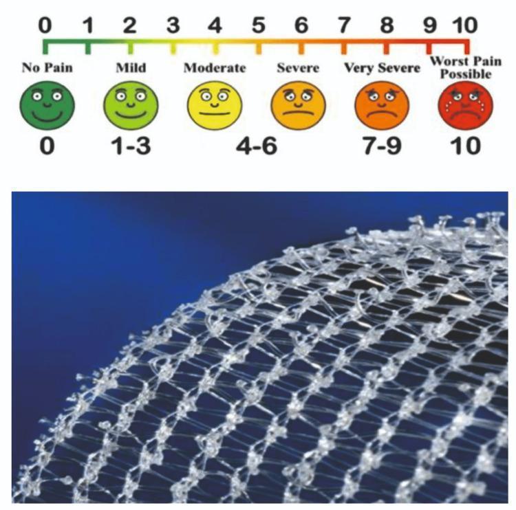
Pain Image:
Numeric Rating Scale (NRS) with faces - 0 se 10 tak pain assess karo
PAIN SCALES:
| Scale | Used For | How |
|---|
| NRS (0-10) | Adults who can speak | "0 se 10 mein kitna dard?" |
| VAS | Research, some adults | 10cm line pe mark karo |
| Wong-Baker Faces | Children 3yr+, language barrier | Faces dikhao |
| FLACC | Infants, unconscious | Observe 5 behaviors |
PQRST Pain Assessment (Must Know!):
🧠 Mnemonic: "PQRST" = Like ECG waveform!
| Letter | Stands For | Question to Ask |
|---|
| P | Provocation/Palliation | Kya karne se barhta/ghatta hai? |
| Q | Quality | Kaisa dard hai? (throbbing, stabbing, burning) |
| R | Region/Radiation | Kahan hai? Kahin aur bhi jaata hai? |
| S | Severity | 0-10 mein kitna? |
| T | Timing | Kab se hai? Continuous ya intermittent? |
SOCRATES (Advanced Pain Assessment):
| S | Site | Kahan hai dard? |
|---|
| O | Onset | Kab shuru hua? |
| C | Character | Kaisa dard hai? |
| R | Radiation | Kahin aur bhi jaata hai? |
| A | Associated symptoms | Aur kya ho raha hai? |
| T | Timing | Kitni der se? |
| E | Exacerbating/Relieving | Kya karne se badh/ghatta hai? |
| S | Severity | Score kitna? |
📚 MODULE 3: GCS - GLASGOW COMA SCALE
Definition:
GCS = Ek score system jo batata hai ki patient kitna conscious (hosh mein) hai
3 cheezein check karte hain: Aankhein, Bolna, Haath pair hilana
GCS SCORING TABLE (MUST MEMORIZE!):
Eye Opening (E) - Max 4:
| Score | Response | Hindi |
|---|
| 4 | Spontaneous | Apne aap khulti hain |
| 3 | To voice | Aawaz pe khulti hain |
| 2 | To pain | Dard pe khulti hain |
| 1 | None | Bilkul nahi khulti |
Verbal Response (V) - Max 5:
| Score | Response | Hindi |
|---|
| 5 | Oriented | Sahi sahi bolta hai (naam, date, jagah) |
| 4 | Confused | Bolta hai par confused |
| 3 | Inappropriate words | Bekar ke word bolata hai |
| 2 | Incomprehensible | Sirf awaazein |
| 1 | None | Kuch nahi |
Motor Response (M) - Max 6:
| Score | Response | Hindi |
|---|
| 6 | Obeys commands | "Haath hilao" pe hilata hai |
| 5 | Localizes pain | Dard ki jagah ko haath lagata hai |
| 4 | Withdraws | Dard se haath hatata hai |
| 3 | Abnormal flexion | Dekorticate posture |
| 2 | Abnormal extension | Decerebrate posture |
| 1 | None | Kuch nahi |
GCS TOTAL SCORE INTERPRETATION:
| Score | Meaning | Action |
|---|
| 15/15 | Fully conscious ✅ | Normal |
| 13-14 | Mild brain injury | Close monitoring |
| 9-12 | Moderate brain injury | ICU, interventions |
| ≤ 8 | Severe / Coma | 🚨 Intubation! Airway protect karo! |
| 3 | Deep coma / Brain death | Worst possible |
🧠 Magic Formula:
GCS = E + V + M (sum all three)
Minimum = 3 (not zero!)
Maximum = 15 (fully conscious)
🧠 Story to Remember GCS:
"Ek patient hospital aaya (E=4, aankhein khuli), nurse ne poocha naam (V=5, sahi bola), nurse ne bola haath hilao (M=6, hilaya) = GCS 15 = Perfect!"
📚 MODULE 4: AVPU SCALE
Definition (Simple):
AVPU = Quick, fast assessment of consciousness - GCS se bhi tezi se!
Emergency mein use hota hai - sirf 4 categories
| Letter | Meaning | Hindi | GCS Equivalent |
|---|
| A | Alert | Poori tarah hosh mein | ~15 |
| V | Voice | Aawaz pe respond karta hai | ~12-13 |
| P | Pain | Sirf dard pe respond karta hai | ~8 |
| U | Unresponsive | Kuch bhi nahi | 3 |
🧠 Mnemonic: "A Very Pretty Unicorn" = Alert, Voice, Pain, Unresponsive
📚 MODULE 5: CRT - CAPILLARY REFILL TIME
Definition:
CRT = Ek baar ungali dabaao aur choddo - kitne second mein rang waapis aata hai
Normal = < 2 seconds (rang foran waapis aata hai)
How to Check:
- Patient ki nail pe 5 seconds tak dabao
- Choddo - time count karo
- Normal = < 2 sec (pink waapis aata hai)
- Abnormal = > 2 sec = poor circulation (shock!)
Normal: < 2 seconds
Abnormal: > 2 seconds = 🚨 check for shock, dehydration, poor perfusion
🧪 MODULE 6: LAB VALUES - QUICK REFERENCE
COMPLETE BLOOD COUNT (CBC):
| Parameter | Normal (Adult) | Critical Low | Critical High |
|---|
| Hb (Male) | 13-17 g/dL | < 7 (transfuse!) | > 20 |
| Hb (Female) | 12-15 g/dL | < 7 (transfuse!) | > 20 |
| WBC | 4000-11000/µL | < 2000 (infection risk!) | > 30,000 (leukemia?) |
| Platelets | 1.5-4 lakh/µL | < 20,000 (bleeding risk!) | > 10 lakh |
| HCT (Male) | 40-52% | < 20% | > 60% |
ELECTROLYTES:
| Electrolyte | Normal | Critical Low | Critical High |
|---|
| Na⁺ (Sodium) | 135-145 mEq/L | < 120 (seizure!) | > 160 (confusion) |
| K⁺ (Potassium) | 3.5-5.0 mEq/L | < 2.5 (arrhythmia!) | > 6.5 (heart stop!) |
| Ca²⁺ (Calcium) | 8.5-10.5 mg/dL | < 7 (tetany!) | > 13 (coma) |
| Mg²⁺ (Magnesium) | 1.5-2.5 mEq/L | < 1.0 | > 4.0 (respiratory depression) |
KIDNEY FUNCTION:
| Test | Normal | Critical |
|---|
| Creatinine | 0.6-1.2 mg/dL | > 10 = dialysis |
| BUN/Urea | 7-25 mg/dL | > 100 |
| eGFR | > 60 mL/min | < 15 = kidney failure |
BLOOD SUGAR:
| Test | Normal | Diabetic |
|---|
| FBS | 70-100 mg/dL | > 126 |
| PPBS | < 140 mg/dL | > 200 |
| RBS | < 200 mg/dL | > 200 |
| HbA1c | < 5.7% | > 6.5% |
Critical Values:
- Hypoglycemia: < 50 mg/dL = 🚨 EMERGENCY (give glucose STAT!)
- Hyperglycemia: > 500 mg/dL = 🚨 DKA risk
🚨 MODULE 7: EMERGENCY DRUG BASICS
"Sabse Important Emergency Medicines"
DRUG 1: ADRENALINE (Epinephrine)
| Category | Details |
|---|
| Generic Name | Adrenaline / Epinephrine |
| Brand (India) | Epinephrine injection (Adrenalin) |
| Drug Class | Sympathomimetic (Alpha + Beta agonist) |
| Action | Dil tez chalata hai, BP badhata hai, bronchi kholta hai |
| Indications | Anaphylaxis, Cardiac Arrest, Severe Asthma |
Doses:
| Indication | Dose | Route |
|---|
| Anaphylaxis | 0.5 mg (0.5 ml of 1:1000) | IM (thigh - lateralis) |
| Cardiac Arrest | 1 mg (10 ml of 1:10,000) | IV every 3-5 min |
| Pediatric Arrest | 0.01 mg/kg | IV |
🏥 Real Example:
Patient ko penicillin injection ke baad suddenly BP gir gayi, throat swell karne lagi, saans nahi aa rahi. Yeh ANAPHYLAXIS hai. STAT Adrenaline 0.5mg IM (thigh mein) do!
🧠 Memory Trick:
"A for Adrenaline = A for Anaphylaxis, A for Arrest" - teen A together!
⚠️ Nursing Responsibility:
- Never dilute 1:1000 for IV use (give 1:10,000 IV)
- Repeat every 5-15 min if needed in anaphylaxis
- Monitor: HR, BP, SpO₂, breathing
- Always have it in crash cart
DRUG 2: ATROPINE
| Category | Details |
|---|
| Generic | Atropine Sulphate |
| Class | Anticholinergic |
| Action | Dil ki gati badhata hai (Parasympathetic block karta hai) |
| Main Use | Bradycardia (dil dheema ho) |
| Indication | Dose | Route |
|---|
| Bradycardia | 0.5-1 mg IV | IV push |
| Organophosphate poisoning | 2-4 mg IV + repeat | IV |
| Pre-anaesthetic | 0.6 mg IM | IM |
🧠 Memory Trick:
"ATROPINE = A = Accelerate" = Heart rate badhao!
DRUG 3: MORPHINE
| Category | Details |
|---|
| Generic | Morphine Sulphate |
| Class | Opioid analgesic (Narcotic) |
| Action | CNS mein pain signal rok deta hai |
| Use | Severe pain (MI, post-op, cancer, trauma) |
| Indication | Dose | Route |
|---|
| Severe pain | 5-10 mg IV/IM | Slow IV over 5 min |
| Morphine infusion | 2-5 mg/hr | IV drip |
⚠️ Side Effects: Respiratory depression, nausea, constipation, hypotension
ANTIDOTE: Naloxone (Narcan) = Morphine ya koi bhi opioid overdose mein
🏥 Monitoring:
- Respiratory rate (har 15 min - must be > 12/min!)
- SpO₂
- BP
- Sedation score
DRUG 4: INSULIN
| Type | Onset | Peak | Duration | Hindi Name |
|---|
| Regular (Actrapid) | 30-60 min | 2-4 hr | 6-8 hr | Tezi se kaam karne wala |
| NPH (Humulin N) | 1-2 hr | 6-12 hr | 18-24 hr | Madhyam kaam karne wala |
| Glargine (Lantus) | 1-2 hr | No peak | 24 hr | Dhire dhire kaam wala |
| Lispro/Aspart | 15 min | 1-2 hr | 3-5 hr | Sabse tez |
CRITICAL NURSING RULES FOR INSULIN:
- ✅ Always SC route (unless DKA - then IV drip)
- ✅ ALWAYS check blood sugar BEFORE giving
- ✅ If sugar < 70 = do NOT give, give glucose instead
- ✅ Two nurse verification for insulin dose
- ✅ Rotate injection sites
- ✅ Store in refrigerator (2-8°C)
🧠 Memory Trick:
"Insulin Injected In SC Sites - Abdomen, Arms, Anterior thigh, Ass" (4 A sites!)
DRUG 5: PARACETAMOL (PCM)
| Category | Details |
|---|
| Generic | Paracetamol / Acetaminophen |
| Brand (India) | Crocin, Calpol, Dolo, Fepanil |
| Class | Antipyretic + Analgesic |
| Action | Fever kam karta hai, mild-moderate pain khatam karta hai |
| Age | Dose | Route |
|---|
| Adult | 500-1000mg q4-6h | PO/IV |
| Child | 10-15 mg/kg q4-6h | PO/PR |
| Max daily | 4g/day adult (2g in liver disease) | - |
⚠️ DANGER: Overdose = Liver failure! Antidote = N-Acetyl Cysteine (NAC)
📋 MODULE 8: NURSING DOCUMENTATION
SBAR Format (Must Know for Viva!):
🧠 SBAR = Situation, Background, Assessment, Recommendation
SBAR EXAMPLE:

S (Situation):
"Patient Mr. Ramesh, 55yr, Bed No. 12, Medicine Ward.
Patient ka BP suddenly drop hua - abhi 80/50 mmHg hai."

B (Background):
"Patient hypertension ke liye admit hai.
2 din pehle antihypertensive start hua tha."

A (Assessment):
"Patient pale, sweating, confused lag raha hai.
BP 80/50, HR 110, SpO2 94%.
Mujhe lagta hai patient shock mein ja raha hai."

R (Recommendation):
"Doctor, aapko abhi patient dekhna chahiye.
Kya IV fluids dun? Koi orders hain?"
SOAP Notes:
| Letter | Stands For | What to Write |
|---|
| S | Subjective | Patient kya keh raha hai (complaints, feelings) |
| O | Objective | Nurse kya dekh rahi hai (vitals, signs, lab values) |
| A | Assessment | Diagnosis ya nursing problem |
| P | Plan | Kya karna hai (interventions, goals) |
📝 INTAKE-OUTPUT (I/O) Chart:
Intake (Andar kya gaya):
- Oral fluids
- IV fluids
- NGT feeds
- Blood products
Output (Bahar kya aaya):
- Urine (most important!)
- Stool
- Vomiting
- Drain output
- Blood loss
Normal Urine Output = 0.5 ml/kg/hr
(70kg adult = ~35 ml/hr minimum)
Oliguria (kam urine) = < 0.5 ml/kg/hr = Kidney problem warning!
🏥 QUICK REFERENCE - 5 MINUTE REVISION
⚡ THE GOLDEN TABLE - Hospital ke Sabse Important Numbers
| Parameter | NORMAL | EMERGENCY |
|---|
| BP | 120/80 mmHg | < 90/60 ya > 180/110 |
| HR | 60-100 bpm | < 40 ya > 150 |
| RR | 12-20/min | < 8 ya > 30 |
| Temp | 36.5-37.5°C | < 35 ya > 40°C |
| SpO₂ | 95-100% | < 90% |
| Blood Sugar | 70-140 mg/dL | < 50 ya > 400 |
| GCS | 15 | ≤ 8 (intubate!) |
| Urine Output | 0.5 ml/kg/hr | < 0.3 ml/kg/hr |
| Hb | 12-17 g/dL | < 7 (transfuse!) |
| K⁺ | 3.5-5.0 mEq/L | < 2.5 ya > 6.5 |
🎓 MODULE QUIZ TIME!
"Pehle Quiz Do, Phir Agle Module Mein Jayenge!"
✏️ NCLEX-STYLE MCQs - PART 1:
Q1. A nurse is preparing to administer a drug labeled "TDS." Which schedule is correct?
- A) Once daily in morning
- B) Twice daily - morning and night ✗
- C) Three times daily - 8AM, 2PM, 8PM ✅
- D) Four times daily
Q2. Patient's SpO₂ drops to 87%. What is the FIRST nursing action?
- A) Call doctor immediately
- B) Apply oxygen via nasal prongs at 2-4 L/min ✅
- C) Position patient upright
- D) Do ABG
Q3. A patient's GCS is: E2, V3, M4. What is the total GCS?
- A) 7
- B) 9 ✅ (2+3+4=9)
- C) 11
- D) 13
Q4. Doctor writes "Inj. Insulin 10 units SC AC." When do you give it?
- A) After food
- B) At bedtime
- C) Before food ✅
- D) Any time
Q5. Normal urine output for a 70kg adult should be at least:
- A) 10 ml/hour
- B) 20 ml/hour
- C) 35 ml/hour ✅ (0.5 x 70)
- D) 100 ml/hour
✏️ VIVA QUESTIONS:
- What does STAT mean? Give a clinical example.
- Difference between SOS and PRN?
- What are the 6 vital signs?
- How do you confirm NGT placement before feeding?
- GCS of 8 - what do you do first?
- Normal BP for a 10-year-old child?
- What is CRT and how do you check it?
- What is KVO and when is it used?
- Why do we not use cold water for tepid sponging?
- PQRST - explain each letter with clinical example.
✏️ PRACTICAL QUESTIONS (OSCE Style):
Q1. Patient ka BP lene ke baad doctor ne poocha "Aapne cuff kahan lagaya?" - Explain correct BP measurement technique step by step.
Q2. "Patient 45 years, post-op Day 1 after appendectomy. He reports pain score 8/10. Aap kya karenge?" - Write full nursing assessment using PQRST + SBAR.
Q3. "GCS check karna hai unconscious patient ka." - Demonstrate step by step how to calculate GCS.
📖 ONE-PAGE REVISION CHEAT SHEET
╔══════════════════════════════════════════════════════════════════════╗
║         NURSING CLINICAL FOUNDATION - REVISION CARD                ║
╠══════════════════════════════════════════════════════════════════════╣
║ TIMING: STAT=Now | OD=1x | BD=2x | TDS=3x | QID=4x | HS=Bedtime   ║
║ WHEN:   AC=Before food | PC=After food | PRN/SOS=When needed        ║
╠══════════════════════════════════════════════════════════════════════╣
║ ROUTES: PO=Oral | IV=Vein | IM=Muscle | SC=Under skin | ID=Skin    ║
╠══════════════════════════════════════════════════════════════════════╣
║ VITALS (ADULT NORMALS):                                             ║
║ BP: 120/80 | HR: 60-100 | RR: 12-20 | Temp: 37°C | SpO2: 95-100% ║
╠══════════════════════════════════════════════════════════════════════╣
║ EMERGENCIES:                                                         ║
║ BP<90/60 = SHOCK | SpO2<90% = O2 NOW | GCS≤8 = INTUBATE           ║
║ Sugar<50 = GLUCOSE | K+>6.5 = CARDIAC ARREST RISK!                ║
╠══════════════════════════════════════════════════════════════════════╣
║ GCS = Eye(4) + Verbal(5) + Motor(6) = MAX 15                       ║
║ AVPU = Alert > Voice > Pain > Unresponsive                         ║
╠══════════════════════════════════════════════════════════════════════╣
║ PAIN: PQRST = Provocation/Quality/Region/Severity/Timing           ║
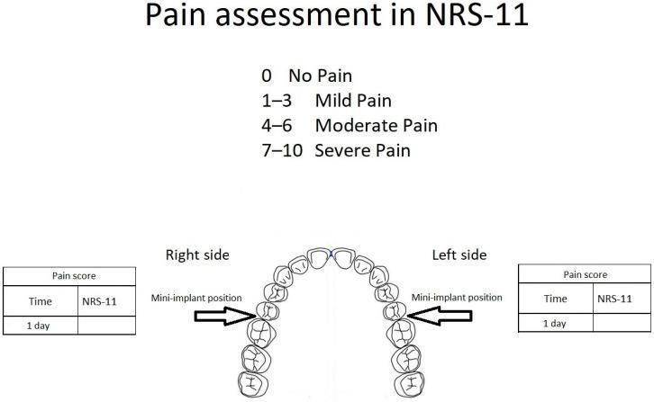
║ NRS: 0=No pain | 1-3=Mild | 4-6=Moderate | 7-10=Severe            ║
╠══════════════════════════════════════════════════════════════════════╣
║ TOP EMERGENCY DRUGS:                                                 ║
║ Adrenaline: Anaphylaxis 0.5mg IM / Arrest 1mg IV                  ║
║ Atropine: Bradycardia 0.5-1mg IV                                   ║
║ PCM: Fever 500-1000mg PO/IV (max 4g/day)                          ║
║ Morphine: Severe pain 5-10mg IV (watch RR!)                       ║
╠══════════════════════════════════════════════════════════════════════╣
║ SBAR: Situation > Background > Assessment > Recommendation          ║
║ I/O: Normal urine = 0.5 ml/kg/hr (Oliguria = <0.5)               ║
╚══════════════════════════════════════════════════════════════════════╝
🎯 WHAT'S COMING NEXT:
Upcoming Modules:
- MODULE 9: Hand Hygiene + Infection Control (WHO 5 moments)
- MODULE 10: Complete Emergency Drugs (Amiodarone, Dopamine, MgSO4, etc.)
- MODULE 11: Common Ward Diseases (Hypertension, Diabetes, Pneumonia, etc.)
- MODULE 12: IV Cannulation + Injections (Complete Practical Guide)
- MODULE 13: Blood Transfusion (Procedure + Reactions + Nursing)
- MODULE 14: ECG Basics for Nurses
- MODULE 15: Sepsis, Shock, Cardiac Arrest - Emergency Management
💡 Professor's Closing Note:
Aaj aapne seekha: Medical terminology, Vital signs, GCS, Pain assessment, Lab values, Emergency drugs basics, aur Documentation. Yeh sab DAILY hospital use mein aata hai.
Daily revision rule: Is module ko raat sone se pehle 5 min review karo. Kal subah quiz dene ki koshish karo bina notes dekhe.
Jab bhi hospital posting mein jao: Vitals chart dekho, medicines ka timing samjho, documentation padhne ki koshish karo. Sab kuch familiar lagega!
Next module ke liye ready ho? Reply karo: "Ready for Module 2" ya koi specific topic ka naam batao jahan aur detail chahiye! 🏥