
Mumps hastaligi nedir
mumps hastalığı kabakulak belirtiler tedavi
mumps parotitis swollen parotid gland child
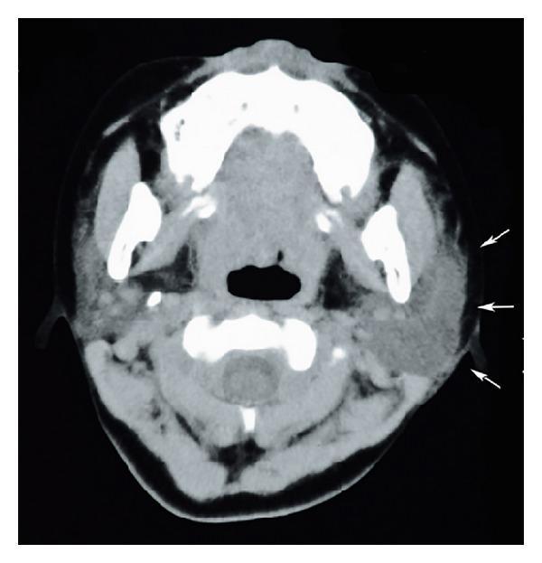
This diagnostic image is an axial computed tomography (CT) scan of the neck at the level of the mandible, demonstrating clinical findings of sialadenitis. The primary focus is the left parotid gland, which is significantly enlarged and swollen compared to the contralateral side. The left parotid exhibits a heterogeneous parenchymal density and ill-defined margins, indicated by white arrows, which suggest active inflammation and adjacent soft tissue edema. In contrast, the right parotid gland appears relatively normal in size with more uniform density. Key anatomical structures visible include the mandibular rami, the base of the tongue, the oropharynx, and the cervical vertebrae. The image serves as an educational example of inflammatory pathology of the salivary glands, highlighting the use of cross-sectional imaging to assess glandular swelling, tissue plane blurring, and potential complications such as abscess formation in cases of acute sialadenitis or mumps-related parotitis.
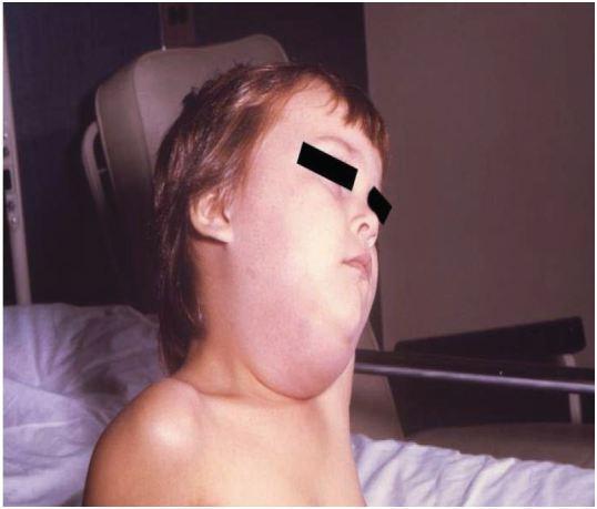
Clinical photograph of a pediatric patient presenting with marked, unilateral swelling of the right parotid region, characteristic of acute mumps parotitis. The image shows a prominent, non-fluctuant bulge extending from the preauricular area, anterior to and below the ear, down to the angle of the mandible and upper cervical region. This localized edema results in the loss of the normal mandibular line and causes the earlobe to be displaced upward and outward. The overlying skin appears taut and mildly erythematous, indicating significant inflammatory glandular enlargement. This finding is the pathognomonic clinical sign of mumps virus infection (Paramyxoviridae family). The visual evidence supports educational recognition of salivary gland pathology and infectious disease manifestations in children. Differential diagnoses for such clinical presentations include bacterial parotitis, salivary calculi, or other viral etiologies like influenza or Epstein-Barr virus.
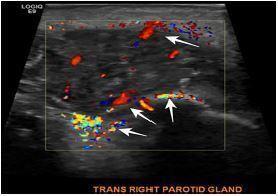
This diagnostic image is a transverse color Doppler ultrasound of the right parotid gland. The image depicts a swollen parotid gland characterized by a heterogeneous, mottled echotexture compared to normal surrounding soft tissue. A color Doppler window overlaying the gland demonstrates significant hypervascularity, evidenced by prominent red and blue signals throughout the parenchyma. Multiple white arrows specifically highlight these areas of increased blood flow. The lack of distinct fluid collections or shadowing suggests the absence of a mature abscess or sialolithiasis. Clinically, these findings—swelling and hypervascularity—are indicative of acute parotitis. The image is labeled at the bottom as 'TRANS RIGHT PAROTID GLAND' and serves as an educational example of inflammatory changes in salivary gland tissue as visualized by point-of-care ultrasound (POCUS).
Mumps (Kabakulak) Hastalığı
Tanım
- Cummings Otolaryngology Head and Neck Surgery ve Goldman-Cecil Medicine
Epidemiyoloji
- İnsanlar tek doğal konaktır; hayvan rezervuarı, böcek vektörü ya da taşıyıcı bilinmemektedir.
- Hastalık çok bulaşıcıdır; enfekte bireylerin solunum damlacıkları (tükürük, burun, idrar salgıları) ile yayılır.
- Vakaların %85'i 15 yaş altı çocuklarda görülür.
- İnkübasyon süresi 12-25 gün arasındadır.
- Bireyler semptomlar başlamadan 2 gün önce ile başlangıçtan 5 gün sonra en bulaşıcı dönemdedir; asemptomatik vakalar da hastalığı bulaştırabilir.
- Ilıman iklimlerde ilkbaharda pik yapar.
Klinik Tablo
Prodromal Dönem (Parotis şişmesinden önce)
- Düşük dereceli ateş
- Baş ağrısı
- Miyalji (kas ağrısı)
- Halsizlik ve yorgunluk
- İştahsızlık
- Artralji
Parotis Tutulumu
- Kulak önü bölgede ağrılı şişlik (bilateral: %75 vakada her iki yanak)
- Kulak ağrısı (otalji)
- Çiğneme ve yutma güçlüğü (trismus, disfaji)
- Kulak kepçesinin dışa/yukarı itilmesi
- Deri gergin ve parlak görünümde; ancak kızarıklık veya ısı artışı yoktur
- Bir yanaktan başlayıp 1-5 gün içinde diğer yanağa geçebilir
Tanı
- Viral seroloji ile doğrulanır:
- S (soluble/çözünür) antikorları: Virüsün nükleoprotein çekirdeğine karşı oluşur; en erken görünen antikorlardır, 10 güne-2 haftada pik yapar, 8-9 ayda kaybolur - aktif enfeksiyonu gösterir.
- V (viral) antikorları: Yüzey hemaglütinine karşı oluşur; geç çıkar ama yıllarca kalır.
- Antikor titresinde 4 kat artış akut enfeksiyon için tanısaldır.
- Lökopeni ve serum amilaz yüksekliği görülebilir.
- Görüntülemede Doppler ultrasonografide parotis bezinde hipervaskülarite saptanır.
Komplikasyonlar
| Komplikasyon | Açıklama |
|---|---|
| Orşit | Ergen/yetişkin erkeklerde en sık; nadiren infertiliteye yol açar |
| Aseptik menenjit | En sık viral menenjit nedenlerinden biridir |
| Pankreatit | Karın ağrısı, bulantı ile seyreder |
| Nefrit | Böbrek tutulumu |
| Sensorinöral işitme kaybı | Kalıcı olabilir |
Tedavi
- Yatak istirahati
- Ağız hijyeni
- Yeterli sıvı alımı
- Ağrı ve ateş için parasetamol / ibuprofen
- Çiğnemeyi azaltan yumuşak diyet
- Salgıyı azaltmak için beslenme düzenlemesi
Korunma: Aşılama
- Canlı-attenue Jerry Lynn suşu aşısı (MMR: Kızamık-Kabakulak-Kızamıkçık)
- 12 aylıktan itibaren subkutan uygulanır; 4-6 yaşta ikinci doz
- Oluşan antikorlar en az 5 yıl kalır
- Önemli: İki doz MMR aşısı almış bireylerde de salgın döneminde vakalar görülebilir; bu nedenle aşı öyküsü tanıyı dışlamaz
- Bağışıklığı baskılanmış hastalarda ve neomisin alerjisi olanlarda kontrendikedir