
Operations of phlegmin and abscesses of neck
Reading File
Reading File
Reading File
Finding Sources
Finding Sources
Searching PubMed
deep neck infection AND drainage
Searching the Web
deep neck space infections surgical drainage airway management guidelines
Reading File
Reading File
Reading File
Reading Article
PMID: 38616333
Reading File
Operations for phlegmon and abscess of the neck
Deep neck infection may be either:
- Phlegmon: diffuse infected edema and inflamed tissue, without a pus cavity. It is usually not drainable.
- Abscess: a localized collection of pus. It needs source control, usually by aspiration or incision and drainage.
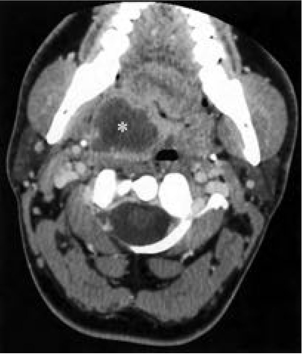
CT with IV contrast maps the involved fascial spaces, but it may not always distinguish phlegmon from pus reliably. An abscess often appears as a low-density, rim-enhancing collection. Cummings Otolaryngology Head and Neck Surgery, p. 1886.
1. Initial principles
-
Airway comes first
- Look for stridor, dyspnea, drooling, voice change, inability to lie flat, severe trismus, floor-of-mouth elevation, or rapidly increasing swelling.
- Secure the airway early in a controlled setting if it is threatened. Avoid repeated traumatic attempts at intubation.
- Prepare for surgical airway when intubation is unsafe or unsuccessful.
-
Resuscitation and investigations
- Admit, keep NPO, IV fluids, analgesia.
- CBC, inflammatory markers, blood cultures if septic.
- Contrast-enhanced CT neck, extending to chest when retropharyngeal or danger-space spread is suspected.
- Send aspirated pus or operative specimen for Gram stain, aerobic and anaerobic culture, and sensitivity.
-
Antibiotics
- Start IV antibiotics covering oral streptococci, anaerobes, and staphylococci.
- Modify after culture results and local resistance patterns.
- Address the source, especially infected teeth, tonsil, salivary gland disease, foreign body, or infected cyst.
2. Treatment of phlegmon
Primary treatment is non-operative, provided there is no airway threat or definite drainable collection:
- IV antibiotics
- Close airway observation
- NPO initially if surgery may become necessary
- Repeat examination and imaging if deterioration occurs
- Treat the primary source, for example dental extraction when appropriate
Operate/explore despite phlegmon if any of the following occurs:
- Threatened airway
- Gas in tissues or air-fluid level
- Clinical sepsis or rapid progression
- Evolving abscess
- No response after about 48-72 hours of IV antibiotics
K. J. Lee’s Essential Otolaryngology, p. 710.
3. Indications for operative drainage of a neck abscess
- A definite abscess in a deep cervical fascial space
- Airway compromise or impending compromise
- Large, multiloculated, or spreading collection
- Gas-forming infection or necrotizing infection
- Failure of medical therapy after 48-72 hours
- Mediastinal extension, neurovascular complication, or sepsis
The operative objectives are to drain pus, obtain cultures, irrigate the involved space, establish dependent drainage, and eliminate the primary focus of infection. K. J. Lee’s Essential Otolaryngology, p. 711.
4. Operative options
| Type/location | Usual drainage route | Key principle |
|---|---|---|
| Small suppurative lymph node | Needle aspiration or small incision | Culture specimen; avoid unnecessary wide dissection |
| Peritonsillar abscess | Needle aspiration or transoral incision and drainage | Drain intraorally; observe airway and bleeding |
| Dental/alveolar or buccal space abscess | Intraoral drainage plus extraction of offending tooth | Remove dental source |
| Submandibular/submental abscess or Ludwig angina | Usually external dependent cervical drainage when pus is present | Airway management and dental source control are priorities |
| Parapharyngeal abscess | Transoral drainage only if medially located and accessible; otherwise transcervical drainage | Avoid carotid sheath and cranial nerve injury |
| Retropharyngeal abscess | Often transoral drainage; external route in selected lateral, extensive, or complicated disease | Assess for danger-space/mediastinal extension |
| Multispace, deep, or recurrent abscess | Transcervical exploration with blunt opening of involved fascial planes | Break loculi, irrigate, place drains, re-image if not improving |
5. General operative sequence
This procedure should be performed by an experienced ENT or head-and-neck surgical team, with anesthesia support.
- Secure or plan the airway before positioning or induction.
- Position for access and prepare both oral cavity and neck when needed.
- Choose the least morbid route that provides safe, complete dependent drainage.
- Make incision in a skin crease or use transoral access as indicated by the involved space.
- Use careful blunt dissection to enter the collection and avoid major vessels and nerves.
- Obtain pus for culture before irrigation.
- Break down loculations, irrigate, and inspect for communication with other spaces.
- Remove the source, such as extraction of an infected tooth or removal of foreign body.
- Place a drain or loose packing when ongoing drainage is needed. Do not tightly close a contaminated cavity.
- Continue IV antibiotics, monitor airway and sepsis, and re-explore or repeat imaging if clinical improvement does not occur.
6. Postoperative care
- Close airway monitoring, sometimes ICU care
- IV antibiotics initially, then culture-directed therapy
- Daily wound and drain assessment
- Monitor for mediastinitis, septic thrombophlebitis of internal jugular vein, carotid complications, aspiration, and recurrent collection
- Repeat CT if persistent fever, swelling, leukocytosis, or poor drain output
- Remove drains only after output and clinical signs have settled
A 2024 systematic review found that, in selected deep-neck abscesses, ultrasound-guided drainage had a shorter hospital stay than conventional incision and drainage, without a statistically significant difference in recurrence. This is an option only when the collection is accessible and the airway is secure, not for unstable or extensive infection (systematic review, PMID 38616333).
Clinical warning: Deep neck swelling with breathing difficulty, stridor, drooling, voice change, rapidly progressing swelling, or inability to swallow is an emergency requiring immediate ENT/anesthesia assessment.
This is a shared conversation. Sign in to Orris to start your own chat.