
Hernia
types of hernia inguinal femoral umbilical diagram anatomy
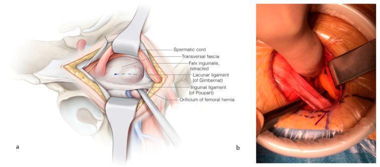
This composite visual contains an anatomical illustration (a) and a corresponding clinical intraoperative photograph (b) detailing the surgical repair of a femoral hernia. Panel (a) provides a labeled anatomical diagram of the right inguinal-femoral region. It depicts the spermatic cord and falx inguinalis retracted cranially, exposing the orificium of the femoral hernia. Key landmarks are identified: the transversalis fascia (opened), the inguinal ligament of Poupart (ventral border), and the lacunar ligament of Gimbernat (medial border). An instrument is shown passing through the femoral orifice to demonstrate the pathology. Panel (b) is a clinical photograph of the surgical field during an open repair. It demonstrates the clinical application of the anatomy shown in panel (a), with retractors maintaining exposure. A surgical instrument is seen incising the lacunar ligament to enlarge the femoral space for mesh placement. The image illustrates the relationship between the inguinal ligament, femoral vessels, and the hernia orifice. The content is intended for intermediate to advanced surgical education, specifically regarding open femoral hernia repair and the anatomy of the femoral canal.
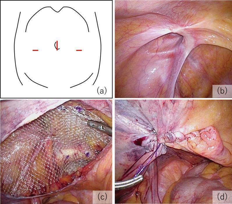
A multi-panel educational figure illustrating the key procedural steps of a Transabdominal Preperitoneal (TAPP) inguinal hernia repair. (a) Schematic diagram of the abdomen showing the laparoscopic port arrangement: a 12 mm umbilical port and two 5 mm lateral ports. (b) Intraoperative laparoscopic photograph showing the internal inguinal anatomy and the hernial orifice, classified as EHS-L2 (European Hernia Society classification for a medium indirect lateral hernia). (c) Surgical view demonstrating mesh fixation; a semi-transparent, macroporous polypropylene mesh is positioned in the preperitoneal space to cover the defect and is secured with visible surgical tacks. (d) Final procedural step showing the closure of the peritoneal flap using continuous suturing to exclude the mesh from the peritoneal cavity, preventing contact with abdominal viscera. The series emphasizes proper anatomical orientation, mesh placement, and peritoneal closure in minimally invasive hernia surgery.
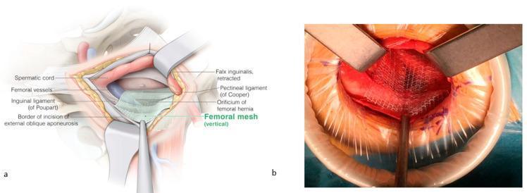
This composite image illustrates the surgical repair of a femoral hernia using a mesh-based approach. Figure (a) is an anatomical diagram of the inguinal and femoral regions during surgery. Key structures identified include the spermatic cord (superior-medial), femoral vessels (lateral), inguinal ligament of Poupart (ventral), and the pectineal ligament of Cooper (dorsal). The diagram shows the placement of a vertical femoral mesh covering the orificium of the femoral hernia, secured over Cooper’s ligament while maintaining an overlay medially and laterally over the femoral vessels. The falx inguinalis is shown in a retracted state. Figure (b) is an intraoperative clinical photograph corresponding to the diagram, showing the surgical field with metal retractors and a circular wound protector. A white, woven surgical mesh is visible, positioned over the femoral orifice and held in place with instruments. The image demonstrates the 'Lichtenstein-like' open technique for femoral hernia reinforcement, highlighting the spatial relationship between the prosthetic mesh and pelvic floor ligaments to prevent recurrence.
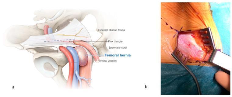
This dual-panel image provides an anatomical and clinical overview of a femoral hernia and its surgical exposure. Panel (a) is a detailed anatomical diagram of the right inguinal and femoral regions. It illustrates a femoral hernia as a distinct bulge emerging medially to the femoral vessels (artery and vein) and caudal to the inguinal canal. Labeled structures include the external oblique fascia, the 'pink triangle' (an area formed by the diversion of fascial vessels toward the external inguinal orifice), the spermatic cord, and femoral vessels. Panel (b) is a clinical photograph showing the intraoperative view after the incision of the external oblique fascia. The surgical field is exposed using three metal claw-style retractors, revealing the underlying fascia and fatty tissue in the femoral region. Sutures are visible along the superior aspect of the skin incision. This content is designed for medical education regarding hernia pathology and surgical repair, demonstrating the critical landmarks needed to differentiate femoral hernias from inguinal hernias and to safely navigate the femoral canal during surgery.
Hernia - A Comprehensive Overview
Definition
- Maingot's Abdominal Operations
Anatomy of the Inguinal Canal
| Wall | Structure |
|---|---|
| Anterior | External oblique aponeurosis |
| Posterior | Transversalis fascia + transversus abdominis muscle |
| Superior | Internal oblique + transversus abdominis |
| Inferior | Inguinal (Poupart's) ligament |
- Schwartz's Principles of Surgery, 11th Ed.
Classification
By Location
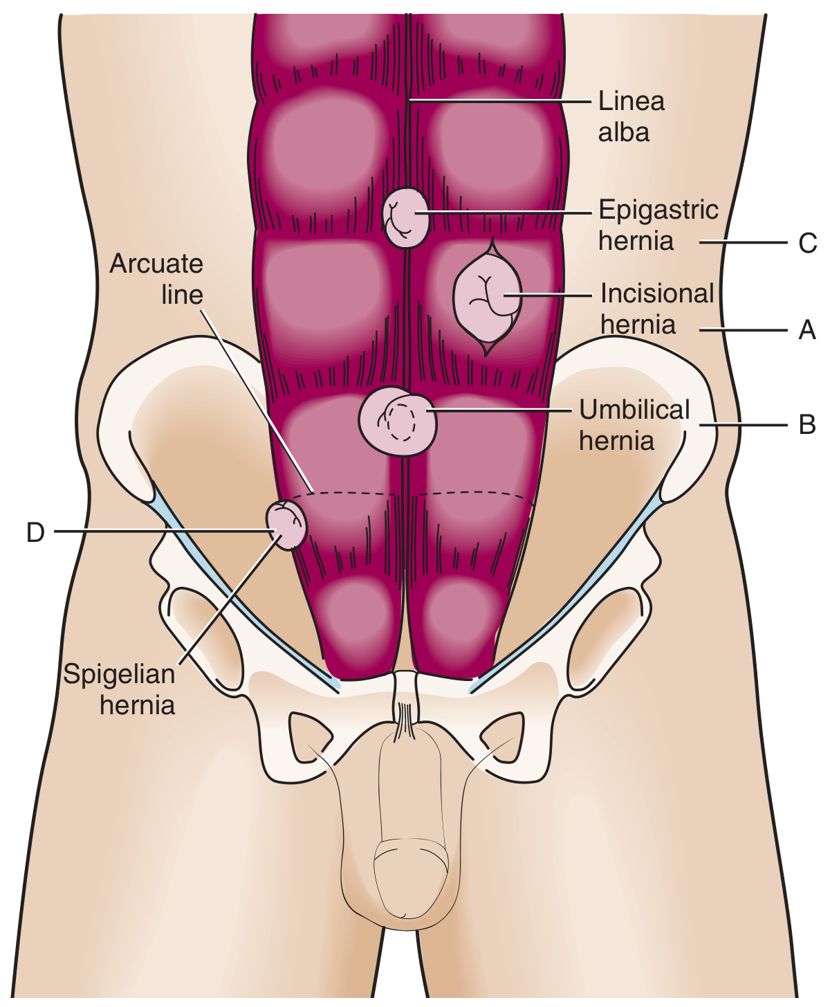
1. Inguinal Hernia (most common, ~75% of all hernias)
| Feature | Indirect | Direct |
|---|---|---|
| Location | Lateral to inferior epigastric vessels | Medial to inferior epigastric vessels (within Hesselbach's triangle) |
| Origin | Deep (internal) inguinal ring | Weakness in inguinal floor |
| Etiology | Congenital (patent processus vaginalis) | Acquired |
| More common in | All ages, especially children; right side more than left | Older adults; males |
| Scrotal extension | Yes, can descend into scrotum | Rarely extends to scrotum |
| Reduces with internal ring pressure | Yes | No |
- Maingot's Abdominal Operations; Roberts and Hedges' Clinical Procedures in Emergency Medicine
2. Femoral Hernia
-
Protrudes inferior to the inguinal ligament, through the femoral canal - the potential space medial to the femoral vein and lateral to the lacunar (Gimbernat's) ligament
-
Appears as a medial thigh mass below the groin crease
-
More common in women
-
High-risk: up to 40-45% present with incarceration or strangulation because the femoral ring is rigid and narrow
-
Accounts for <10% of all groin hernias but is disproportionately dangerous
-
Maingot's; Roberts and Hedges'
3. Incisional Hernia
- Occurs through a previous surgical incision site
- Affects up to 1 in 5 patients following abdominal surgery
- Risk factors: wound infection, obesity, poor healing, steroids
- Carries a recurrence rate of 20-50% after repair
- Incarceration risk is inversely proportional to defect size (smaller defects = higher risk)
4. Umbilical Hernia
- Traverses the fibromuscular ring of the umbilicus
- Children: usually congenital, often resolves spontaneously by age 5; surgery if >2 cm, persists beyond 5 years, or becomes incarcerated
- Adults: acquired (associated with obesity, ascites, pregnancy); more prone to incarceration and strangulation in adults than children
- Accounts for ~15% of emergency hernia surgery; up to 60% show strangulation at the time of surgery
5. Epigastric Hernia
- Through the linea alba, between the xiphoid and umbilicus
- Found in 4% of pediatric patients evaluated for hernias; usually small in adults
- Located in the epigastric region
6. Spigelian Hernia
- Through the spigelian fascia (aponeurosis of the transversus abdominis lateral to the rectus sheath)
- Typically in patients aged 40-70 years
- Incarceration rates up to 20% (often with omentum)
- Ultrasound is a valuable adjunct for diagnosis
7. Internal Hernias
- Congenital types: paraduodenal (most common congenital type) or through the foramen of Winslow
- Iatrogenic (most common overall): after Roux-en-Y gastric bypass, colectomy, or Whipple procedure - bowel herniates through mesenteric defects
- Obturator hernia: bowel through the obturator canal - always requires urgent surgical repair due to high risk of strangulation; no role for conservative management
Reducibility - Key Terminology
| Term | Definition |
|---|---|
| Reducible | Hernia contents can be returned to normal position spontaneously or manually |
| Incarcerated | Cannot be reduced; does not automatically imply ischemia or emergency |
| Strangulated | Ischemia or obstruction results from compression at the hernia neck - a surgical emergency |
- Schwartz's Principles of Surgery; Sleisenger & Fordtran's GI and Liver Disease
- Femoral: ~40%
- Umbilical: up to 60% at time of emergency surgery
- Incisional: ~2%
Diagnosis
- Palpate the inguinal canal by inverting scrotal skin in males (narrower in females)
- Ask patient to cough or perform Valsalva maneuver
- Differentiate indirect from direct: indirect hernia strikes the fingertip, direct strikes the volar pad of the finger; pressure over the internal ring reduces indirect but not direct
- CT scan: required in obese patients or when exam is equivocal; diagnostic for incarcerated spigelian and other occult hernias
- Ultrasound: helpful for spigelian hernias and inguinal testes
Treatment
Principles
- Elective repair is associated with far less morbidity than emergency repair
- Strangulation is a surgical emergency requiring urgent intervention
- Manual reduction of incarcerated hernia may be attempted (without signs of strangulation) and is most successful for groin and umbilical hernias - must still be followed by expeditious surgical repair
Surgical Approaches for Inguinal Hernia
| Approach | Description |
|---|---|
| Open (Lichtenstein) | Tension-free mesh repair via groin incision; gold standard open technique |
| Laparoscopic TAPP | Transabdominal preperitoneal - mesh placed via laparoscope from inside abdomen, peritoneum then closed |
| Laparoscopic TEP | Totally extraperitoneal - preperitoneal space developed without entering peritoneum |
| Robotic | Better ergonomics and visualization; rapidly being adopted |
- Schwartz's Principles of Surgery, 11th Ed.
- Bassini (1884/1889): Revolutionized hernia repair with anatomical dissection and posterior canal reinforcement; <5 recurrences in >250 patients
- McVay/Lotheissen: Cooper's ligament repair for both inguinal and femoral defects
- Usher: Introduced modern synthetic polyethylene mesh
Special Considerations
-
Children with groin hernia: May present atypically with vomiting, lethargy, or irritability - always consider incarceration
-
Hernia and bowel obstruction: Hernia accounted for 44.5% of all operations for acute small bowel obstruction (SBO) in one large California dataset; incarcerated hernia causes intestinal ischemia in up to 75% and necrosis in >25% of SBO cases
-
Contraindication to manual reduction: Severe tenderness, skin erythema, signs of strangulated bowel
-
Sleisenger & Fordtran's GI and Liver Disease, 10th Ed.