
Hemodynamic disorders. Thrombosis. Etiopathogenesis and morphology. In detail
Reading File
Reading File
Finding Sources
Searching PubMed
"thrombosis"[MeSH Terms] AND "pathogenesis"
Reading File
Finding Sources
Reading File
Reading File
Reading File
Reading File
Reading File
Reading File
Reading File
Reading File
Searching Images
deep vein thrombosis gross specimen thrombus lines of Zahn laminated
Hemodynamic Disorders: Thrombosis (Etiopathogenesis and Morphology)
1. Context: hemodynamic disorders
Hemodynamic disorders are disturbances of blood volume, flow, or vessel integrity. They include:
- Edema
- Hyperemia and congestion
- Hemorrhage
- Hemostasis and thrombosis
- Embolism
- Infarction
- Shock
Thrombosis is the pathological form of hemostasis. It is the inappropriate formation of a blood clot within an intact vessel or heart chamber during life.
Normal hemostasis versus thrombosis
- Hemostasis is a localized, regulated response to vessel injury. It has five steps:
- Brief arteriolar vasoconstriction.
- Primary hemostasis: platelets adhere via von Willebrand factor (vWF) bound to exposed subendothelial collagen, then activate and aggregate.
- Secondary hemostasis: tissue factor (TF) triggers the coagulation cascade, thrombin is generated, and fibrinogen becomes fibrin.
- Clot stabilization and retraction.
- Counter-regulation, which limits the clot to the injury site.
- Thrombosis is the same machinery activated at the wrong time or place, or in excess.
Normal anti-thrombotic defenses (in my own words, as the retrieved text was truncated here)
- Endothelium: prostacyclin (PGI2), nitric oxide, and adenosine diphosphatase (ADPase) inhibit platelets.
- Thrombomodulin, the protein C/S system, heparin-like molecules with antithrombin III, and tissue factor pathway inhibitor (TFPI) inhibit coagulation.
- t-PA drives fibrinolysis.
2. Etiopathogenesis: the Virchow triad
Thrombosis is driven by one or more of three primary abnormalities (the Virchow triad): (1) endothelial injury, (2) stasis or turbulent blood flow, and (3) hypercoagulability. These may act independently or in combination. - Robbins, Cotran & Kumar Pathologic Basis of Disease, Thrombosis
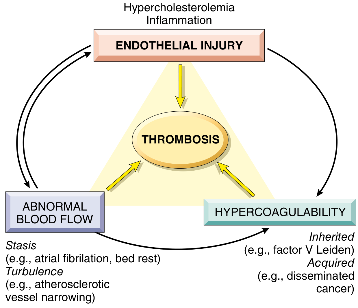
Endothelial integrity is the most important factor. Endothelial injury can alter local flow and coagulability, and abnormal flow can in turn injure endothelium.
A. Endothelial injury (dominant in arteries and the heart)
- Endothelial injury that activates platelets almost always underlies thrombus formation in the heart and arteries. High flow rates there impede clot formation.
- Cardiac and arterial clots are platelet-rich. Platelet adherence and activation is a prerequisite under high shear stress. This is the rationale for aspirin and other antiplatelet drugs in coronary disease and acute MI.
- Severe injury exposes vWF and tissue factor. Examples are ruptured atherosclerotic plaque, trauma, vasculitis, and a surgical or catheter injury.
- Endothelial activation or dysfunction does not need anatomic denudation. It is a shift in endothelial gene expression toward a prothrombotic state. Causes include:
- Physical injury and infectious agents
- Abnormal flow
- Inflammatory mediators
- Hypercholesterolemia and homocystinemia
- Cigarette smoke toxins
- Prothrombotic changes include:
- Procoagulant: thrombomodulin, protein C, and TFPI are downregulated, and tissue factor is upregulated.
- Antifibrinolytic: t-PA is reduced.
- Platelet-activating: PGI2 and NO are reduced, and vWF is increased.
B. Abnormal blood flow (stasis and turbulence)
- Turbulence is important in arteries and the heart. It causes endothelial injury or dysfunction and creates countercurrents that form local pockets of stasis.
- Stasis is the major factor in venous thrombi.
- Normal flow is laminar: platelets travel centrally, separated from the endothelium by a slower plasma layer. Stasis and turbulence therefore:
- Promote endothelial activation (more procoagulant activity and leukocyte adhesion).
- Disrupt laminar flow and bring platelets into contact with endothelium.
- Prevent washout and dilution of activated clotting factors, and prevent inflow of clotting factor inhibitors.
- Clinical settings:
- Ulcerated atherosclerotic plaques expose vWF and TF and cause turbulence.
- Aneurysms cause local stasis.
- MI causes noncontractile myocardium or a ventricular aneurysm, giving mural thrombi.
- Mitral stenosis with a dilated left atrium and atrial fibrillation is a prime site.
- Hyperviscosity (polycythemia vera) and sickled red cells slow small-vessel flow.
C. Hypercoagulability (important in venous thrombosis)
It is an abnormally high tendency of blood to clot, usually from altered coagulation factors. It is divided into primary (genetic) and secondary (acquired) causes (Table 4.2).
Primary (genetic)
- Factor V Leiden
- It is an Arg506Gln substitution that makes factor V resistant to inactivation by activated protein C.
- About 2% to 15% of people of European ancestry carry it. The frequency approaches 60% in people with recurrent DVT.
- It is autosomal dominant. Heterozygotes have a 4- to 8-fold increased risk of venous thrombosis, and homozygotes about 12-fold.
- Prothrombin G20210A mutation
- It is in the 3'-untranslated region and raises prothrombin levels.
- It is found in 1% to 2% of the population. Risk is 2- to 4-fold in heterozygotes and 7-fold in homozygotes.
- Rare: antithrombin III, protein C, or protein S deficiency. These present with venous thrombosis and recurrent thromboembolism from adolescence or early adulthood.
- Very rare: fibrinolysis defects and homozygous homocystinuria (cystathionine beta-synthase deficiency).
- Increased factors VIII, IX, XI, or fibrinogen are also listed, with unknown genetics.
- Heterozygous Factor V Leiden or prothrombin variants alone carry only moderately increased risk. Risk rises sharply with a second hit such as pregnancy, immobilization, or prolonged flights. Inherited thrombophilia should be considered in any patient under 50 with thrombosis, even when acquired risk factors are present.
Secondary (acquired)
- Strong risk factors:
- Prolonged bed rest or immobilization
- MI
- Atrial fibrillation
- Tissue injury (surgery, fracture, burns)
- Cancer
- Prosthetic cardiac valves
- DIC
- Heparin-induced thrombocytopenia (HIT)
- Antiphospholipid antibody syndrome (APS)
- Other risk factors:
- Cardiomyopathy
- Nephrotic syndrome (loss of antithrombin III in urine)
- Pregnancy and postpartum state
- Oral contraceptives (increased hepatic synthesis of coagulation factors and reduced anticoagulant synthesis)
- Sickle cell anemia
- Smoking
- Cancer (Trousseau syndrome, or migratory thrombophlebitis) acts through tumor-associated inflammation, tissue factor release, and procoagulants such as mucin from tumor cells.
- Trauma, surgery, and burns cause immobilization, vascular insult, procoagulant release from injured tissue, increased hepatic factor synthesis, and decreased t-PA.
- Age: advanced age independently increases DVT risk.
Heparin-induced thrombocytopenia (HIT)
- Heparin displaces platelet factor 4 (PF4) from endothelium and forms PF4/heparin complexes.
- Heparin binding changes PF4 conformation and creates a neoantigen, and IgG antibodies form against it.
- The immune complexes bind platelet Fc receptors and activate platelets.
- Activated platelets express procoagulant molecules, which causes thrombosis.
- Macrophages clear antibody-coated platelets, which causes thrombocytopenia.
- Thrombosis occurs in about 50% of cases and affects both veins and arteries. Sequelae include skin necrosis, limb gangrene, stroke, and MI.
- Diagnosis needs anti-PF4/heparin antibodies.
- Low-molecular-weight heparin causes HIT less often. Direct factor Xa or thrombin inhibitors avoid the risk.
- Vaccine-induced thrombotic thrombocytopenia (VITT) is a rare anti-PF4 syndrome after some adenoviral-vector COVID-19 vaccines. It often involves cerebral venous sinuses.
Antiphospholipid antibody syndrome (APS)
- It is an autoimmune disorder with antiphospholipid antibodies plus venous or arterial thrombosis and/or pregnancy complications (recurrent miscarriage, fetal death, premature birth).
- Primary APS (about 50%) has no other autoimmune disease. Secondary APS is associated with autoimmune disease, most often SLE.
- The target antigens are cardiolipin and beta-2 glycoprotein I. The antibodies activate endothelium, neutrophils, monocytes, and platelets. Complement is activated and fibrinolysis is inhibited.
- Early fetal loss comes mainly from interference with trophoblast growth and a failure of placentation, not thrombosis. Late complications (preeclampsia, preterm birth) come from both trophoblast dysfunction and complement-mediated inflammation.
- Clinical features include:
- Recurrent DVT and PE
- Pulmonary hypertension
- Stroke
- Bowel infarction
- Renovascular hypertension
- Valve vegetations
- Thrombocytopenia
- Laboratory findings:
- The "lupus anticoagulant" prolongs PTT in vitro, yet the patient is hypercoagulable in vivo.
- Biologic false-positive syphilis tests occur, because the VDRL antigen contains cardiolipin.
- Antibodies are present in 5% to 15% of normal people, so a "second hit" such as infection, smoking, or pregnancy is probably required.
- Treatment is anticoagulation.
3. Morphology
General features
- Thrombi can form anywhere in the cardiovascular system.
- Arterial and cardiac thrombi begin at sites of turbulence or endothelial injury. Venous thrombi begin at sites of stasis.
- They are focally attached to the vessel wall at the point of origin.
- Arterial thrombi grow retrograde (against flow). Venous thrombi extend in the direction of flow. Both therefore propagate toward the heart.
- The propagating tail is poorly attached and is the part that fragments and embolizes.
Lines of Zahn
- These are grossly and microscopically visible laminations: pale layers of platelets and fibrin alternating with darker layers rich in red cells.
- They indicate that the thrombus formed in flowing blood, so they distinguish antemortem thrombi from bland, nonlaminated postmortem clots.
Types by location
| Type | Features |
|---|---|
| Mural thrombi (heart chambers, aortic lumen) | Cardiac: abnormal contraction (arrhythmia, dilated cardiomyopathy, MI) or endomyocardial injury (myocarditis, catheter trauma). Aortic: ulcerated atherosclerotic plaque or aneurysmal dilation. Prone to embolization. |
| Arterial thrombi | Often occlusive. Most common sites in decreasing order: coronary, cerebral, femoral arteries. They form a friable meshwork of platelets, fibrin, red cells, and degenerating leukocytes (white or "pale" thrombi). Usually superimposed on ruptured plaque. Other causes: vasculitis, trauma. |
| Venous thrombi (phlebothrombosis) | Almost invariably occlusive, forming a long luminal cast. Contain many enmeshed red cells and relatively few platelets, so they are called red or stasis thrombi. Firm, focally attached, with lines of Zahn. Lower-limb veins account for about 90%. Other sites: upper extremity, periprostatic plexus, ovarian and periuterine veins, dural sinuses, portal and hepatic veins. |
| Valvular vegetations | Thrombi on heart valves. Infective: bacteria or fungi on damaged valves (infective endocarditis). Sterile: nonbacterial thrombotic endocarditis in hypercoagulable states, and Libman-Sacks endocarditis in SLE. |
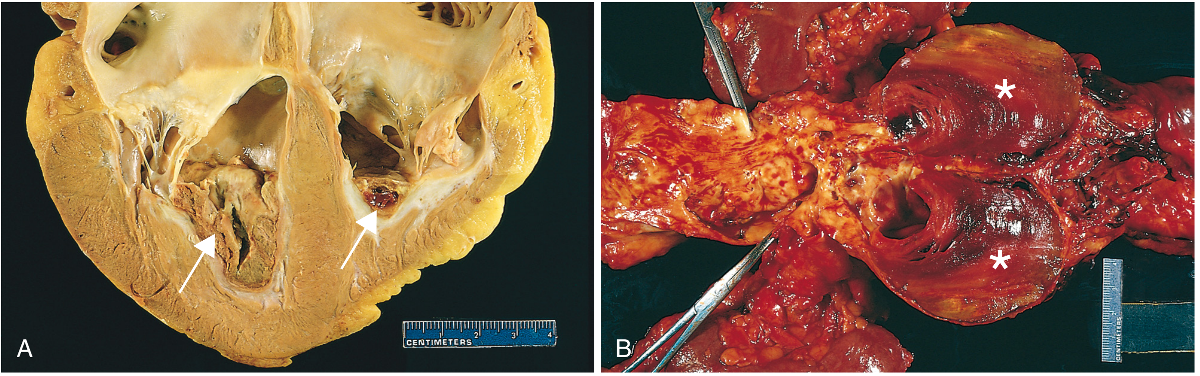
Mural thrombi. (A) Thrombus in the left and right ventricular apices over a white fibrous scar. (B) Laminated thrombus in a dilated abdominal aortic aneurysm, with friable mural thrombi on advanced atherosclerosis. - Robbins, Cotran & Kumar Pathologic Basis of Disease, Fig. 4.13
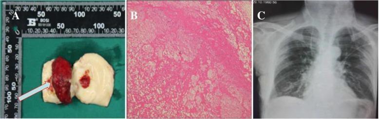
Gross specimen and H&E histology (panel B) of a thrombus showing laminated layers of fibrin and trapped red cells (lines of Zahn).
Antemortem thrombus versus postmortem clot
| Feature | Thrombus | Postmortem clot |
|---|---|---|
| Consistency | Firm, friable | Gelatinous |
| Attachment | Attached to vessel wall | Usually not attached |
| Surface and color | Dull, rough; lines of Zahn | Smooth, glistening; dark red dependent part with yellow "chicken fat" upper part |
| Laminations | Present | Absent |
4. Fate of the thrombus
Over days to weeks, a thrombus undergoes some combination of four events:
- Propagation: it accumulates more platelets and fibrin and enlarges.
- Embolization: it dislodges and travels to other sites in the vasculature.
- Dissolution: fibrinolysis can rapidly shrink and remove recent thrombi. Older thrombi have more cross-linked fibrin and resist lysis. This is why t-PA works mainly in the first hours of an acute coronary thrombosis.
- Organization and recanalization: endothelial cells, smooth muscle cells, and fibroblasts grow in. Capillary channels form and partly restore the lumen. Eventually only a fibrous lump may remain in the vessel wall.
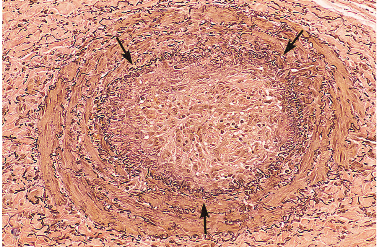
Thrombosed artery on elastic stain. The original lumen, outlined by the internal elastic lamina, is filled with organized thrombus. - Fig. 4.14
Other outcomes: the center of a thrombus may undergo enzymatic digestion by lysosomal enzymes from trapped leukocytes and platelets. In bacteremia, thrombi can become infected and erode the vessel wall, producing a mycotic aneurysm.
5. Clinical consequences
Thrombi cause harm by obstructing a vessel or by embolizing.
- Venous (DVT):
- Local effects are painful congestion and edema distal to the obstruction.
- The main danger is pulmonary embolism.
- Predisposing factors: bed rest and immobilization (loss of the calf muscle pump), congestive heart failure, trauma, surgery, burns, pregnancy and the postpartum period, cancer (Trousseau syndrome), and advanced age.
- Arterial and cardiac:
- The chief problem is usually occlusion of a critical vessel (coronary or cerebral), causing infarction.
- Embolization to the brain, kidneys, or spleen is common because of their rich blood supply.
- Atherosclerosis is the major cause. MI causes ventricular mural thrombi. Rheumatic mitral disease with atrial fibrillation causes atrial thrombi.
- DIC: widespread microcirculatory thrombosis in conditions such as obstetric complications and advanced malignancy.
- Microthrombi cause organ dysfunction, especially of the brain, lungs, heart, and kidneys.
- Platelets and clotting factors are consumed (consumptive coagulopathy), and fibrinolysis is activated. Bleeding can then follow, as hemorrhagic stroke or hypovolemic shock.
6. Quick summary for revision
- Virchow triad: endothelial injury, abnormal flow, hypercoagulability.
- Arteries and heart: endothelial injury and turbulence give platelet-rich pale thrombi (the aspirin target).
- Veins: stasis and hypercoagulability give red, fibrin- and RBC-rich thrombi (the anticoagulant target).
- Lines of Zahn are the hallmark of antemortem thrombi.
- The commonest inherited thrombophilia is Factor V Leiden. The next is the prothrombin G20210A mutation.
- The four fates are propagation, embolization, dissolution, and organization with recanalization.
Recent literature check: a PubMed search of the last 2 years returned systematic reviews on red blood cells in thrombosis (PMID 40154506), the pathophysiology of thrombophilia (PMID 40157494), and factors associated with thrombosis in Behçet syndrome (PMID 40288105). None contradicts the textbook account above. The red cell review suggests red cells take an active part in clot formation and stability, which adds detail to the traditional view of red cells as passive constituents.
This is a shared conversation. Sign in to Orris to start your own chat.