
Clavipectoral fascia explain
clavipectoral fascia anatomy diagram
An anatomical diagram illustrating the surgical planes and fascial relationships during the Kocher maneuver, a mobilization technique used in abdominal surgery. The illustration depicts a cross-sectional view of the right upper quadrant. Key structures include the ascending colon and duodenum, both shown in cross-section. The lobulated pancreas is visible medially, adjacent to the duodenum. The diagram highlights critical retroperitoneal fascial layers: Toldt’s fascia (retrocolic fusion fascia), Treitz’s fascia (retroduodenopancreatic fusion fascia), and Fredet’s fascia (preduodenopancreatic fascia). A specific 'dissection plane' is marked with parallel lines and directional arrows, indicating the surgical path between the fusion fascia of Toldt and the fusion fascia of Treitz. This plane allows for the medial mobilization of the duodenum and pancreatic head to expose the underlying inferior vena cava and right kidney, the latter of which is shown in cross-section at the base of the diagram. This illustration is an educational tool for oncogynecologists and general surgeons to understand retroperitoneal anatomy during cytoreductive surgery.
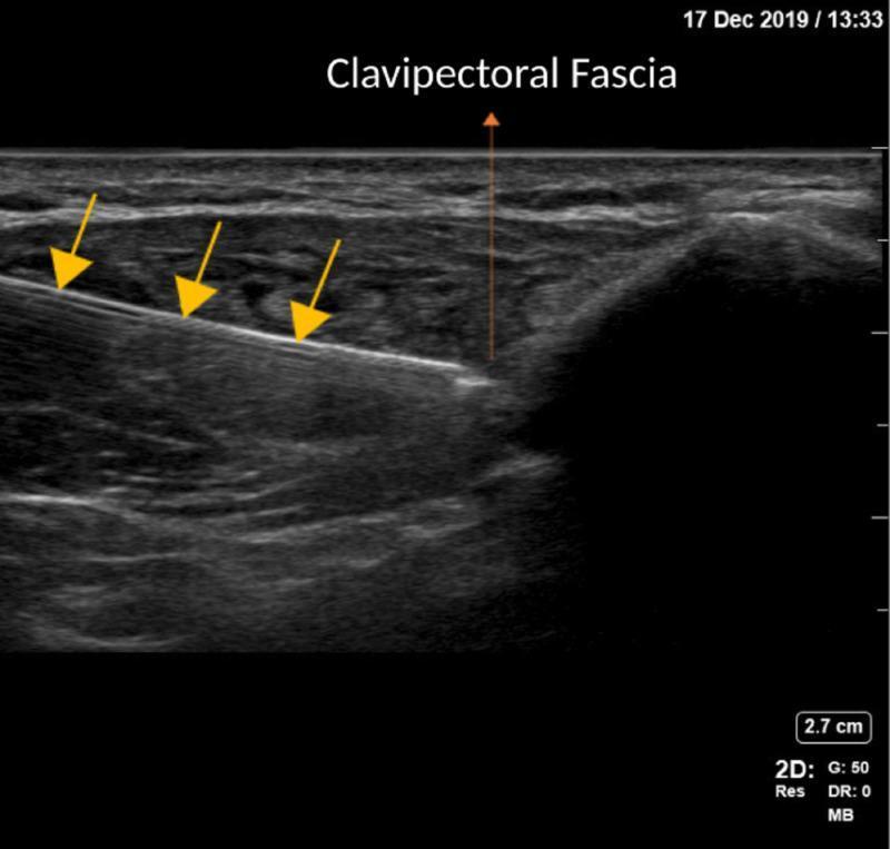
This diagnostic ultrasound image demonstrates the performance of a Clavipectoral Fascial Plane Block (CPB) using an in-plane needle technique. The imaging shows a cross-section of the anterior chest region near the clavicle. A highly echogenic (hyperechoic), linear needle is clearly visualized, highlighted by three yellow arrows, as it advances in a caudal-to-cephalad direction. The tip of the needle is positioned just superficial to the clavipectoral fascia, which is identified with an orange arrow. The anatomical landscape includes relatively hypoechoic superficial layers and deeper, more heterogeneous muscle tissue. The image illustrates the precise placement of a needle for anesthetic delivery within the fascial plane. This visual serves as a pedagogical resource for regional anesthesia, highlighting ultrasound-guided procedural landmarks, in-plane needle tracking, and the specific fascial anatomy required for effective CPB execution.
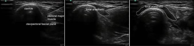
This composite of three diagnostic ultrasound images demonstrates the procedural technique for a Clavipectoral Fascial Plane Block (CPB). The primary anatomical landmark is the clavicle, visualized as a prominent, curvilinear hyperechoic structure with characteristic posterior acoustic shadowing. Overlying the clavicle is the pectoral major muscle, exhibiting a heterogeneous fibrillar echotexture. The clavipectoral fascia is identified as a hypoechoic interface located between the muscle and the clavicular periosteum. The series illustrates the sequential injection of local anesthetic, which appears as a dynamic, expanding hypoechoic (anechoic) fluid collection. The anesthetic is seen tracking along the clavipectoral fascial plane, effectively coating the anterior surface of the clavicle medially and laterally. This regional anesthesia technique is used for perioperative pain management in clavicular fractures or surgeries, targeting the sensory nerves that pass through this fascial space while avoiding deep structures like the pleura or brachial plexus.
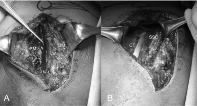
This composite clinical photograph (Figures 10A and 10B) illustrates the surgical anatomy of the deltopectoral approach during a shoulder arthroplasty procedure. Figure 10A demonstrates the deltopectoral interval, showing the anatomical separation between the pectoralis major (PM) muscle medially and the deltoid muscle (DM) laterally. The cephalic vein is visible within this interval, serving as a critical landmark for surgical dissection. Metal retractors are used to expose the deeper muscular layers. Figure 10B reveals the underlying structures after further dissection and incision of the clavipectoral fascia. Labeled features include the conjoint tendon (CT), positioned laterally, and the subscapularis tendon (SSC), which is situated more medially and demonstrates a characteristically interwoven fibrous texture. This visual guide serves an educational role for orthopedic surgical training, highlighting key anatomical landmarks and tissue planes required for safe access to the glenohumeral joint during prosthetic replacement.
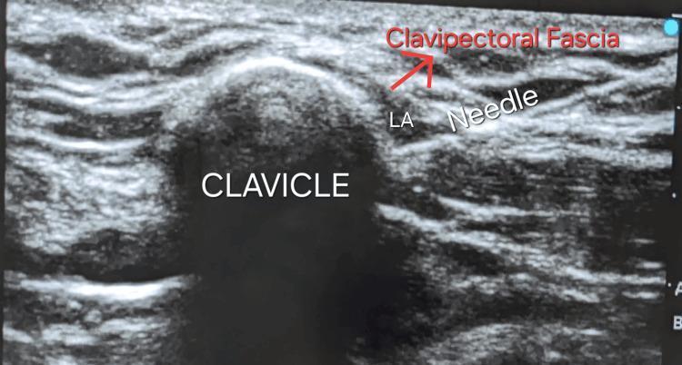
This diagnostic ultrasound image demonstrates the performance of a clavipectoral fascial plane block for regional anesthesia. The central landmark is the clavicle, identified as a hyperechoic, semi-circular cortical surface with a prominent posterior acoustic shadow. Superficial to the bone, the clavipectoral fascia appears as a distinct layer of organized, hyperechoic linear bands. A hyperechoic needle is visualized using an in-plane approach, with its tip positioned within the fascial plane. An anechoic space labeled 'LA' indicates the administration of local anesthetic, which is seen separating the fascial layers. This imaging illustrates the precise needle-to-target relationship required for effective anesthesia in the management of clavicular fractures. The anatomical context focuses on the pectoral region, utilizing the high-frequency linear probe to distinguish soft tissue layers from bony landmarks.
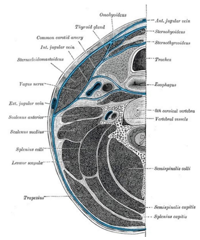
This medical illustration presents a transverse anatomical diagram of the neck at the level of the sixth cervical vertebra (C6), focusing on the compartmentalization provided by the deep cervical fascia (highlighted in blue). The diagram labels several critical structures within these fascial planes. The visceral compartment contains the trachea, esophagus, and thyroid gland. Lateral to the viscera, the carotid sheath is shown enveloping the common carotid artery, internal jugular vein, and vagus nerve. Muscular structures are identified including the sternocleidomastoid and trapezius (investing layer), the strap muscles (omohyoid, sternohyoid, sternothyroid), and the prevertebral/paravertebral muscles (scalenus anterior and medius, longus colli, semispinalis, and splenius groups). Vascular landmarks like the external and anterior jugular veins and vertebral vessels are also depicted. This diagram is designed for educational purposes to demonstrate the spatial relationships of cervical anatomy and the potential pathways for the spread of deep neck space infections.
Clavipectoral Fascia
Definition and Position
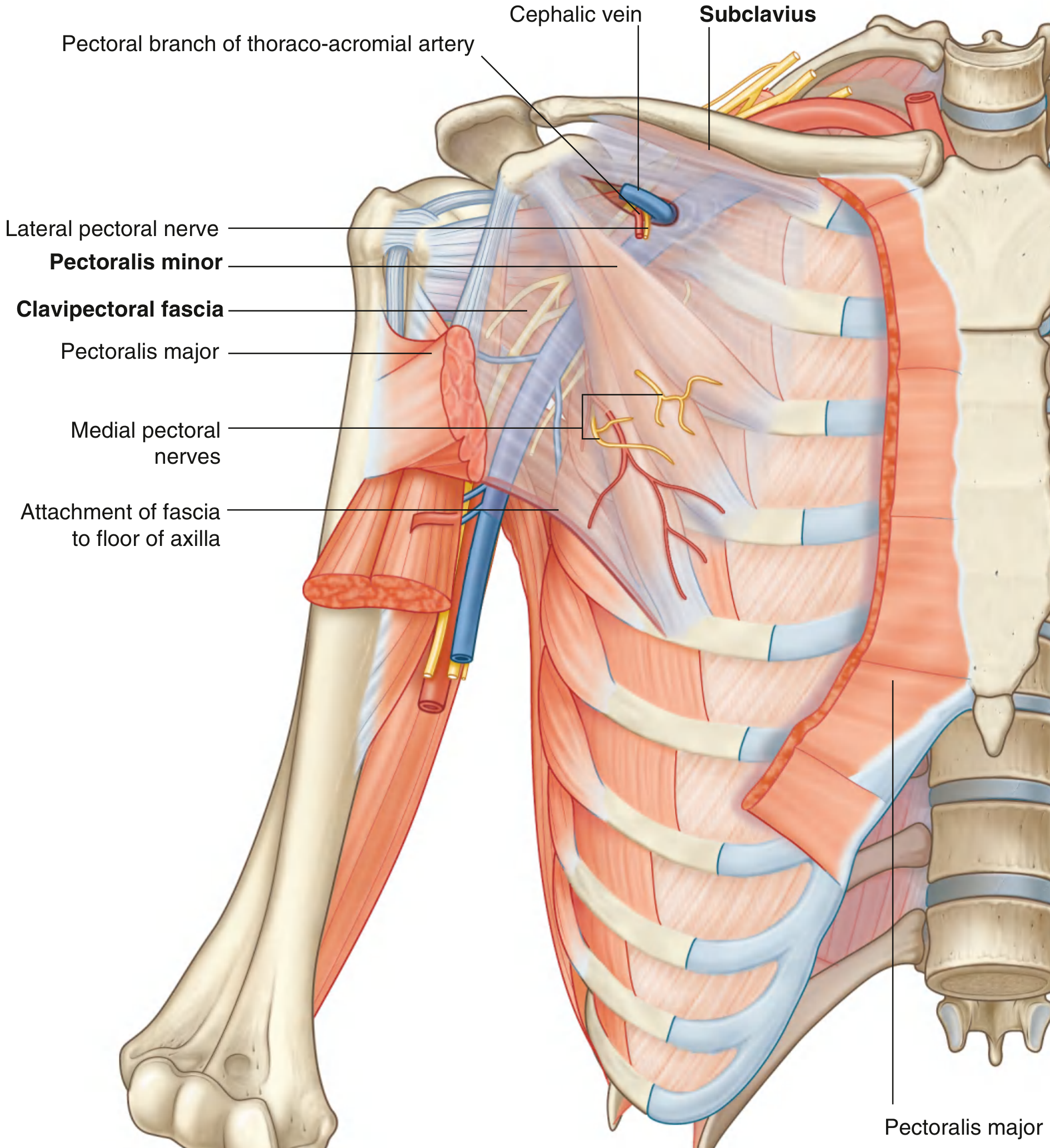
- Gray's Anatomy for Students, p. 845: "A continuous layer of deep fascia, the clavipectoral fascia, encloses the subclavius and pectoralis minor and attaches to the clavicle above and to the floor of the axilla below."
Attachments
| Level | Attachment |
|---|---|
| Superior | Clavicle (encloses subclavius above) |
| Intermediate | Spans the gap between subclavius and pectoralis minor |
| Inferior | Becomes continuous with the axillary fascia (floor of axilla) |
Parts / Subdivisions
1. Upper Part (superior to subclavius)
- Attaches to the clavicle on either side of the subclavius muscle, enclosing it.
2. Costocoracoid Membrane
- The portion of the clavipectoral fascia lying between the subclavius and pectoralis minor muscles.
- It is pierced by:
- Cephalic vein - drains upward to join the axillary vein
- Thoraco-acromial artery - branches to supply the pectoral region, deltoid, acromioclavicular joint, and clavicle
- Lateral pectoral nerve - primarily supplies the pectoralis major
3. Suspensory Ligament of the Axilla
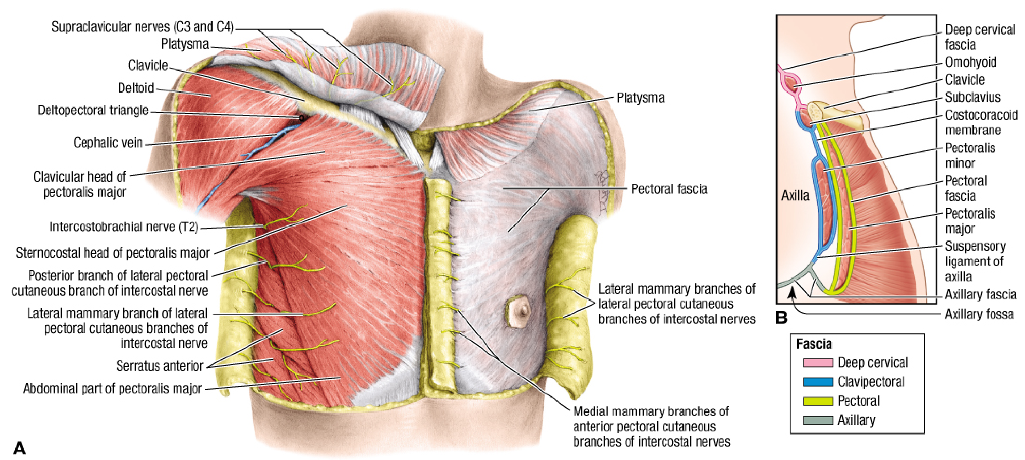
- The portion inferior to pectoralis minor, connecting downward to the axillary fascia (floor of the axilla).
- When the arm is abducted, the pectoralis minor pulls this portion upward, which in turn elevates the axillary fascia and overlying skin, forming the characteristic hollow (fossa) of the axilla.
Sagittal Section Diagram (Fischer's Mastery of Surgery)
Structures Piercing the Clavipectoral Fascia
- Cephalic vein - passes upward through the deltopectoral groove (the clavipectoral triangle bounded by the deltoid, pectoralis major, and clavicle) and drains into the axillary vein
- Thoraco-acromial artery - emerges and divides into pectoral, deltoid, clavicular, and acromial branches
- Lateral pectoral nerve - supplies pectoralis major
- Lateral thoracic artery - leaves the axilla passing through the fascia inferior to pectoralis minor
Relation to the Subclavian Vein
The Clavipectoral Triangle
- Medially - pectoralis major
- Laterally - deltoid
- Superiorly - clavicle
Clinical Relevance
| Context | Relevance |
|---|---|
| Deltopectoral approach (shoulder surgery) | The fascia is incised after exposing the deltopectoral interval to reach the glenohumeral joint and subscapularis |
| Axillary lymph node dissection / sentinel node biopsy | The fascia is divided and subsequently reapproximated with interrupted sutures |
| Clavipectoral fascial plane block (CPB) | A regional anesthesia technique where local anesthetic is injected into this fascial plane under ultrasound guidance - used for perioperative pain management in clavicular fractures |
| Subclavian vein catheterization | Understanding the fascia's relation to the vein aids safe central line insertion |
| Apical lymph nodes | Level III (apical) axillary nodes lie posterior to the subclavius, which is enveloped by this fascia; access requires division of pectoralis minor |
Summary Layers (Anterior to Posterior)
- Skin and superficial fascia
- Pectoralis major
- Pectoral fascia (investing layer over pectoralis major)
- Clavipectoral fascia - deep to pectoralis major; encloses subclavius and pectoralis minor
- Axillary contents (fat, vessels, brachial plexus)