
Small incision cataract surgery in ak khurana in short
small incision cataract surgery SICS scleral tunnel steps
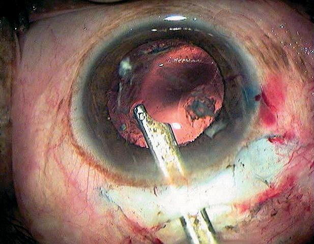
This clinical photograph captures a surgical step during Small Incision Cataract Surgery (SICS). The image shows an operative field of the human eye under surgical illumination. A metallic Simcoe cannula is inserted through a corneal-scleral tunnel incision into the anterior chamber. The cannula tip is positioned near the capsular opening, where it is being used to aspirate residual white lens cortex material. The posterior capsule is visible behind the iris, showing signs of a surgical opening. The surrounding conjunctiva and sclera exhibit expected surgical trauma, including minor subconjunctival hemorrhage and hyperemia from tissue manipulation. The cornea remains relatively transparent, providing a clear view of the intraocular structures and the irrigation-aspiration process. This visual demonstrates the essential technique of cortical cleanup to ensure a clear visual axis before intraocular lens (IOL) implantation in ophthalmological surgery.
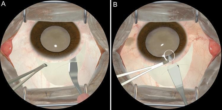
This surgical diagram displays a surgeon's-eye view of a simulated Manual Small Incision Cataract Surgery (MSICS), specifically focusing on the creation of a scleral tunnel. The visual consists of two panels, A and B, rendered via a virtual reality simulator. In both frames, the eye is held open by a wire lid speculum, revealing the conjunctiva, vascularized sclera, a brown iris, and a centrally located lens. Panel A illustrates the tunnel dissection phase, where a crescent knife is utilized to create a lamellar pocket within the sclera. Panel B demonstrates the subsequent step of entering the anterior chamber; a keratome is shown penetrating the previously dissected tunnel into the eye, with a white circle highlighting the point of entry. The primary educational focus is the visualization of precise instrument angles and anatomical positioning required for successful scleral tunnel construction during cataract procedures.
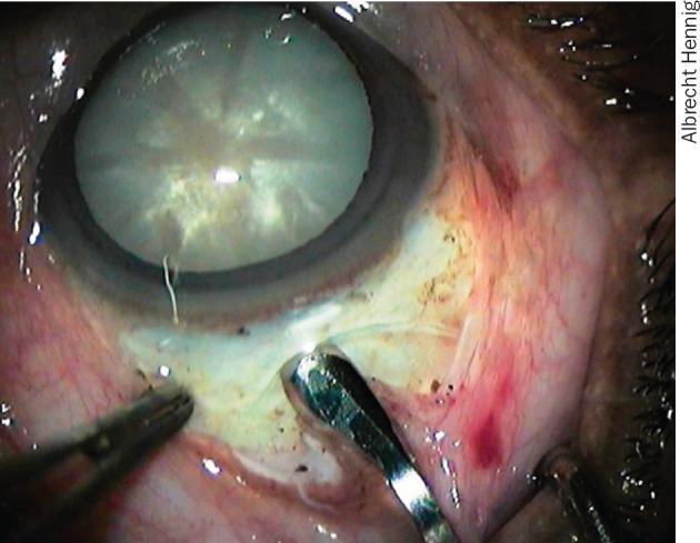
This clinical photograph captures a surgical step during an ophthalmological procedure, likely Manual Small Incision Cataract Surgery (MSICS). The image demonstrates the stabilization of the globe using metallic toothed forceps applied to the superior sclera. A 'frown-shaped' scleral tunnel incision is visible, with surgical instruments being used to create or manage the tunnel architecture. The central eye feature is a mature cataract, characterized by a dense, opaque, milky-white crystalline lens visible through the pupil. The conjunctiva and episclera show mild surgical trauma and injection (redness) typical of the intraoperative phase. The educational focus of the image is on the technique of firm scleral fixation required to ensure a stable operative field during the construction of the incision. Key anatomical structures shown include the limbus, sclera, cornea, and the opacified lens. This material is relevant for ophthalmic surgical training and understanding cataract extraction techniques.
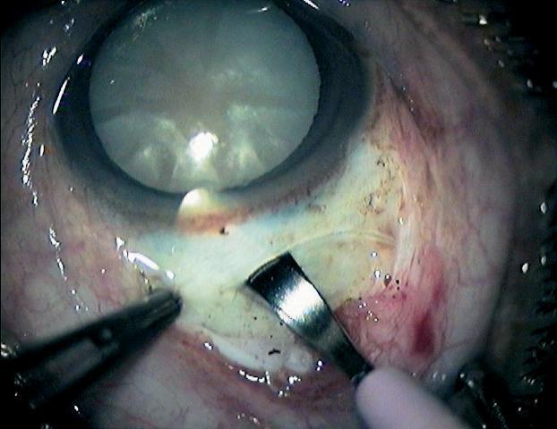
This clinical photograph captures a surgical field during an ophthalmic procedure, specifically the construction of a sclerocorneal tunnel incision for manual small incision cataract surgery (MSICS). The image shows the eye of a patient with a mature, white, opaque cataract visible through the dilated pupil. Surgical instruments are in active use: toothed forceps are seen stabilizing the sclera on the left, while a metal crescent knife is performing a partial-thickness lamellar dissection into the sclera near the limbus. The depth of the tunnel is being monitored by the visibility of the crescent blade through the overlying scleral fibers, a critical step to ensure a self-sealing wound and avoid perforation. The surrounding conjunctiva shows mild surgical hyperemia. This image serves as a teaching tool for ocular surgery, illustrating proper instrument positioning and tissue handling for creating the entry point into the anterior chamber during cataract extraction.
Small Incision Cataract Surgery (SICS) - AK Khurana
Overview
Steps of SICS (as in AK Khurana)
1. Incision Construction
- A conjunctival flap (fornix-based or limbus-based) is raised superiorly.
- A frown-shaped (or straight) scleral groove is made 1-2 mm behind the limbus, about 6-7 mm wide, to half the scleral thickness.
- A sclero-corneal tunnel is dissected forward using a crescent knife into clear cornea (2 mm anterior to limbus).
- The internal opening is wider than the external, creating a self-sealing valve mechanism.
2. Entry into Anterior Chamber
- A keratome (angled blade) is used to enter the anterior chamber through the tunnel floor, producing a triplanar entry.
- Viscoelastic is injected to maintain the anterior chamber depth.
3. Capsulotomy
- A continuous curvilinear capsulorhexis (CCC) is performed, or alternatively, a can-opener capsulotomy.
- Trypan blue dye can be used to stain the anterior capsule (especially for white/mature cataracts).
4. Hydrodissection and Hydrodelineation
- Hydrodissection: BSS is injected under the anterior capsule to free cortical-capsular attachments.
- Hydrodelineation: separates the epinucleus from the nucleus (optional but useful).
5. Nucleus Delivery (Key Step - Unique to SICS)
- Viscoexpression: viscoelastic injected behind nucleus to prolapse it into anterior chamber, then expressed out through tunnel.
- Irrigating vectis: a wire loop with irrigation side port is passed behind the nucleus; nucleus rides out on fluid current.
- Sinskey hook + Vectis (sandwich technique): nucleus sandwiched between hook anteriorly and vectis posteriorly.
- Phaco-sandwich: nucleus emulsified in AC manually using two instruments.
6. Cortical Aspiration
- Residual cortex is aspirated using a Simcoe cannula (manual irrigation-aspiration).
- The I/A tip is passed through the side port and into the capsular bag.
7. IOL Implantation
- A rigid PMMA IOL (typically 5.5-6.5 mm optic) is implanted into the capsular bag.
- The tunnel incision is self-sealing, so no sutures are required in most cases.
8. Wound Closure
- The conjunctival flap is reposited and sealed with cautery or a suture.
- A sub-conjunctival injection of antibiotic + steroid is given at the end.
Advantages of SICS
| Feature | Benefit |
|---|---|
| Self-sealing tunnel | No sutures needed - less astigmatism |
| No phaco machine | Low cost, suitable for camps |
| Works for dense/mature/hard cataracts | Better than phaco for grade IV-V nucleus |
| Faster learning curve | Easier to train |
| Good visual outcomes | Comparable to phaco (vision recovers in days) |
Disadvantages / Complications
- More astigmatism than phaco (but less than conventional ECCE)
- Larger incision (6-7 mm) than phaco (2-3 mm)
- Nucleus delivery can be tricky (Descemet's membrane tear risk)
- Not ideal for small pupils or shallow anterior chamber
- Risk of vitreous loss with very hard cataracts
Comparison: SICS vs. Phacoemulsification
| Parameter | SICS | Phaco |
|---|---|---|
| Incision size | ~6-7 mm | 2-3 mm |
| Sutures | Usually none | None |
| IOL type | PMMA (rigid) | Foldable |
| Astigmatism | Moderate | Minimal |
| Cost | Low | High |
| Dense cataracts | Excellent | Difficult |
| Technology needed | Minimal | Expensive machine |
Key Points for Exams (AK Khurana Style)
- SICS incision is sclero-corneal tunnel - begins 2 mm behind limbus, ends 2 mm in front.
- The external opening is narrower than the internal opening - this is the basis of self-sealing.
- Nucleus is delivered whole (not fragmented) - this is the major difference from phaco.
- Simcoe cannula is used for cortical aspiration.
- SICS is the preferred technique for high-volume/camp surgery in India.
- Trypan blue (0.1%) is used for staining the anterior capsule in white cataract.