
Vascular ring
Reading File
Searching sources...
Finding Sources
Searching PubMed
vascular ring congenital aortic arch anomaly
Finding Sources
Finding Sources
Good - I have multiple sources. Let me now read the most detailed sections.
Reading File
Reading File
Reading File
Reading File
Reading File
Reading File
Reading File
Loading Image
Loading Image
I now have comprehensive information from multiple authoritative textbooks. Let me compose the full answer.
Vascular Ring
Definition
A vascular ring is a congenital anomaly of aortic arch development in which the esophagus and trachea are completely surrounded by vascular structures (patent vessels, fibrous ligamenta, or both). This distinguishes it from a pulmonary artery (PA) sling, where the left PA passes posterior to the trachea but anterior to the esophagus - an incomplete encirclement.
- Braunwald's Heart Disease, p. 900
Embryology
Six paired pharyngeal arch arteries form early in fetal life. Several regress fully or partially; others become the aortic arch, brachiocephalic branches, proximal pulmonary arteries, and ductus arteriosus. Abnormal patterns of regression lead to vascular rings. The distal right dorsal aorta normally regresses; failure to do so produces a double aortic arch. Involution of the left fourth arch produces a right aortic arch.
Classification
Complete Vascular Rings
These completely encircle the trachea and esophagus.
| Type | Mechanism | Notes |
|---|---|---|
| Double aortic arch | Failure of the distal right dorsal aorta to regress; both arches persist | Most common symptomatic type; right arch dominant in most cases; left and right carotid + subclavian arteries arise from respective arches |
| Right aortic arch + aberrant left subclavian artery + left ligamentum arteriosum | Left fourth arch involutes; apex is right of trachea; aberrant LSA courses retroesophageally; ligamentum completes the ring | Associated with Kommerell diverticulum - a dilated remnant at the LSA origin from the proximal descending aorta |
| Right aortic arch + mirror-image branching + retroesophageal ligamentum | Only partial resorption of distal left fourth arch; Kommerell diverticulum gives rise to the ligamentum arteriosum | Ligamentum passes posteriorly to join the left PA, completing the ring |
| Left aortic arch + right descending aorta + right ligamentum | Extremely rare | |
| Left aortic arch + right descending aorta + atretic right aortic arch | Extremely rare |
Partial Rings / Near-Rings
These cause compression without anatomically completing a ring.
| Type | Mechanism | Clinical Note |
|---|---|---|
| Innominate artery compression syndrome | Innominate artery originates left of midline; courses anterior to trachea causing compression | Presents in first 1-2 years with respiratory symptoms |
| Aberrant right subclavian artery | Most common arch anomaly (~0.5-1% of population); courses retroesophageally from the descending aorta | Usually asymptomatic; can cause dysphagia lusoria in 7th-8th decade when vessel becomes ectatic and calcified |
| PA sling (pulmonary artery sling) | Left PA branches from right PA; passes behind the right main bronchus between trachea and esophagus | Associated with complete tracheal rings in 30-40%, causing tracheal stenosis |
Clinical Presentation
Complete rings - symptoms generally appear within weeks to months of life:
- Respiratory: Stridor, non-productive "seal bark" or brassy cough, apnea, frequent respiratory infections, wheezing
- Respiratory symptoms dominate early; dysphagia becomes prominent once solid foods are introduced
- Double aortic arch presents earlier than right arch with retroesophageal left ligamentum
- Commonly misdiagnosed as asthma, tracheomalacia, or reflux
Adults with vascular rings most commonly present with dysphagia; respiratory symptoms (wheezing, stridor, dyspnea on exertion) occur but are less frequent. Some are discovered incidentally (e.g., right aortic arch seen on chest X-ray).
- Mulholland and Greenfield's Surgery, p. 4455-4456
- Braunwald's Heart Disease, p. 900
Diagnostic Imaging
| Modality | Role | Findings |
|---|---|---|
| Chest X-ray (CXR) | First-line | Right aortic arch, widened superior mediastinum, indistinct aortic knuckle, tracheal displacement/compression, hyperinflation or atelectasis |
| Barium esophagogram | Diagnostic in most cases | Double arch: bilateral + posterior constant esophageal indentations (right indentation slightly higher); retroesophageal subclavian: oblique posterior defect |
| Echocardiography | Useful in neonates/infants; limited by acoustic windows | Can demonstrate arch sidedness, double arch; absence of normal innominate bifurcation suggests aberrant subclavian artery |
| CT angiography (CTA) | Gold standard for anatomy; best for airway | Fully delineates all components of the ring; reveals airway compression; ductal ligament inferred (not directly seen unless calcified) |
| MR angiography (MRA) | Also diagnostic; no radiation | Can visualize airway compression; preferred for follow-up |
| Bronchoscopy | Tracheal anatomy and degree of compression | Important for PA sling and assessing tracheal rings |
- Harriet Lane Handbook, p. 876
- Grainger & Allison's Diagnostic Radiology, p. 452-453
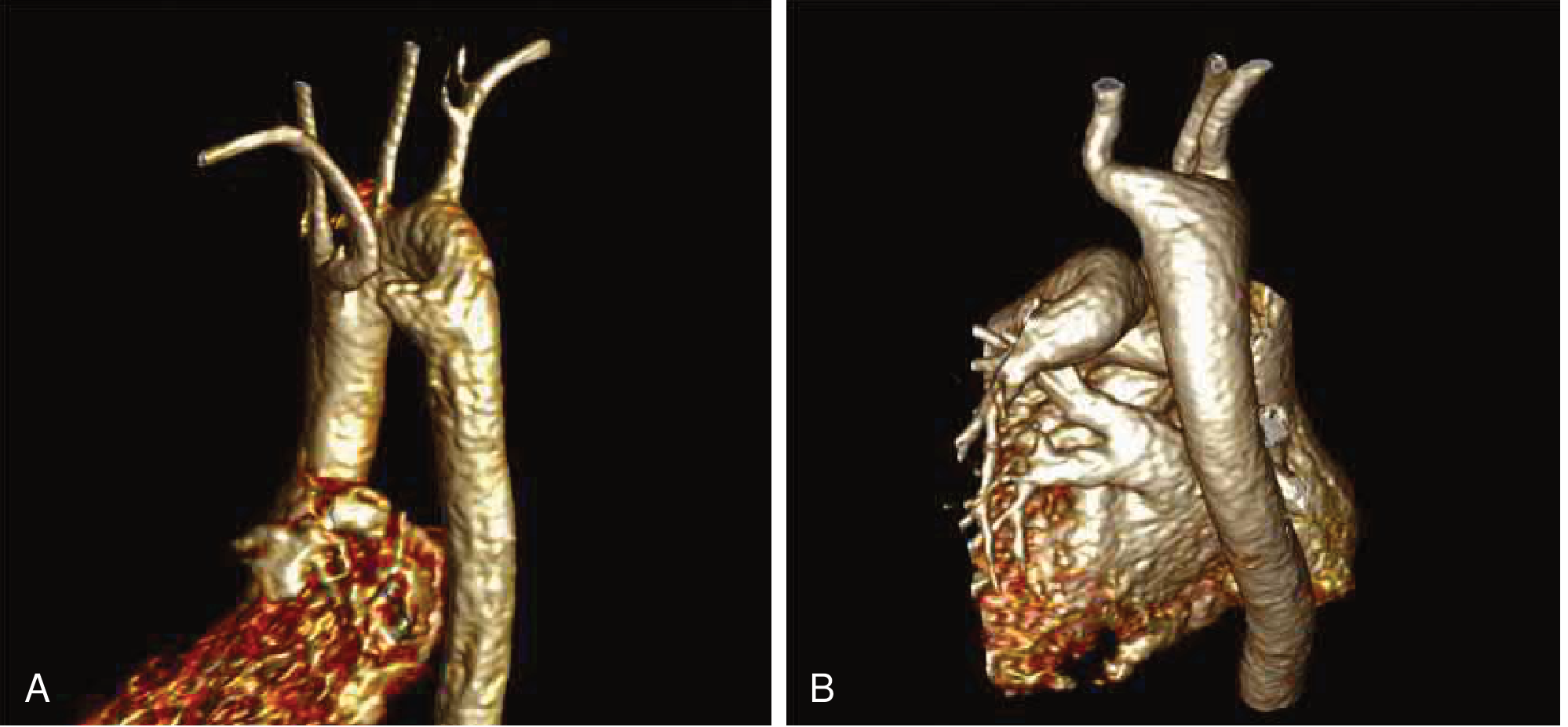
3D gadolinium-enhanced MR angiograms. A: double aortic arch with atresia of the distal left arch. B: right aortic arch with Kommerell diverticulum and aberrant left subclavian artery. (Braunwald's Heart Disease)
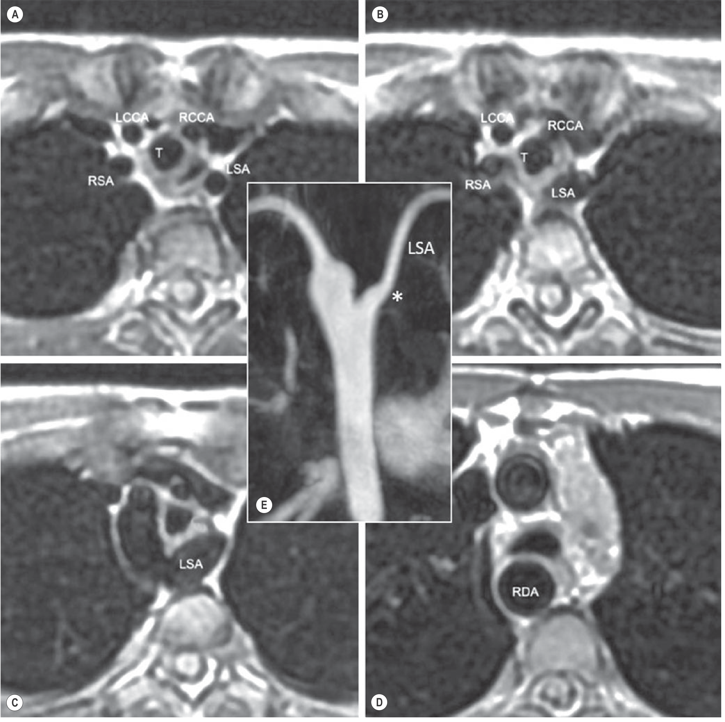
MRI black-blood fast spin-echo axial images of right aortic arch with aberrant LSA (left subclavian artery). The mild basal convexity () of the aberrant LSA suggests traction by the ligamentum arteriosum. T = trachea; LCCA = left common carotid; RCCA = right common carotid; RSA = right subclavian; RDA = right descending aorta. (Grainger & Allison)*
Treatment
Indications
All symptomatic complete and partial vascular rings are repaired surgically. Asymptomatic complete rings are also generally repaired given the risk of symptoms with growth.
Complete Rings
Double aortic arch:
- Left posterolateral thoracotomy (3rd intercostal space)
- Ligation and division of the ductus/ligamentum
- Ligation and division of the nondominant (usually atretic left) arch segment
- Dissection around trachea and esophagus to lyse adhesions
Right aortic arch + retroesophageal left ligamentum:
- Same approach - division of ligamentum and lysis of adhesions
- If Kommerell diverticulum is present and causes residual compression, it may be resected or suspended posteriorly
Kommerell diverticulum (with right aortic arch + aberrant LSA) - more extensive options:
- Translocation of aberrant subclavian artery to the ipsilateral carotid + diverticulum resection (must also divide ductal ligament to release ring)
- Hybrid repair: surgical subclavian translocation + endovascular exclusion of diverticulum (stent graft + embolization)
- Rationale: the diverticulum can continue compressing the airway after simple ring release, and can become aneurysmal or rupture
Partial Rings
| Type | Surgery |
|---|---|
| Innominate artery compression | Mild: observation (may resolve with growth). Severe (apnea, significant stridor, recurrent infections): simple division, reimplantation to ascending aorta, or suspension to sternum |
| Aberrant right subclavian artery | Division via left posterolateral thoracotomy; rarely reimplantation/grafting needed |
| PA sling | Reimplantation of left PA to main PA; tracheal stenosis from complete rings addressed concurrently (tracheoplasty) |
- Mulholland and Greenfield's Surgery, p. 4456-4457
- Braunwald's Heart Disease, p. 900
Key Points to Remember
- Most common complete ring: double aortic arch (right arch usually dominant)
- Second most common: right aortic arch + aberrant left subclavian artery + left ligamentum arteriosum
- Most common arch anomaly overall: aberrant right subclavian artery (~0.5-1%; usually incidental; rarely causes dysphagia lusoria in adults)
- Vascular rings are often misdiagnosed as asthma or reflux in children
- CTA/MRA is the definitive diagnostic test
- Kommerell diverticulum can persist as a compressing structure and carries aneurysm/rupture risk even after ring division
- PA sling is associated with complete tracheal rings in 30-40% of cases - a separate and important comorbidity
Recent literature note: A 2026 review (PMID 40701783) discusses evolving diagnostic approaches for vascular rings, reflecting improving detection rates with widespread use of CTA and echocardiography in the current era.
This is a shared conversation. Sign in to Orris to start your own chat.