
Cholecystectomy procedure
"cholecystectomy"[MeSH Terms]
SAGES guidelines laparoscopic cholecystectomy safe cholecystectomy critical view of safety
laparoscopic cholecystectomy critical view of safety cystic duct cystic artery hepatocystic triangle surgical diagram
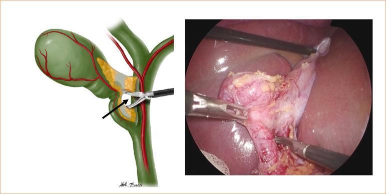
This composite image features an anatomical diagram and a corresponding laparoscopic photograph illustrating the 'critical view of safety' during a cholecystectomy. The diagram on the left shows the gallbladder, cystic duct, cystic artery, and common bile duct with an arrow pointing to a gap created by a Maryland-type dissector. The right panel displays a real-world surgical field of view. It depicts the gallbladder infundibulum being retracted and dissected. A Maryland-type laparoscopic forceps is visible, performing blunt dissection to separate the cystic duct from the cystic artery within Calot’s triangle (the hepatocystic triangle). The gallbladder tissue shows signs of inflammation and surface vascularization, consistent with acute cholecystitis. The procedure focuses on clearing the hepatocystic triangle of fat and fibrous tissue to clearly identify the two, and only two, structures entering the gallbladder, a vital step to prevent iatrogenic bile duct injuries. This resource is intended for surgical residents and medical students studying minimally invasive hepatobiliary surgery.
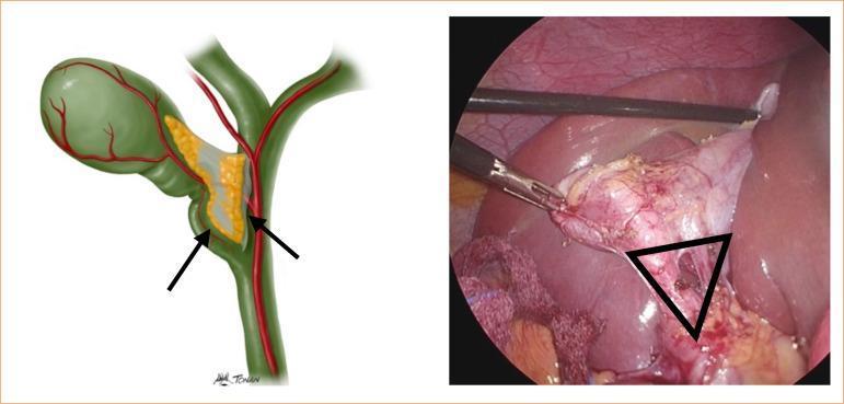
This composite image consists of an anatomical illustration and a corresponding endoscopic photograph demonstrating the landmarks of Calot's triangle (hepatocystic triangle) during a laparoscopic cholecystectomy. The anatomical diagram on the left illustrates the gallbladder, cystic duct, and common hepatic duct (marked with arrows), with the cystic artery passing through the triangle. The laparoscopic view on the right displays the surgical field with laparoscopic graspers retracting a distended, erythematous gallbladder. A black triangular overlay identifies Calot’s triangle, bounded superiorly by the inferior margin of the liver, laterally by the cystic duct, and medially by the common hepatic duct. The operative photograph shows evidence of inflammatory changes, including tissue hypervascularity and edema, which can obscure clear identification of biliary structures. This visual is designed to teach the 'critical view of safety' by emphasizing the anatomical boundaries required to prevent iatrogenic bile duct injuries during minimally invasive hepatobiliary surgery.
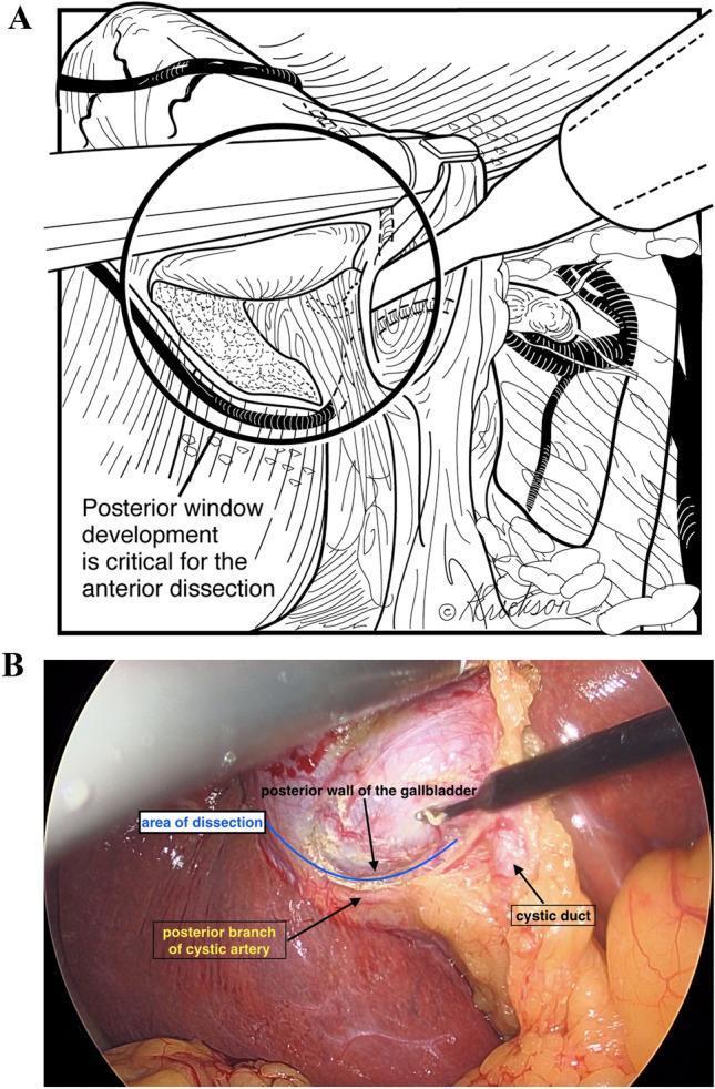
This composite image illustrates a critical step in laparoscopic cholecystectomy, specifically the development of the 'posterior window.' Panel A is an anatomical line diagram highlighting that posterior window development is essential for safe anterior dissection. Panel B is a corresponding intra-operative endoscopic photograph showing the surgical field in the hepatocystic triangle (Triangle of Calot). The photo identifies key structures: the posterior wall of the gallbladder, the cystic duct, and the posterior branch of the cystic artery. A surgical instrument is seen performing dissection in the avascular plane between these structures, labeled as the 'area of dissection.' This step facilitates the circumferential mobilization of the gallbladder infundibulum while protecting the biliary and vascular anatomy. The visualization emphasizes the importance of identifying the posterior cystic artery branch relative to the gallbladder wall to avoid inadvertent injury to the right hepatic artery or common bile duct during the attainment of the Critical View of Safety.
Cholecystectomy

Laparoscopic procedure: operative outline
-
Preparation
- General anesthesia, prophylactic measures as appropriate, sterile preparation and draping.
- Patient is placed supine, usually with reverse Trendelenburg and left tilt to expose the right upper abdomen.
-
Access and ports
- Pneumoperitoneum is created with carbon dioxide.
- Typically, four ports are inserted: umbilical camera port, epigastric working port, and two right upper quadrant ports.
-
Exposure
- The fundus of the gallbladder is retracted upward toward the diaphragm.
- The infundibulum is retracted laterally and inferiorly to open the hepatocystic triangle, also called Calot triangle.
-
Safe identification of anatomy
- Fat and fibrous tissue are carefully cleared from the hepatocystic triangle.
- Before dividing structures, the surgeon obtains the critical view of safety:
- The hepatocystic triangle is cleared.
- The lower one-third of the gallbladder is separated from the liver bed.
- Only two structures are seen entering the gallbladder: the cystic duct and cystic artery.
This safety step is intended to prevent common bile duct injury, as emphasized in the SAGES safe cholecystectomy guideline. -
Division of cystic structures
- The cystic artery is clipped and divided.
- The cystic duct is clipped and divided. Intraoperative cholangiography or other biliary imaging may be used when ductal anatomy is unclear or common bile duct stones are suspected.
-
Gallbladder removal
- The gallbladder is dissected from the liver bed with electrosurgery.
- Hemostasis and bile leakage are checked.
- The gallbladder is placed in a retrieval bag and removed, commonly through the umbilical port.
-
Completion
- Ports are removed under vision, pneumoperitoneum released, and skin wounds closed.
- A drain is not routinely necessary but may be placed selectively.
If the critical view cannot be achieved
Key risks
- Bile leak
- Bleeding
- Injury to the common bile duct or nearby bowel
- Retained common bile duct stones
- Infection, wound complications, venous thromboembolism
- Conversion from laparoscopic to open surgery