
Pathogenesis and classification of necrosis md pathology exam robbins based answer
Reading File
Reading File
Finding Sources
Finding Sources
Finding Sources
Searching PubMed
necrosis AND cell death
Searching the Web
Robbins Basic Pathology necrosis mechanisms morphologic patterns coagulative liquefactive caseous fat fibrinoid gangrenous
Reading File
Reading File
Finding Sources
Finding Sources
Reading File
Reading File
Reading File
Reading File
Necrosis: Pathogenesis and Classification
Definition
Necrosis is the morphologic expression of irreversible cell injury and cell death in living tissue. It results from severe damage that disrupts cell membranes, causes enzyme leakage and digestion of the cell, and provokes inflammation in adjacent tissue.
It is always pathologic, unlike apoptosis, which may be physiologic or pathologic.
Robbins & Kumar Basic Pathology, Ch. 1, pp. 19-22.
Robbins & Kumar Basic Pathology, Ch. 1, pp. 19-22.
Pathogenesis of necrosis
Causes
Major causes include:
-
Ischemia and hypoxia
- Most common cause in many organs.
- Ischemia is more severe than hypoxia because it also reduces nutrient delivery and prevents removal of toxic metabolites.
- Examples: myocardial infarction, renal infarction, limb gangrene.
-
Chemical and toxic injury
- Poisons, drugs, industrial chemicals, and microbial toxins can directly injure membranes, mitochondria, DNA, or proteins.
-
Physical agents
- Trauma, burns, radiation, electric shock, extremes of temperature.
-
Infections
- Bacterial and fungal infections often produce necrosis through toxins and leukocyte-mediated enzymatic destruction.
-
Enzyme leakage
- Example: pancreatic lipases released in acute pancreatitis digest peripancreatic fat, producing fat necrosis.
-
Immune-mediated injury
- Immune complexes and antibodies can damage blood-vessel walls, producing fibrinoid necrosis.
Sequence of events
Severe injury
→ ATP depletion and mitochondrial dysfunction
→ failure of ion pumps, especially Na⁺/K⁺-ATPase
→ Na⁺ and water influx causing cellular and organelle swelling
→ increased cytosolic Ca²⁺
→ activation of phospholipases, proteases, endonucleases, and ATPases
→ plasma, mitochondrial, lysosomal, and organellar membrane damage
→ leakage of intracellular proteins and lysosomal enzymes
→ enzymatic digestion of the cell
→ release of intracellular contents and DAMPs
→ acute inflammatory reaction.
→ ATP depletion and mitochondrial dysfunction
→ failure of ion pumps, especially Na⁺/K⁺-ATPase
→ Na⁺ and water influx causing cellular and organelle swelling
→ increased cytosolic Ca²⁺
→ activation of phospholipases, proteases, endonucleases, and ATPases
→ plasma, mitochondrial, lysosomal, and organellar membrane damage
→ leakage of intracellular proteins and lysosomal enzymes
→ enzymatic digestion of the cell
→ release of intracellular contents and DAMPs
→ acute inflammatory reaction.
Key irreversible changes
Robbins emphasizes three central features of irreversible injury:
- Inability to reverse mitochondrial dysfunction and restore oxidative phosphorylation/ATP production.
- Severe membrane damage, including plasma and organellar membranes.
- Irreversible DNA and chromatin damage.
Lysosomal rupture releases hydrolytic enzymes that digest cellular constituents. Leakage of cellular contents into extracellular tissue triggers inflammation.
Robbins & Kumar Basic Pathology, pp. 19-21.
Robbins & Kumar Basic Pathology, pp. 19-21.
Morphology of necrotic cells
Cytoplasmic changes
- Increased eosinophilia: cytoplasm becomes more pink on H&E stain because of loss of RNA and denaturation of cytoplasmic proteins.
- Cytoplasm may become glassy, homogeneous, and vacuolated.
- Membrane disruption causes leakage of cell contents.
- Cellular outlines may remain temporarily visible in coagulative necrosis.
Nuclear changes
All result from DNA and chromatin breakdown:
-
Pyknosis
- Nuclear shrinkage with increased basophilia.
- Nucleus becomes small, dark, and shrunken.
-
Karyorrhexis
- Fragmentation of the pyknotic nucleus.
-
Karyolysis
- Fading and dissolution of the nucleus due to DNase activity.
Sequence:
Pyknosis → karyorrhexis → karyolysis
Pyknosis → karyorrhexis → karyolysis
In 1-2 days, the nucleus may disappear completely.
Robbins & Kumar Basic Pathology, pp. 20-21.
Robbins & Kumar Basic Pathology, pp. 20-21.
Classification: Morphologic patterns of necrosis
These patterns are morphologic descriptions. They often indicate the underlying cause, but do not themselves define the molecular mechanism.
| Type | Basic mechanism/morphology | Typical sites and examples |
|---|---|---|
| Coagulative | Protein denaturation predominates over enzymatic digestion. Tissue architecture is preserved temporarily. | Ischemic infarction in solid organs except brain, such as heart, kidney, spleen |
| Liquefactive | Enzymatic digestion predominates, converting tissue into viscous liquid. | Brain infarct; bacterial and fungal infections, abscess |
| Gangrenous | Clinical term, usually ischemic necrosis of a limb or bowel. | Dry gangrene, wet gangrene |
| Caseous | Amorphous granular, friable, cheese-like necrotic material with complete loss of architecture. | Tuberculosis; some fungal infections |
| Fat | Destruction of adipocytes, commonly by pancreatic lipases; fatty acids bind calcium. | Acute pancreatitis, traumatic injury to fat |
| Fibrinoid | Bright pink, fibrin-like material in vessel walls due to immune complex deposition and plasma protein leakage. | Vasculitis, severe hypertension, transplant rejection |
1. Coagulative necrosis
Definition: A form of necrosis in which tissue architecture remains preserved for several days despite cell death.
Pathogenesis:
Ischemic injury denatures structural proteins and intracellular enzymes. Because proteolysis is inhibited, dead cells are not immediately digested.
Ischemic injury denatures structural proteins and intracellular enzymes. Because proteolysis is inhibited, dead cells are not immediately digested.
Morphology:
- Firm tissue.
- Preserved cell outlines or "ghost cells."
- Eosinophilic, anucleate cells.
- Later removed by leukocytes and macrophages.
Examples:
- Myocardial infarction.
- Renal infarction.
- Splenic infarction.
- Adrenal infarction.
Exception: Ischemic injury in the brain produces liquefactive, not coagulative, necrosis.
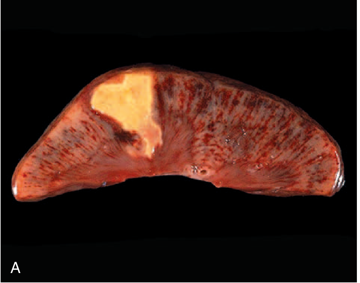
Robbins & Kumar Basic Pathology, p. 21.
2. Liquefactive necrosis
Definition: Necrosis in which enzymatic digestion completely liquefies dead cells and tissue.
Pathogenesis:
Hydrolytic enzymes, especially from neutrophils and macrophages, digest the tissue rapidly.
Hydrolytic enzymes, especially from neutrophils and macrophages, digest the tissue rapidly.
Morphology:
- Soft, liquid, viscous mass.
- Tissue architecture is completely lost.
- In bacterial infection, the creamy-yellow material is pus.
- A localized collection of pus is an abscess.
Examples:
- Cerebral infarction.
- Bacterial abscess.
- Fungal infection.
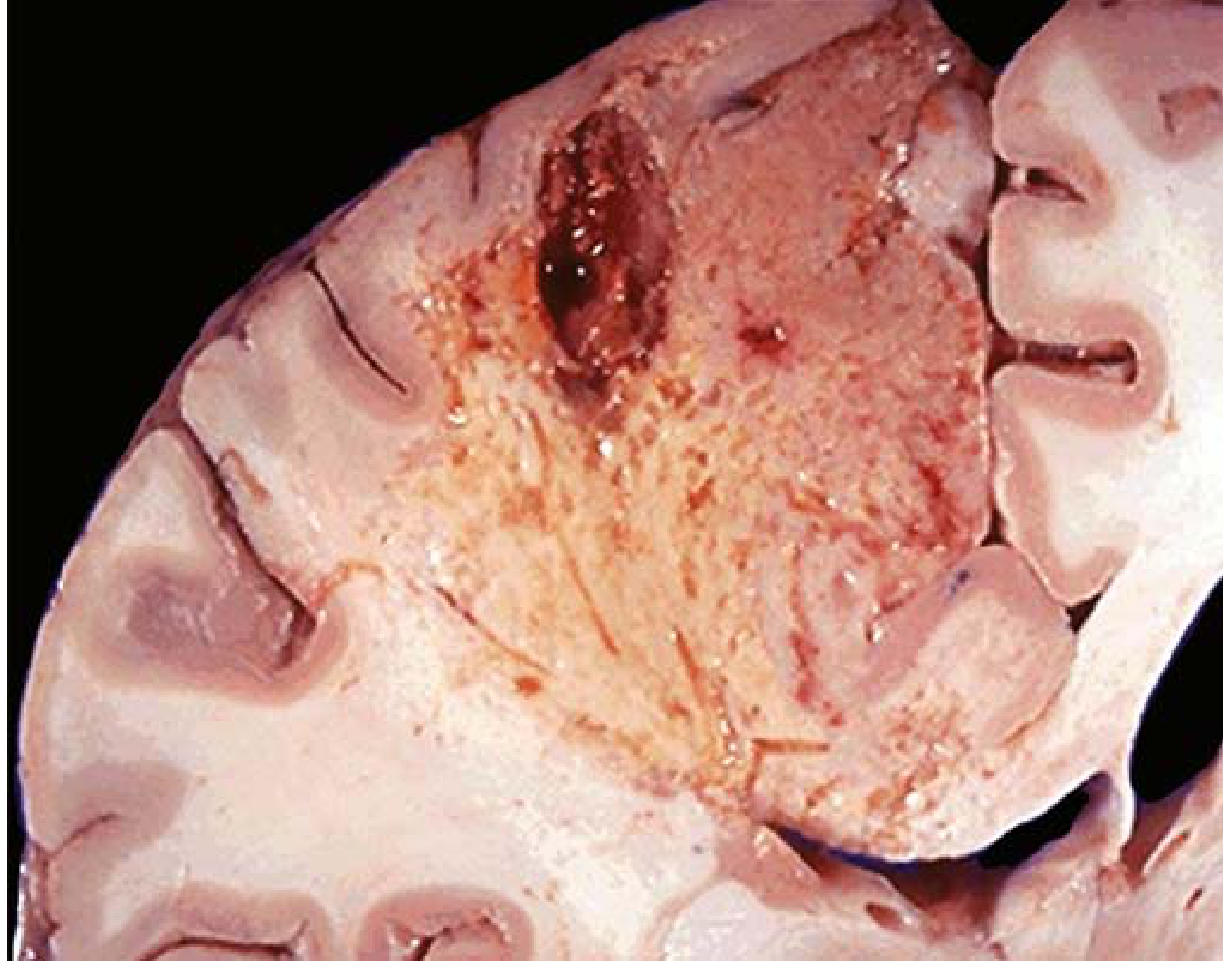
Robbins & Kumar Basic Pathology, p. 21.
3. Gangrenous necrosis
Definition: A clinical term, not a distinct microscopic pattern of cell death. It usually refers to ischemic necrosis of a limb, bowel, or other large tissue area.
Dry gangrene
- Due to arterial occlusion and ischemia.
- Usually represents coagulative necrosis.
- Tissue becomes dry, black, shriveled, and mummified.
Wet gangrene
- Dry gangrene with superadded bacterial infection.
- Bacteria and recruited leukocytes cause enzymatic tissue digestion.
- Thus, it has a liquefactive component.
- Tissue becomes swollen, soft, moist, foul-smelling, and may lead to sepsis.
Examples:
- Diabetic foot with arterial insufficiency.
- Ischemic bowel with bacterial infection.
4. Caseous necrosis
Definition: A distinctive form of necrosis with a soft, friable, yellow-white, "cheese-like" gross appearance.
Morphology:
- Amorphous granular eosinophilic debris on microscopy.
- No recognizable cellular outlines.
- Tissue architecture is completely obliterated.
- Often surrounded by epithelioid macrophages, giant cells, and lymphocytes, forming a granuloma.
Examples:
- Tuberculosis, especially pulmonary tuberculosis.
- Certain fungal infections.
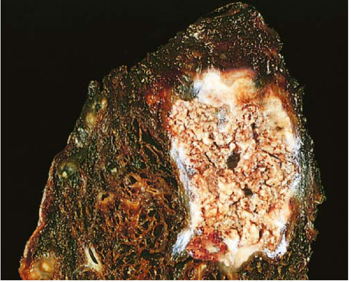
Robbins & Kumar Basic Pathology, p. 21.
5. Fat necrosis
Definition: Focal destruction of adipose tissue.
Enzymatic fat necrosis
Most commonly occurs in acute pancreatitis.
Mechanism:
- Pancreatic lipases escape from injured acinar cells/ducts.
- Lipases digest triglycerides in adipocytes.
- Free fatty acids are released.
- Fatty acids combine with calcium to form calcium soaps, called saponification.
Morphology:
- Gross: chalky-white deposits in peripancreatic or mesenteric fat.
- Microscopy: shadowy outlines of dead fat cells, basophilic calcium deposits, and inflammatory infiltrate.
Traumatic fat necrosis
- Can occur in adipose tissue after trauma, classically breast tissue.
Robbins & Kumar Basic Pathology, pp. 21-22.
6. Fibrinoid necrosis
Definition: A special form of vascular wall necrosis recognized microscopically as bright eosinophilic, amorphous, fibrin-like material.
Pathogenesis:
- Immune complexes deposit in vessel walls.
- Plasma proteins, including fibrin, leak into the damaged vessel wall.
- The combination of immune complexes and plasma proteins gives the vessel wall a bright pink appearance on H&E staining.
Examples:
- Immune-mediated vasculitis.
- Malignant or severe hypertension.
- Vascular lesions in transplant rejection.
Robbins & Kumar Basic Pathology, p. 22.
Necrosis versus apoptosis: key exam table
| Feature | Necrosis | Apoptosis |
|---|---|---|
| Nature | Always pathologic | Physiologic or pathologic |
| Cell size | Cell swelling | Cell shrinkage |
| Cell membrane | Disrupted | Intact, with blebbing |
| Cellular contents | Leak into extracellular space | Enclosed in apoptotic bodies |
| Inflammation | Common | Usually absent |
| Nuclear change | Pyknosis → karyorrhexis → karyolysis | Nuclear fragmentation |
| Mechanism | Severe irreversible injury, membrane failure, enzymatic digestion | Regulated, caspase-mediated cell death |
Robbins & Kumar Basic Pathology, p. 20.
Short conclusion for exams
Necrosis is irreversible, pathologic cell death caused by severe injury. The central events are ATP depletion, mitochondrial failure, calcium influx, membrane damage, lysosomal enzyme leakage, cellular digestion, and inflammation. The important morphologic patterns are coagulative, liquefactive, gangrenous, caseous, fat, and fibrinoid necrosis.
High-yield associations:
- Myocardial/renal infarct: coagulative necrosis
- Brain infarct and abscess: liquefactive necrosis
- Tuberculosis: caseous necrosis
- Acute pancreatitis: enzymatic fat necrosis with saponification
- Vasculitis/severe hypertension: fibrinoid necrosis
- Ischemic limb: gangrene
Make one page revision note
Reading File
Searching PubMed
necrosis AND cell death
NECROSIS: One-Page Revision Note
Definition
Necrosis = irreversible, pathologic cell death in living tissue due to severe injury.
Features: cell swelling, membrane rupture, enzymatic digestion, leakage of cell contents, and inflammation.
Features: cell swelling, membrane rupture, enzymatic digestion, leakage of cell contents, and inflammation.
Pathogenesis: sequence
Severe injury
(ischemia/hypoxia, toxins, infection, trauma, immune injury)
↓
Mitochondrial dysfunction and ATP depletion
↓
Failure of Na⁺/K⁺ pump → Na⁺ and water influx → cell swelling
↓
↑ Cytosolic Ca²⁺ → activation of phospholipases, proteases, endonucleases, ATPases
↓
Damage to plasma membrane, mitochondria, ER, and lysosomes
↓
Lysosomal enzyme release → autodigestion of cell
↓
Leakage of intracellular contents/DAMPs
↓
Acute inflammation and removal of debris
(ischemia/hypoxia, toxins, infection, trauma, immune injury)
↓
Mitochondrial dysfunction and ATP depletion
↓
Failure of Na⁺/K⁺ pump → Na⁺ and water influx → cell swelling
↓
↑ Cytosolic Ca²⁺ → activation of phospholipases, proteases, endonucleases, ATPases
↓
Damage to plasma membrane, mitochondria, ER, and lysosomes
↓
Lysosomal enzyme release → autodigestion of cell
↓
Leakage of intracellular contents/DAMPs
↓
Acute inflammation and removal of debris
Hallmarks of irreversible injury
- Failure to restore mitochondrial oxidative phosphorylation and ATP production
- Severe plasma and organelle membrane damage
- Irreversible DNA/chromatin damage
Morphology
Cytoplasmic changes
- Increased eosinophilia: pink cytoplasm on H&E
- Swelling, vacuolation, membrane disruption
- Leakage of cellular enzymes and proteins
Nuclear changes
Pyknosis → Karyorrhexis → Karyolysis
- Pyknosis: nuclear shrinkage, dark basophilic nucleus
- Karyorrhexis: nuclear fragmentation
- Karyolysis: fading/dissolution of nucleus due to DNase action
Classification of necrosis
| Type | Main feature | Typical example |
|---|---|---|
| Coagulative | Architecture preserved temporarily; firm tissue; ghost cells | Ischemic infarcts in solid organs except brain: heart, kidney, spleen |
| Liquefactive | Enzymatic digestion makes tissue liquid/viscous | Brain infarct; bacterial or fungal abscess |
| Gangrenous | Clinical term for ischemic necrosis of limb/bowel | Dry gangrene = coagulative; wet gangrene = infection + liquefaction |
| Caseous | Soft, friable, yellow-white "cheese-like"; no architecture | Tuberculosis; some fungal infections |
| Fat | Lipase-mediated adipocyte destruction; calcium soap formation | Acute pancreatitis, trauma to fat |
| Fibrinoid | Bright pink fibrin-like vessel-wall necrosis | Vasculitis, severe hypertension, transplant rejection |
High-yield points
- All ischemic infarcts are coagulative except brain, which undergoes liquefactive necrosis.
- Pus/abscess = liquefactive necrosis caused by neutrophil enzymes.
- TB = caseous necrosis with granulomatous inflammation.
- Acute pancreatitis = enzymatic fat necrosis + saponification + chalky-white calcium deposits.
- Dry gangrene = coagulative necrosis due to ischemia.
- Wet gangrene = dry gangrene with bacterial infection and liquefaction.
- Fibrinoid necrosis involves blood vessels and is seen in immune-mediated vascular injury.
Necrosis vs apoptosis
| Feature | Necrosis | Apoptosis |
|---|---|---|
| Cell size | Swelling | Shrinkage |
| Membrane | Ruptured | Intact |
| Contents | Leak out | Packed into apoptotic bodies |
| Inflammation | Present | Absent/minimal |
| Significance | Always pathologic | Physiologic or pathologic |
Mnemonic for types:
Cells Lose Good Control, Finally Failing
= Coagulative, Liquefactive, Gangrenous, Caseous, Fat, Fibrinoid.
Cells Lose Good Control, Finally Failing
= Coagulative, Liquefactive, Gangrenous, Caseous, Fat, Fibrinoid.
Robbins & Kumar Basic Pathology, Ch. 1, pp. 19-22.
This is a shared conversation. Sign in to Orris to start your own chat.