I now have all the content and images needed for a comprehensive answer. Let me compile it.
Histology of the Kidney
The kidney is composed of ~1 million nephrons per kidney, each responsible for filtration, reabsorption, and secretion. Histologically, the kidney is divided into an outer cortex and an inner medulla (with outer and inner zones), and terminates at renal papillae that drain into minor calyces.
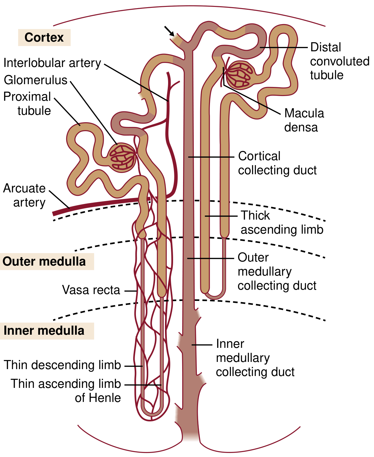
Nephron Organization Diagram
Fig. 1.3 from the NKF Primer on Kidney Diseases, 8e - each nephron consists of a glomerulus, proximal convoluted tubule, thin limbs, thick ascending limb, macula densa, distal convoluted tubule, and collecting duct.
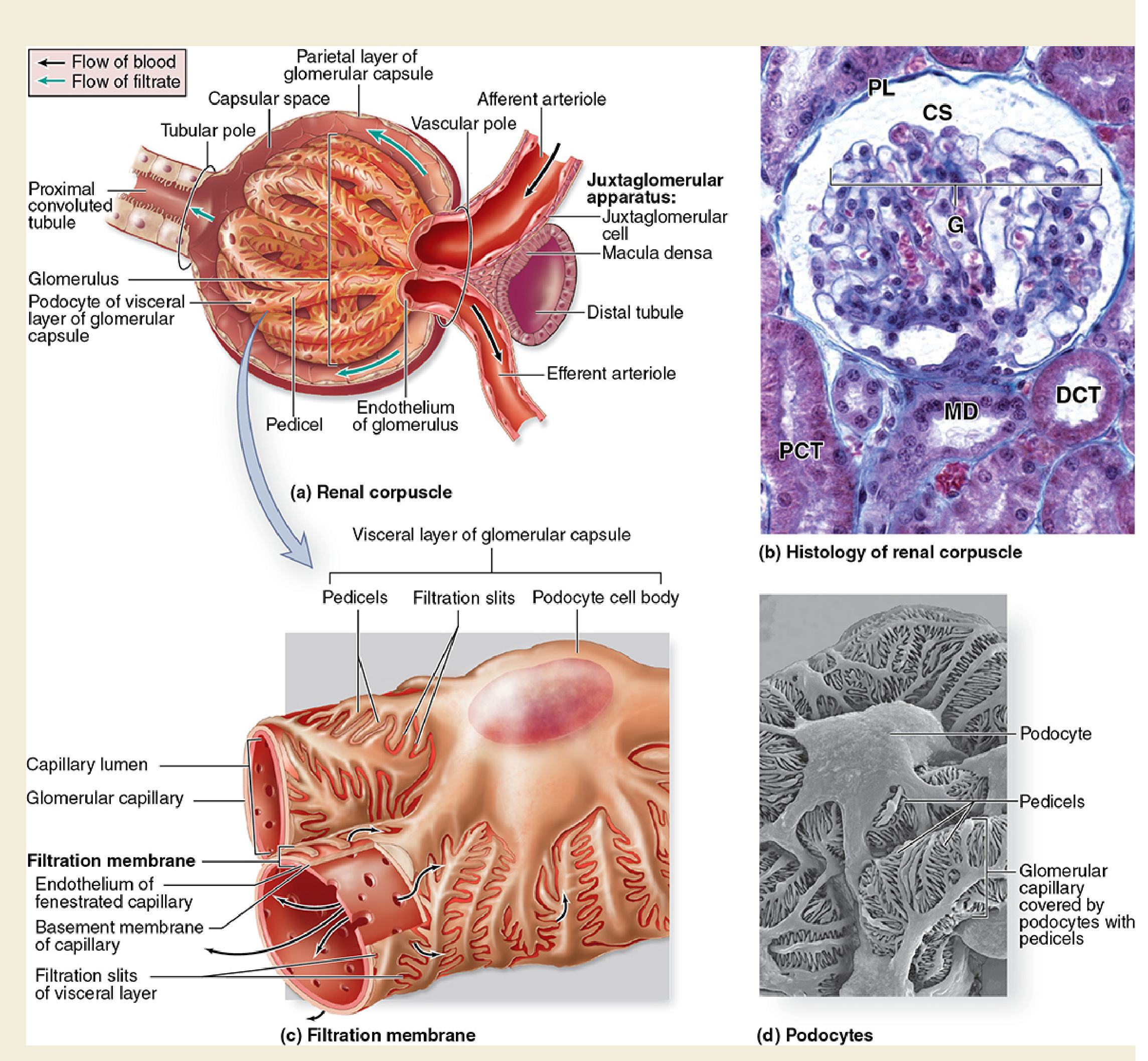
1. Renal Corpuscle (Malpighian Corpuscle)
Each nephron begins in a renal corpuscle (~200 µm diameter), located exclusively in the cortex.
Components:
| Structure | Details |
|---|
| Glomerulus | Tuft of fenestrated capillaries; interposed between afferent and efferent arterioles |
| Bowman's (Glomerular) Capsule | Double-walled epithelial cup surrounding the glomerulus |
| Parietal layer | Simple squamous epithelium forming the outer wall |
| Visceral layer | Composed of podocytes - complex branching cells with foot processes (pedicels) that wrap around capillaries |
| Capsular (urinary) space | Space between the two layers; receives the ultrafiltrate |
| Vascular pole | Where afferent arteriole enters and efferent arteriole exits |
| Tubular pole | Where the PCT begins; parietal squamous epithelium transforms into cuboidal PCT epithelium |
Glomerular Filtration Barrier (3 layers):
- Fenestrated endothelium - pores 70-100 nm; bears negative charge (glycocalyx); prevents cells from passing
- Glomerular Basement Membrane (GBM) - fused basal laminae of endothelium + podocytes; type IV collagen, laminin, fibronectin; 250-400 nm thick; main size-selective barrier
- Podocyte pedicels with slit diaphragms - interdigitating foot processes bridged by nephrin; charge-selective barrier
The barrier is net negatively charged, which restricts passage of albumin and other anionic proteins. Under normal conditions, protein excretion is only 40-80 mg/day (mostly uromodulin/Tamm-Horsfall protein).
Mesangium:
- Mesangial cells (modified smooth muscle) and mesangial matrix occupy the spaces between capillary loops
- Provide structural support, phagocytose debris, and can regulate capillary blood flow
2. Juxtaglomerular Apparatus (JGA)
Located at the vascular pole where the afferent arteriole meets the distal tubule:
| Cell type | Location | Function |
|---|
| Juxtaglomerular (granular) cells | Wall of afferent arteriole | Synthesize and secrete renin |
| Macula densa | Thick wall of distal tubule adjacent to glomerulus | Senses NaCl concentration in tubular fluid; signals JG cells |
| Lacis (extraglomerular mesangial) cells | Between macula densa and glomerulus | Signaling intermediary |
3. Proximal Convoluted Tubule (PCT)
- Location: Cortex
- Epithelium: Simple cuboidal to low columnar
- Key features:
- Prominent brush border (microvilli) on luminal surface - increases surface area ~40-fold
- Abundant mitochondria (basal striations)
- Eosinophilic (pink) cytoplasm
- Lumens often appear narrow or occluded due to the tall cells and microvilli
- Basal infoldings interdigitate with adjacent cells
- Function: Reabsorbs ~67% of filtered water and electrolytes, all glucose, amino acids, vitamins, and small proteins; secretes H+, NH4+, and organic anions/cations (drug metabolism)
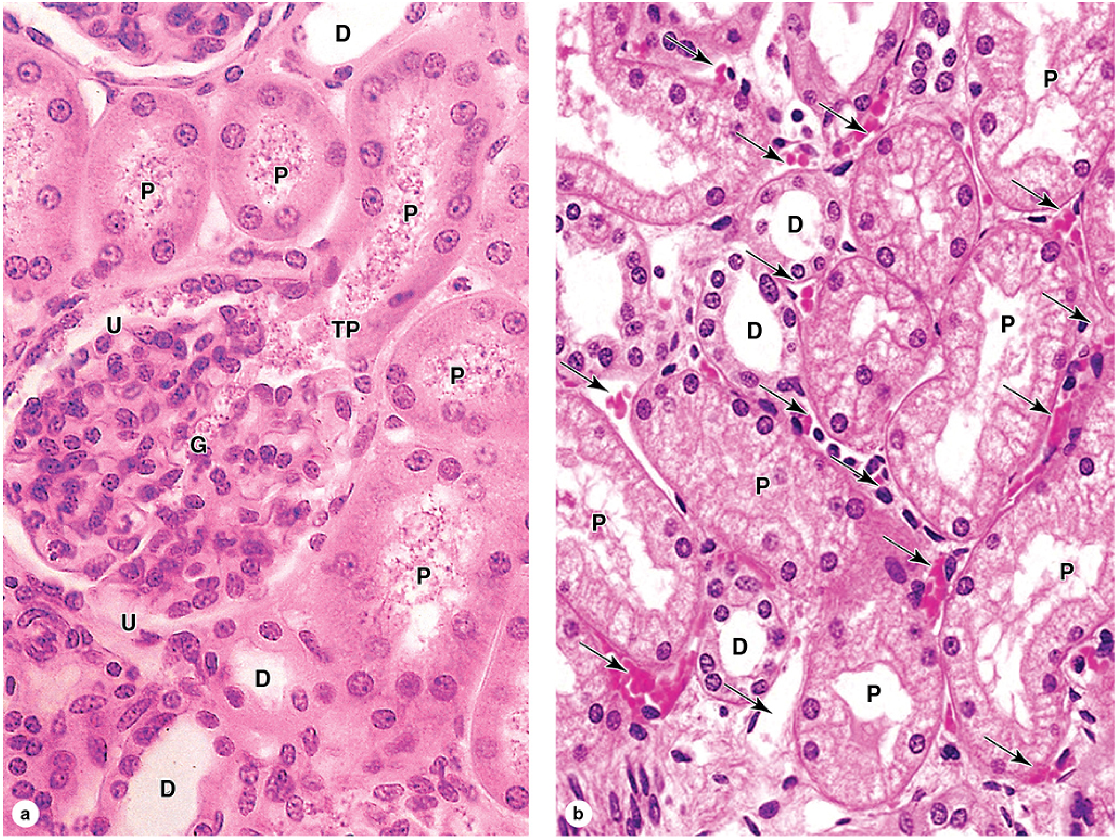
On H&E, PCT cells are larger and pinker than DCT cells, with indistinct cell borders and a fuzzy luminal border (brush border).
4. Loop of Henle
Descends from the cortex into the medulla. Two types of nephrons:
- Cortical nephrons - short loops, reach outer medulla only
- Juxtamedullary nephrons - long loops, reach inner medulla/papillary tip (important for concentration)
| Segment | Epithelium | Location | Key Feature |
|---|
| Thin descending limb | Simple squamous | Outer + inner medulla | Permeable to water; impermeable to solutes |
| Thin ascending limb | Simple squamous | Inner medulla | Permeable to Na+/Cl-; impermeable to water |
| Thick ascending limb (TAL) | Simple cuboidal, no microvilli, many mitochondria | Outer medulla + medullary rays | Contains NKCC2 (Na-K-2Cl cotransporter); actively reabsorbs electrolytes; impermeable to water - creates medullary gradient |
The thin limbs are easily confused with capillaries on H&E; they are distinguished by the absence of red blood cells and a thin, flat epithelium.
5. Distal Convoluted Tubule (DCT)
- Location: Cortex
- Epithelium: Simple cuboidal
- Key features:
- Cells are smaller than PCT cells
- No prominent brush border (smooth luminal surface)
- Cell borders more distinct
- Lumens appear wider and more open than PCT
- Fewer and shorter microvilli; basolateral folds present
- Function: Fine-tuning of electrolyte reabsorption (NaCl via NCC transporter); aldosterone-sensitive segment; 10-12 mm long
On H&E, DCT cells are paler and smaller with more visible lumens compared to PCT.
Summary: How to Distinguish PCT vs DCT on H&E
| Feature | PCT | DCT |
|---|
| Cell size | Larger | Smaller |
| Brush border | Prominent (fuzzy lumen) | Absent (clear lumen) |
| Lumen | Narrow, often occluded | Wide, open |
| Cytoplasm | Deeply eosinophilic | Paler |
| Location | Cortex (predominant) | Cortex (near glomerulus) |
6. Collecting Duct System
- Location: Medullary rays (cortex) → outer medulla → inner medulla → papilla
- Two cell types:
| Cell type | Histology | Function |
|---|
| Principal cells | Most abundant; cuboidal to columnar; pale cytoplasm; distinct cell membranes | ADH-regulated water reabsorption (aquaporin-2); aldosterone-regulated Na+/K+ exchange |
| Intercalated cells | Fewer; scattered; slightly darker cytoplasm; more mitochondria | Acid-base balance (H+ secretion type A; HCO3- secretion type B); K+ reabsorption |
- Collecting ducts converge to form papillary ducts (of Bellini) that open at the papillary apex (area cribrosa)
- Epithelium transitions from cuboidal to columnar as ducts enlarge toward the papilla
7. Renal Interstitium and Vasculature
- Cortex: Minimal interstitial connective tissue; peritubular capillaries are dense
- Medulla: More abundant interstitium with interstitial cells; contains proteoglycans important for the concentration gradient
- Vasa recta: Straight capillaries descending alongside the loops of Henle; hairpin turn configuration maintains the osmotic gradient; supplied by efferent arterioles of juxtamedullary glomeruli
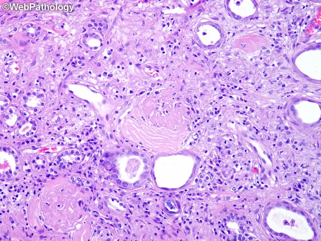
Normal Kidney H&E - Cortex
Quick Reference: What's in Cortex vs Medulla
| Structure | Cortex | Medulla |
|---|
| Renal corpuscles (glomeruli) | ✓ | ✗ |
| PCT | ✓ | ✗ |
| DCT | ✓ | ✗ |
| Macula densa | ✓ | ✗ |
| Thin limbs of Henle | ✗ | ✓ |
| TAL (bulk) | Medullary rays | Outer medulla |
| Collecting ducts | Medullary rays | ✓ |
| Vasa recta | ✗ | ✓ |
Sources: Junqueira's Basic Histology: Text and Atlas, 17e, Ch. 19; NKF Primer on Kidney Diseases, 8e, Ch. 1; Brenner and Rector's The Kidney, Ch. 2.