
Pathogenesis of septic shock md pathology exam question and give Robbins based answer
Reading File
Reading File
Finding Sources
Finding Sources
Finding Sources
Searching PubMed
septic shock AND pathophysiology
Searching the Web
septic shock pathophysiology endothelial dysfunction microvascular thrombosis Surviving Sepsis Campaign
Reading File
Finding Sources
Reading Web Page
https://pmc.ncbi.nlm.nih.gov/articles/PMC7810126
Reading File
Reading File
Reading File
Pathogenesis of Septic Shock
Definition: Septic shock is a form of distributive shock caused by severe microbial infection. It results from a dysregulated systemic inflammatory response causing vasodilatation, increased vascular permeability, endothelial injury, intravascular coagulation, tissue hypoperfusion, and multiorgan dysfunction.
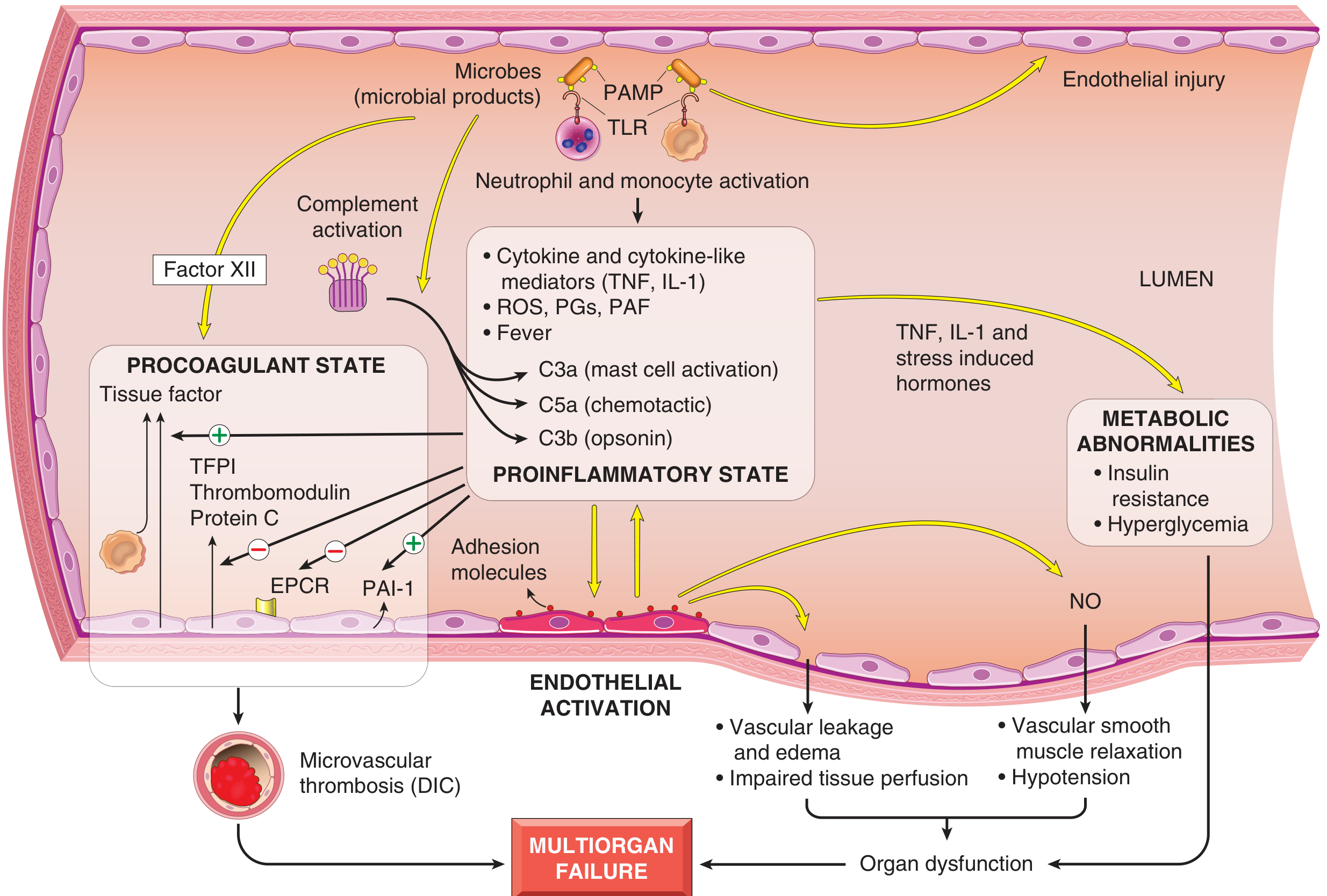
Etiologic trigger
Septic shock is most often caused by gram-positive bacterial infections, followed by gram-negative bacteria and fungi. Microbial products, including bacterial endotoxin and other pathogen-associated molecular patterns (PAMPs), initiate the process.
1. Activation of innate immunity and inflammatory response
Microbial PAMPs are recognized by:
- Toll-like receptors (TLRs)
- G-protein-coupled receptors recognizing bacterial peptides
- C-type lectin receptors, such as dectin receptors recognizing fungal cell-wall components
- Complement system
These activate macrophages, neutrophils, dendritic cells, endothelial cells, and complement. The activated cells release:
- TNF and IL-1
- Type I interferons, IL-12, IL-18
- Chemokines
- Reactive oxygen species
- Prostaglandins and other lipid mediators
- Complement products, especially C3a and C5a
This produces a generalized proinflammatory state. Microbial components may also activate coagulation directly through factor XII. Thrombin in turn amplifies inflammation through protease-activated receptors.
2. Endothelial activation and vascular leakage
Inflammatory cytokines activate and injure endothelial cells. This causes:
- Increased expression of endothelial adhesion molecules, promoting leukocyte adherence and tissue injury
- Disruption of endothelial tight junctions
- Increased capillary permeability
- Leakage of protein-rich fluid into interstitial tissues, causing widespread edema
- Impaired nutrient delivery and removal of metabolic waste
Thus, the effective circulating volume falls and tissue perfusion declines.
3. Vasodilatation and hypotension
Activated endothelial cells and inflammatory cells produce nitric oxide (NO) and other vasodilator mediators.
This leads to:
- Relaxation of vascular smooth muscle
- Marked peripheral vasodilatation
- Venous pooling of blood
- Reduced systemic vascular resistance
- Severe hypotension
Together with plasma leakage, vasodilatation produces inadequate tissue perfusion and cellular hypoxia.
4. Induction of a procoagulant state and DIC
Sepsis shifts the hemostatic balance toward coagulation:
- Cytokines increase tissue factor expression by monocytes and endothelial cells.
- Endothelial anticoagulant mechanisms decrease, including:
- Tissue factor pathway inhibitor
- Thrombomodulin
- Endothelial protein C receptor
- Fibrinolysis is suppressed by increased plasminogen activator inhibitor-1 (PAI-1).
- Sluggish microvascular flow from edema and vascular leakage causes stasis and reduces washout of activated clotting factors.
The result is widespread thrombin generation and deposition of fibrin-rich microthrombi in small vessels. This further reduces tissue perfusion. Severe cases develop disseminated intravascular coagulation (DIC), with both thrombosis and bleeding due to consumption of platelets and coagulation factors.
5. Metabolic abnormalities
Severe sepsis causes:
- Insulin resistance and hyperglycemia
- Cytokine-mediated suppression of insulin release
- Increased gluconeogenesis due to TNF, IL-1, catecholamines, glucagon, growth hormone, and glucocorticoids
- Cellular hypoxia with impaired oxidative phosphorylation
- Increased lactate production and lactic acidosis
Later, relative adrenal insufficiency may occur. Rarely, DIC may cause adrenal hemorrhage and necrosis, as in Waterhouse-Friderichsen syndrome.
6. Myocardial depression and multiorgan dysfunction
High concentrations of cytokines and secondary mediators depress myocardial contractility, reducing cardiac output. Hypotension, edema, and microvascular thrombosis decrease oxygen and nutrient delivery. Oxidative stress also damages mitochondria, impairing cellular oxygen utilization.
The end result is:
Microbial infection → innate immune activation → cytokine storm + endothelial injury → vasodilatation, capillary leak, DIC, metabolic derangement, and myocardial depression → tissue hypoperfusion and cellular hypoxia → multiorgan dysfunction/failure.
Organs commonly affected include kidneys, lungs, brain, heart, and liver. Acute lung injury may manifest as ARDS.
7. Counter-regulatory immunosuppression
After the initial hyperinflammatory phase, some patients develop immunosuppression due to:
- Shift from Th1 to Th2 cytokine responses
- IL-10, soluble TNF receptors, and IL-1 receptor antagonist
- Lymphocyte apoptosis
- Immunosuppressive effects of apoptotic cells
Therefore, patients may alternate between hyperinflammatory and immunosuppressed states.
Flowchart for writing in exams
Microbial PAMPs
↓
TLR/complement/innate immune-cell activation
↓
TNF, IL-1, cytokines, ROS, complement, lipid mediators
↓
Endothelial activation and injury
↓
NO-mediated vasodilatation + vascular leakage + tissue edema
↓
Tissue factor activation + reduced anticoagulants + inhibited fibrinolysis
↓
Microvascular thrombosis/DIC + myocardial depression
↓
Hypotension, hypoperfusion, lactic acidosis
↓
Multiorgan dysfunction and death
↓
TLR/complement/innate immune-cell activation
↓
TNF, IL-1, cytokines, ROS, complement, lipid mediators
↓
Endothelial activation and injury
↓
NO-mediated vasodilatation + vascular leakage + tissue edema
↓
Tissue factor activation + reduced anticoagulants + inhibited fibrinolysis
↓
Microvascular thrombosis/DIC + myocardial depression
↓
Hypotension, hypoperfusion, lactic acidosis
↓
Multiorgan dysfunction and death
Robbins & Kumar Basic Pathology, “Shock” and “Pathogenesis of Septic Shock,” pp. 96-98.
This is a shared conversation. Sign in to Orris to start your own chat.