
What does this line mean that near the cardiac and the peritoneum on the posterior surface is reflected onto the diaphragm as the gastrophrenic ligament?
gastrophrenic ligament stomach peritoneum diaphragm anatomy diagram
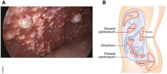
Educational composite image illustrating peritoneal anatomy and metastatic disease. Panel A is a clinical laparoscopic photograph of diaphragmatic peritoneal metastases originating from colorectal cancer. It shows numerous pearly-white, multifocal, nodular lesions of varying sizes (miliary to larger bulbous masses) scattered across the erythematous, vascularized peritoneal surface. Panel B is a medical diagram showing a sagittal view of the abdominal cavity, detailing the anatomical relationship between the parietal peritoneum (lining the abdominal wall and diaphragm) and the visceral peritoneum (enveloping the organs). Labeled structures include the liver, stomach, pancreas, duodenum, colon, small intestine (SI), rectum, bladder, and the omentum—the largest peritoneal fold. This visual comparison aids in understanding the dissemination of gastrointestinal malignancies within the peritoneal space, emphasizing the transition from normal serous membrane anatomy to advanced metastatic disease.
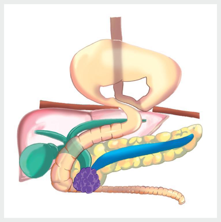
This medical anatomical diagram illustrates a complex surgical and pathological scenario involving the upper gastrointestinal and biliary systems. The image is divided by the diaphragm, showing a large hiatal hernia of the 'upside-down stomach' type, where the majority of the stomach has prolapsed into the thoracic cavity. Below the diaphragm, the abdominal anatomy includes the liver, gallbladder, and pancreas. A significant pathological finding is a purple, nodular mass located in the head of the pancreas, which is shown causing extrinsic compression of the distal common bile duct and the second portion of the duodenum. Additionally, the main pancreatic duct is depicted as dilated (highlighted in blue), indicating obstructive changes proximal to the tumor. The diagram provides clinical context for managing biliary and duodenal obstruction in the presence of severe anatomical distortion. This visual is suitable for gastroenterology and surgical oncology education, focusing on pancreatic malignancy and hiatal hernia complications.
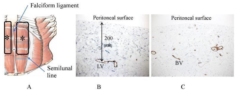
This composite educational image illustrates the vascular anatomy of the human anterior abdominal wall and peritoneum through a combination of an anatomical diagram and immunohistochemical staining. Image A is an anatomical diagram showing the distribution of subperitoneal blood vessels extending from the hypochondrium to the semilunal line, excluding the area of the falciform ligament (marked with an asterisk). Image B is a photomicrograph of D2-40 immunostaining demonstrating a subperitoneal lymphatic vessel (LV) situated approximately 200 μm beneath the peritoneal surface. Image C shows CD31 immunostaining of submesothelial blood vessels (BV) in the same region, highlighting a relatively scarce vascular density and different morphology compared to the lymphatic network. This image serves to educate on the spatial organization of the blood-peritoneal barrier (BPB) and the specific depth and distribution of initial lymphatic vessels, which are critical for understanding peritoneal fluid absorption and the pathways of trans-lymphatic cancer metastasis.
Breaking Down the Statement
Key concepts to understand first
What the sentence means, step by step
Putting it together in plain language
Why it matters clinically
- The gastrophrenic ligament is one of four ligaments that hold the stomach in its normal position inside the abdomen (along with the gastrohepatic, gastroduodenal, and gastrosplenic ligaments).
- It must be divided during fundoplication (anti-reflux surgery) and other upper gastric procedures to mobilize the fundus.
- Disruption of this ligament (along with the phrenoesophageal membrane) is part of the mechanism that allows a hiatal hernia to develop, where the stomach slides or rolls up through the diaphragmatic hiatus into the chest.
- It also forms part of the posterior boundary of the lesser sac (omental bursa).
Summary diagram (conceptual)
DIAPHRAGM (above)
      |
      | ← gastrophrenic ligament (peritoneum reflected here)
      |
POSTERIOR SURFACE of STOMACH FUNDUS/CARDIA