
Recent advances in access cavity designs. Give me 25 marks mds theory answer with diagrams and contents
recent advances access cavity designs endodontics conservative ninja truss 2025
"access cavity" endodontics conservative ninja truss
https://pmc.ncbi.nlm.nih.gov/articles/PMC11991902
access cavity designs endodontics conservative ninja truss traditional diagram
PMID: 34623506
Endodontic access cavity designs comparison diagram showing 6 types side by side in cross-section: 1) Traditional access cavity (TradAC) - wide divergent walls, full deroofing of pulp chamber, removes pericervical dentin; 2) Conservative access cavity (ConsAC) - partial deroofing, convergent walls, preserves pulp horns; 3) Ultraconservative / Ninja cavity (UltraAC) - minimal opening from central fossa, very convergent super narrow entry; 4) Truss access cavity (TrussAC) - two separate small windows with dentinal bridge/truss between them; 5) Caries-driven access cavity - irregular opening following caries path; 6) Restorative-driven access cavity - follows outline of existing restoration. Each design labeled with name and key anatomical landmarks including pulp chamber, canal orifices, pericervical dentin zone (4mm above to 6mm below CEJ highlighted in yellow), marginal ridges. Clean educational dental illustration style, white background, anatomical cross-sections of molar tooth.
Educational dental diagram showing pericervical dentin concept. Cross-section of a molar tooth showing the critical pericervical dentin zone highlighted in orange - defined as dentin 4mm above and 6mm below the cemento-enamel junction (CEJ). Arrows showing stress distribution through pericervical dentin. Labels: enamel, dentin, pulp chamber, root canal, CEJ, pericervical dentin zone, cortical bone level. Comparison showing how traditional access cavity (red shading) removes pericervical dentin vs conservative access cavity (green outline) preserving it. Clean medical textbook illustration style on white background.
Dental diagram comparing guided endodontic access cavity using CBCT and surgical guide. Three panels: Left panel shows CBCT scan cross-section of a calcified molar with superimposed virtual access path planning (blue dotted line). Middle panel shows a 3D-printed surgical stent/guide placed on the tooth occlusal surface directing the bur to correct entry point. Right panel shows the resulting minimal guided access cavity - tiny circular opening precisely centered over canal orifice in a calcified tooth. Labels: CBCT scan, virtual planning, surgical guide, calcified canal, guided access cavity. Clean educational illustration style, labeled diagram on white background, dental anatomy style.
Recent Advances in Access Cavity Designs
Contents / Index
- Introduction
- Historical Perspective and Traditional Access Cavity (TradAC)
- Rationale for Change - Pericervical Dentin Concept
- Classification of Modern Access Cavity Designs
- Conservative Access Cavity (CEC / ConsAC)
- Ultraconservative / Ninja Access Cavity (UEC / UltraAC)
- Truss Access Cavity (TrussAC)
- Caries-Driven Access Cavity (CariesAC)
- Restorative-Driven Access Cavity (RestoAC)
- Guided / Dynamic Navigation Access
- Technological Enablers
- Comparative Evaluation - Advantages and Disadvantages
- Evidence-Based Assessment
- Decision-Making Criteria
- Conclusion
1. Introduction
2. Historical Perspective and the Traditional Access Cavity (TradAC)
- Complete removal of the pulp chamber roof
- Removal of all pulp horns
- Divergent walls (widest at the occlusal surface, narrowest at the orifice level)
- Straight-line access to all canal orifices
- Removal of all coronal interferences
- Extensive removal of sound coronal dentin, especially pericervical dentin
- Weakens the crown-root complex
- Increases risk of cuspal fracture and vertical root fracture
- Makes post-endodontic restoration more complex and costly
3. Rationale for Change - The Pericervical Dentin Concept
Pericervical dentin = dentin located 4 mm above and 6 mm below the cemento-enamel junction (CEJ)
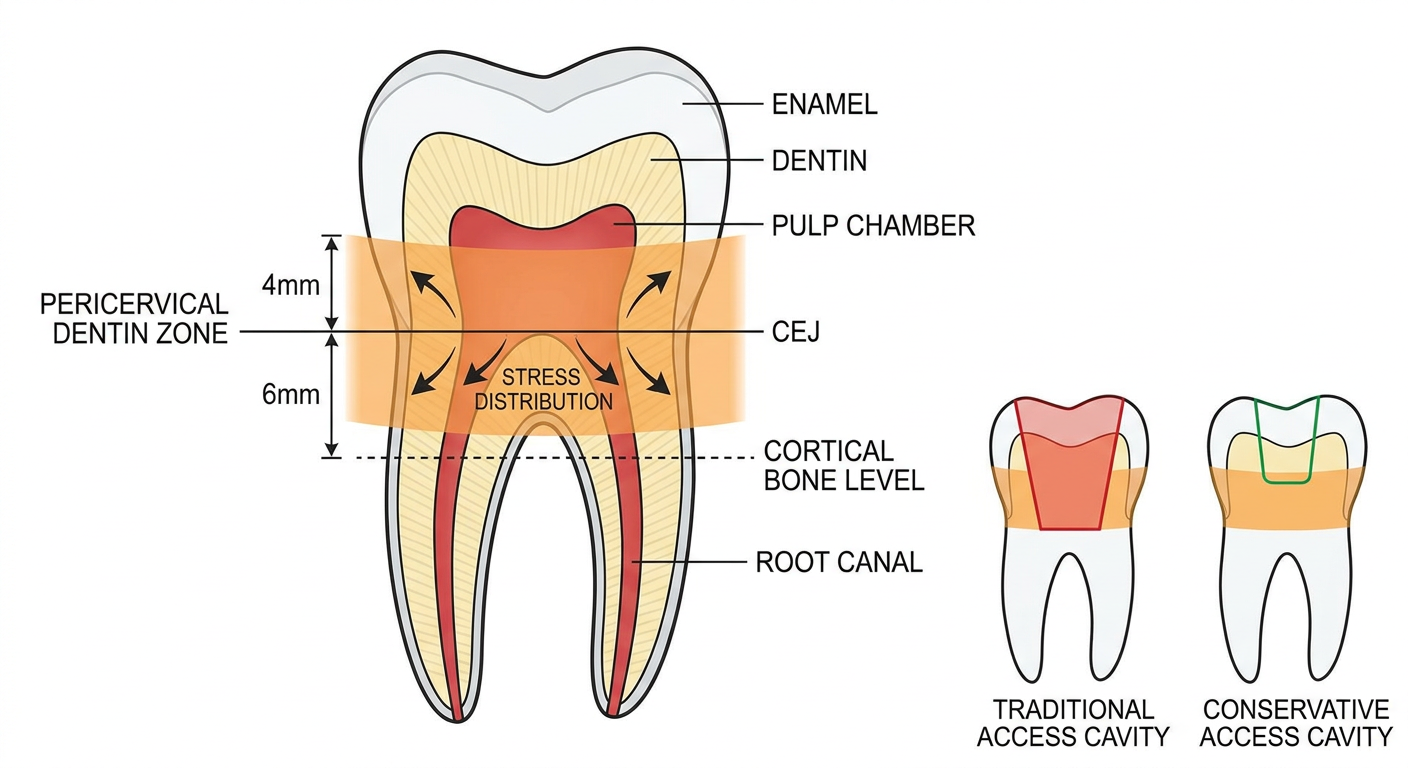
- Acts as the key stress distributor for occlusal forces transmitted from crown to root
- Analogous to the "waist" of an hourglass - critical load-bearing zone
- Loss of PCD disproportionately reduces fracture resistance compared to coronal or apical dentin loss
- Traditional access cavities invariably destroy PCD, especially in premolars and mandibular incisors
- Preservation of PCD is now the primary goal of modern access cavity designs
4. Classification of Modern Access Cavity Designs
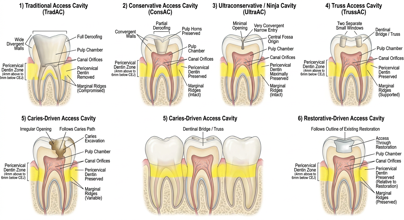
| Acronym | Full Name | Key Feature |
|---|---|---|
| TradAC | Traditional Access Cavity | Full deroofing, divergent walls |
| ConsAC | Conservative Access Cavity | Partial deroofing, convergent walls |
| UltraAC | Ultraconservative / Ninja | Minimal entry, super-convergent |
| TrussAC | Truss Access Cavity | Separate windows with dentinal bridge |
| CariesAC | Caries-Driven Access Cavity | Caries-guided access path |
| RestoAC | Restorative-Driven Access Cavity | Follows existing restoration outline |
5. Conservative Access Cavity (CEC / ConsAC)
- Partial removal of the pulp chamber roof (leaving pulp horn areas intact)
- Slightly convergent walls (occlusal convergence rather than divergence)
- Canal orifices are visible one at a time using different angulation with magnification
- Preserves marginal ridges and pericervical dentin
- Maxillary incisors: Lingual access in the cingulum area, small oval/round preparation
- Maxillary premolars: Oval occlusal preparation, smaller than traditional
- Mandibular molars: Trapezoidal form reduced, preserving oblique ridge
- Maxillary molars: Rhomboidal outline reduced; MB2 canal located with ultrasonics/magnification
- Greater fracture resistance compared to TradAC
- Preserves PCD
- Reduces post-endodontic restoration complexity
- Feasible without high magnification (loupes are adequate)
- Partial roof removal may leave residual pulpal tissue in horns
- Requires angulated viewing to locate all orifices
- Risk of missed canals increases without adequate magnification
- Ultrasonics needed for thorough debridement of pulp horns
6. Ultraconservative / Ninja Access Cavity (UEC / UltraAC)
- Entry point: central fossa (posteriors) or incisal edge (anteriors)
- No extension beyond the initial entry
- Super-convergent preparation - widest occlusally, narrowest at pulpal floor
- Pulp horns and occlusal enamel are preserved
- Canal orifices accessed through the narrow "keyhole"
- Maximum preservation of PCD and occlusal enamel
- Greatest fracture resistance of all designs
- Reduced cuspal flexure under occlusal loading
- May allow bonded restorations without crown coverage in selected cases
- High risk of missed canals - especially MB2, second mesiobuccal in maxillary molars
- Severely restricts instrument angulation and flexibility
- Inadequate debridement of pulp horns - risk of persistent tissue, discoloration
- Requires operating microscope (mandatory, not optional)
- Increased instrumentation errors: ledging, transportation, perforation
- Canal orifices invisible simultaneously - sequential blind exploration
- Not recommended for routine use - reserved for specific indications (calcified canals, esthetic anterior teeth) under magnification
7. Truss Access Cavity (TrussAC)
- Mandibular molars: Two separate cavities - one mesial window over mesial canals (ML + MB), one distal window over distal canal(s)
- Maxillary molars: One window for MB1/DB canals, a separate window for the palatal canal (some add a third for MB2)
- The dentinal ridge/truss between windows is preserved intact
- No attempt to create a unified single access
- Each window independently optimized for its target canals
- Highest preservation of mid-pulpal floor dentin
- Superior fracture resistance compared to TradAC (meta-analysis: Ballester et al., 2021 - Clin Oral Investig)
- Preservation of cingulum, oblique ridges, and marginal ridges
- Reduced post-endodontic restoration burden
- Higher technical complexity - risk of perforation is significant
- Inaccurate entry placement risks trapping pulpal tissue between windows
- Difficult to negotiate canal curvatures from a constrained angulation
- Risk of incomplete debridement of pulpal floor between the windows
- Limited clinical outcome data; predominantly in-vitro evidence
- Not supported by current systematic review evidence for routine use (Ballester et al., 2021, PMID: 34623506)
8. Caries-Driven Access Cavity (CariesAC)
- No additional preparation beyond caries removal
- Access cavity outline is irregular and tooth/lesion specific
- The carious dentin itself guides the cavity path
- All sound dentin including enamel, marginal ridges, and PCD is preserved
- Large carious lesions already encroaching on pulp chamber
- Posterior teeth with extensive caries where traditional access would be redundant
- Teeth with heavily compromised crowns where preserving remaining structure is critical
- Absolute maximum preservation of sound tooth structure
- No additional tooth structure removed beyond what caries has already destroyed
- Biologically and structurally sound rationale in heavily carious teeth
- Highly unpredictable access shape
- May not provide adequate visualization or instrument angulation
- Risk of incomplete roof removal, missed canals
- Restricted to teeth with appropriate caries distribution
9. Restorative-Driven Access Cavity (RestoAC)
- Follows the pre-existing restoration margin and shape
- No extension into sound tooth structure
- Often applies to teeth with large MOD restorations or full-coverage crowns
- Combined restoration removal + access preparation in a single step
- Maximum preservation of residual natural tooth structure
- Clinically efficient when large restorations are already present
- Avoids additional structural compromise
- Access shape entirely dictated by the restoration - may be suboptimal
- Crown perforations in ceramic or metal-ceramic crowns are technically demanding
- May leave portions of old restoration in cavity floor, affecting visibility
10. Guided / Dynamic Navigation Access Cavity
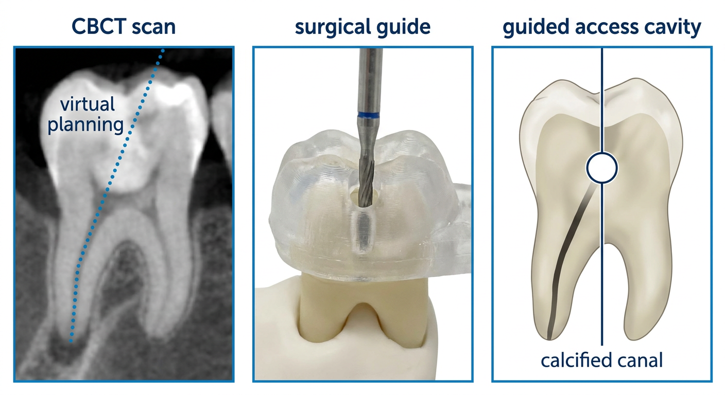
a) Static Guided Endodontics
- CBCT scan of the tooth is obtained pre-operatively
- Virtual access cavity path is planned on 3D reconstruction software
- A 3D-printed surgical stent/guide is fabricated and seated on the tooth
- The access bur is directed through the guide sleeve to the planned entry point
- Enables pinhole access to calcified canals with precision impossible by freehand
b) Dynamic Navigation (Real-Time Guided Endodontics)
- Uses optical tracking systems (similar to surgical navigation in orthopedics)
- Real-time visualization of the bur position relative to the CBCT scan displayed on a monitor
- Allows continuous adjustment during preparation
- Systems: X-Guide (X-NAV Technologies), Navident
- Precision drilling into calcified canals - success rates up to 90% in obliterated cases
- Minimizes perforation risk
- Reduces over-preparation; preserves pericervical dentin optimally
- Enables true minimal access (0.8-1mm entry points reported in literature)
- High equipment cost
- Learning curve for operator
- CBCT radiation exposure
- Static guides may not accommodate intraoperative adjustments
- Limited clinical outcome data
11. Technological Enablers of Modern Access Designs
| Technology | Role in Conservative Access |
|---|---|
| Operating microscope | 10-20x magnification; mandatory for UltraAC; enables visualization of canal orifices through narrow openings |
| Dental loupes | 2.5-4.5x; adequate for ConsAC; improves vision for partial deroofing |
| CBCT | Pre-operative 3D anatomy; identifies calcifications, extra canals, root curvatures; guides access planning |
| Ultrasonic instruments | Endo tips (ET series) for troughing, pulp horn debridement, removal of calcified tissue without expanding access cavity |
| Flexible NiTi files | Reciproc, WaveOne Gold, TruNatomy - designed with smaller pitch/tip to negotiate canals from convergent access cavities |
| LED co-axial illumination | Fibre-optic illumination through microscope; enables transillumination to locate orifices |
| Digital dentistry / CAD-CAM | Fabrication of static navigation guides for guided access |
12. Comparative Evaluation
12a. Fracture Resistance
- TrussAC > ConsAC > TradAC for fracture resistance (in vitro, all marginal ridges intact)
- Key finding (Ballester et al., 2021 - systematic review, PMID 34623506): When one or more marginal ridges are lost, the type of access cavity has NO significant effect on fracture resistance. This means the post-endodontic restoration is more important than the access cavity design for tooth survival.
12b. Cleaning and Disinfection Efficacy
- TradAC consistently shows better cleaning of pulp horns and chamber walls
- ConsAC is equivalent to TradAC when supplemented with ultrasonic activation and microscope
- UltraAC / Ninja cavity: significantly inferior cleaning; residual tissue in pulp horns is a documented risk
- TrussAC: pulpal floor between windows may harbor residual tissue
12c. Risk of Missed Canals
- ConsAC without microscope: Significantly higher risk of missed canals (especially MB2 in maxillary molars) - p < 0.05 (Ballester et al., 2021)
- UltraAC has the highest risk of all designs for missed canals
- Ultrasonic troughing + dental operating microscope are mandatory to mitigate this risk
12d. Treatment Time
- ConsAC, UltraAC: Longer treatment time due to difficulty locating all canal orifices
- TrussAC: Longer; requires two separate preparation sequences
- Guided access: longer setup, shorter drilling time
12e. Clinical Outcome (Long-term)
- No randomized clinical trial data comparing MIAC vs TradAC for clinical success rates (periapical healing)
- Current evidence is predominantly in-vitro; clinical evidence is limited and low-quality (Elmatary et al., 2025 - Br Dent J, PMID: 40217029)
- The International Endodontic Journal consensus (Silva et al., 2022, PMID: 35100441): MIACs show promise but long-term clinical evidence is inadequate to replace TradAC as the universal standard
13. Evidence-Based Assessment
-
Ballester et al. (2021) - 33 studies included. Level of evidence: low. Concluded ConsAC and TrussAC improve fracture resistance only when all marginal ridges are preserved. UltraAC is for limited occasions only. PMID: 34623506
-
Shabbir et al. (2021) - Comprehensive classification and review. TradAC remains the evidence-backed standard; MIAC requires technology-backed application. PMID: 34058252
-
Silva et al. (2022) - International Endodontic Journal review. Calls for universal nomenclature and warns against adopting MIAC without supporting technology. PMID: 35100441
-
Elmatary et al. (2025) - Most recent British Dental Journal review. Confirms advances in magnification and CBCT drive the trend, but warns against social media-driven adoption of Ninja/Truss without adequate evidence. PMID: 40217029
-
Mrinalini et al. (2024) - Review in Cureus. Conservative and truss designs outperform traditional for fracture resistance; no clinical RCT evidence exists. PMID: 39371789
14. Decision-Making Criteria for Access Cavity Selection
1. TOOTH TYPE
   - Anterior tooth with esthetic concerns → ConsAC / UltraAC (with microscope)
   - Molar with all marginal ridges intact → ConsAC or TrussAC (with magnification)
   - Molar with existing MOD restoration → RestoAC or TradAC

2. PULP CHAMBER ANATOMY
   - Large, well-calcified chamber → ConsAC feasible
   - Calcified/obliterated canal → Guided access (CBCT + stent / navigation)

3. AVAILABLE TECHNOLOGY
   - No microscope, no CBCT → TradAC remains safest option
   - Loupes only → ConsAC maximum; not UltraAC or TrussAC
   - Full setup (microscope + CBCT + ultrasonics) → Any MIAC feasible

4. OPERATOR EXPERIENCE
   - Specialist / experienced clinician → All designs applicable
   - General practitioner / student → TradAC or simple ConsAC

5. EXISTING CARIES / RESTORATION
   - Large pre-existing caries → CariesAC
   - Pre-existing large restoration → RestoAC

6. POST-ENDODONTIC RESTORATION PLAN
   - Crown planned regardless → TradAC or ConsAC (access design less critical)
   - Bonded restoration / preservation of tooth → ConsAC / TrussAC to preserve ridges
15. Conclusion
- No single design is universally superior - case selection determines the optimal cavity
- The post-endodontic restoration is more influential on long-term tooth survival than the access cavity design alone
- UltraAC (Ninja) and TrussAC, popularized by social media, carry real clinical risks (missed canals, perforations) and require mandatory use of operating microscope
- The current evidence base is predominantly in-vitro; clinical RCT data is urgently needed
- The pragmatic recommendation (Ballester et al., 2021) remains: "Maintain the access cavity as small as practical without jeopardizing root canal treatment quality"
- Technology is not a substitute for anatomy - conservative access without adequate visualization and instrumentation leads to poorer outcomes than traditional access
Quick Reference: Access Cavity Designs at a Glance
| Design | Deroofing | Walls | PCD Preservation | Magnification Required | Evidence Level |
|---|---|---|---|---|---|
| TradAC | Complete | Divergent | Minimal | Loupes | High (clinical) |
| ConsAC | Partial | Convergent | Moderate | Loupes (minimum) | Moderate |
| UltraAC (Ninja) | Minimal | Super-convergent | High | Microscope (mandatory) | Low |
| TrussAC | Partial (2 windows) | Convergent (each) | High | Microscope | Low |
| CariesAC | Caries-guided | Irregular | Maximum | Loupes | Limited |
| RestoAC | Restoration-guided | Follows restoration | Maximum | Loupes | Limited |
| Guided | Pinhole | Minimal | Maximum | CBCT + Stent/Nav | Emerging |
- Ballester B et al. Current strategies for conservative endodontic access cavity preparation. Clin Oral Investig. 2021. PMID: 34623506
- Elmatary A et al. Endodontic access cavity preparation: challenges and recent advancements. Br Dent J. 2025. PMID: 40217029
- Silva EJNL et al. Present status and future directions - Minimal endodontic access cavities. Int Endod J. 2022. PMID: 35100441
- Shabbir J et al. Access Cavity Preparations: Classification and literature review. J Endod. 2021. PMID: 34058252
- Mrinalini et al. Endodontic Access Cavity Design and Fracture Resistance. Cureus. 2024. PMID: 39371789
- Clark D, Khademi J. Modern molar endodontic access and directed dentin conservation. Dent Clin North Am. 2010.