
1. Define tuberculosis. Discuss in detail the etiopathogenesis, pathology, clinicopathological features, laboratory diagnosis, complications, and recent advances in the diagnosis 2. Define HIV infection and AIDS. Discuss the etiopathogenesis, pathogenesis, pathology, laboratory diagnosis, opportunistic infections, AIDS-defining malignancies, and recent advances in HIV infection.
tuberculosis granuloma caseous necrosis histopathology
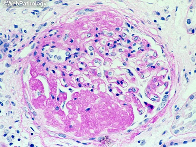
This is a histopathology image obtained with light microscopy from a formalin-fixed paraffin-embedded tissue section stained with Hematoxylin and Eosin. The field demonstrates a granulomatous inflammatory lesion, typically well circumscribed. The central area shows eosinophilic, acellular to granular necrotic material (caseous necrosis), surrounded by a dense mantle of epithelioid histiocytes with elongated, pale-staining nuclei and abundant pale cytoplasm. Multinucleated giant cells of the Langhans type may be present at the periphery. Scattered lymphocytes and occasional plasma cells form a peripheral lymphohistiocytic cuff around the granuloma. The surrounding stroma contains collagen fibers and occasional fibroblasts, with minimal additional inflammatory cells in this field. The morphology is classic for granulomatous inflammation, most commonly associated with infectious etiologies such as Mycobacterium tuberculosis or fungal infections, but can be seen in sarcoidosis or foreign body reactions depending on clinical context. Diagnostic significance: identification of a caseating granuloma raises suspicion for mycobacterial or fungal infection and warrants targeted microbiologic stains (Ziehl-Neelsen for acid-fast bacilli; GMS or PAS for fungi), cultures, and molecular testing. Clinical correlation is essential, including TB exposure history, immune status, and presentation—cough, weight loss, fever, pulmonary nodules or granulomatous lymphadenitis. This image is educational for teaching, differential diagnosis, and research into granulomatous disease pathology.
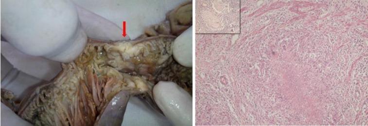
A composite image demonstrating clinical and histopathological findings of abdominal tuberculosis. The left panel is a clinical photograph of a resected small bowel segment during a laparotomy. A prominent red arrow indicates a focal stricture with significant luminal narrowing and mucosal thickening, consistent with granulomatous ileitis. The right panel displays a histopathological section (H&E stain, 100x magnification) showing an epithelioid cell granuloma. Key features include central caseous necrosis and a Langhans giant cell located adjacent to the muscularis mucosae. An inset in the upper left corner of the histopathology panel shows a similar granulomatous reaction within a dissected mesenteric lymph node. These findings collectively illustrate the classic morphological and microscopic features of intestinal tuberculosis, highlighting the progression from macroscopic obstruction to microscopic necrotizing granulomatous inflammation.
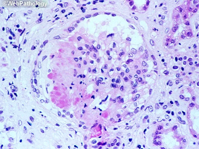
This is a light microscopy image of a formalin-fixed, paraffin-embedded tissue section stained with Hematoxylin and Eosin (H&E). The sample reveals a well-formed granulomatous lesion characterized by a surrounding cuff of lymphocytes and numerous epithelioid histiocytes with elongated, crescent-shaped nuclei. Within the granuloma, multinucleated giant cells (Langhans-type) are visible, and a central amorphous pink necrotic area (caseous necrosis) is evident. The tissue architecture demonstrates organized granulomatous inflammation with a necrotic core, consistent with infectious granulomas. The differential includes mycobacterial disease (tuberculosis) and certain fungal infections (histoplasmosis, coccioidomycosis), though non-necrotizing granulomas can occur in sarcoidosis. The staining highlights chromatin-rich nuclei and cytoplasmic eosinophilia; occasional fibroblasts and mild edema extend into the perigranulomatous region. Clinically, granulomatous tissue with caseation is highly suggestive of TB in the appropriate clinical context and warrants further diagnostic workup (acid-fast bacilli staining, fungal stains, culture, PCR). This image serves as teaching material for histopathology education, differential diagnosis of granulomatous inflammation, and correlation with radiologic and microbiologic findings in suspected infectious etiologies. Additional keywords for indexing: epithelioid macrophages, granuloma, necrosis, caseation, tuberculosis, mycobacteria, fungal infection, lymphohistiocytic rim, tissue biopsy, pathology slide, H&E, histology teaching. Clinical correlation with imaging and sputum analysis improves diagnostic yield.
HIV AIDS pathogenesis CD4 T cell depletion diagram
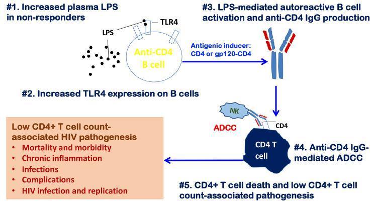
This pathophysiology diagram illustrates the mechanism of autoimmune-mediated CD4+ T cell depletion in people with HIV (PWH). The process is depicted in five numbered stages: (1) Increased plasma lipopolysaccharide (LPS) in non-responders, representing microbial translocation. (2) Upregulation of TLR4 expression on anti-CD4 B cells. (3) LPS-mediated B cell activation leading to the production of anti-CD4 IgG autoantibodies, stimulated by CD4 or gp120-CD4 complexes. (4) These autoantibodies bind to CD4 T cells, recruiting Natural Killer (NK) cells to perform antibody-dependent cell-mediated cytotoxicity (ADCC). (5) Resultant CD4+ T cell death and subsequent low CD4+ T cell counts. A summary box highlights the clinical consequences of this pathway, including chronic inflammation, increased infections, and HIV-associated morbidity and mortality. This diagram serves as an educational model for immune reconstitution failure despite antiretroviral therapy (ART).
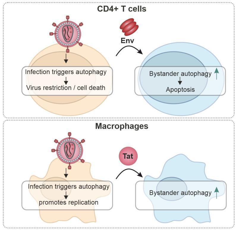
This pathophysiology diagram illustrates the cell-type-specific roles of autophagy during HIV-1 infection in CD4+ T cells and Macrophages. The diagram is divided into two horizontal sections. The top section (CD4+ T cells) shows that direct infection triggers an antiviral autophagy response, leading to virus restriction and cell death. Simultaneously, released viral Envelope (Env) proteins interact with uninfected 'bystander' CD4+ T cells, inducing bystander autophagy and subsequent apoptosis. The bottom section (Macrophages) depicts a contrasting mechanism: in these cells, infection-triggered autophagy is hijacked to promote viral replication rather than restriction. Additionally, secreted Tat proteins are shown modulating autophagy in bystander macrophages. The HIV-1 virion is represented as a spherical particle with surface envelope trimers and a central cone-shaped capsid. The figure highlights the dual nature of autophagy as both a host defense mechanism in lymphoid cells and a proviral pathway in myeloid cells, emphasizing clinical concepts of immune depletion and viral persistence.
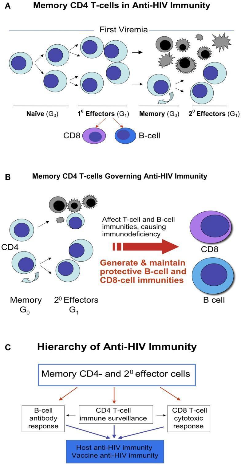
This medical illustration depicts the role and differentiation of memory CD4 T-cells in anti-HIV immunity across three panels. Panel A (Memory CD4 T-cells in Anti-HIV Immunity) shows the lineage from Naïve (G0) CD4 cells following first viremia, differentiating into 1st Effectors (G1). These further progress into Memory (G0) cells or 2nd Effectors (G1), with some cells undergoing apoptosis due to viral infection. It indicates CD4-dependent help for CD8 T-cells and B-cells. Panel B (Memory CD4 T-cells Governing Anti-HIV Immunity) illustrates the recall response where memory CD4 cells differentiate upon re-encountering HIV, highlighting their role in generating and maintaining protective B-cell and CD8-cell immunities, while noting that CD4 depletion causes immunodeficiency. Panel C (Hierarchy of Anti-HIV Immunity) presents a flow diagram positioning Memory CD4 and 2nd effector cells at the apex of a hierarchy. They regulate a B-cell antibody response, CD4 T-cell immune surveillance, and CD8 T-cell cytotoxic response, which collectively constitute host and vaccine anti-HIV immunity. The diagram emphasizes the 'commander-in-chief' role of CD4 memory cells in orchestrating both cellular and humoral immune responses.
Kaposi sarcoma skin lesion HIV AIDS
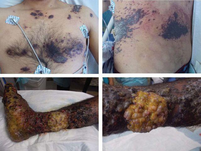
A composite of clinical photographs showing severe cutaneous manifestations of AIDS-associated Kaposi's sarcoma. The upper panels display the patient's torso, featuring widespread, multifocal skin lesions. These lesions vary from small, purplish-red macules to large, confluent plaques. Some plaques exhibit a hyperkeratotic, thickened, and rough texture, primarily distributed across the chest, upper abdomen, and lateral trunk. The lower panels focus on the right lower extremity, demonstrating an advanced, fungating tumor mass on the medial calf. This approximately 10 x 10 cm lesion is lobulated and exophytic, showing mixed yellow purulent exudate and dark necrotic areas. The surrounding skin of the lower leg and foot appears severely edematous with signs of secondary infection, dry gangrene, and extensive ulceration. This imagery illustrates the clinical presentation of disseminated Kaposi's sarcoma in the context of profound immunosuppression and high HIV viral load, highlighting both the classic multifocal plaques and rare, aggressive exophytic variants.
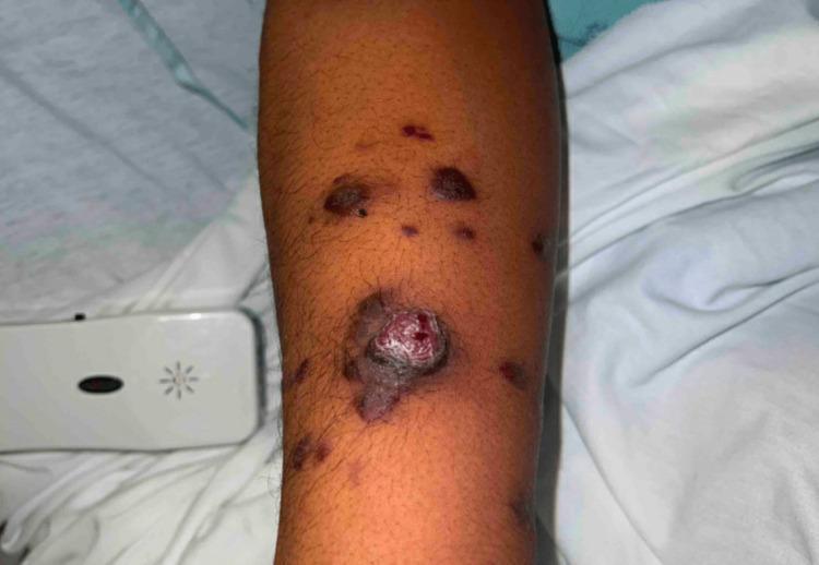
Clinical photograph of the right lower arm demonstrating multiple, multifocal skin lesions characteristic of Kaposi sarcoma (KS). The image shows a spectrum of lesion morphologies, including violaceous to dark-brown papules, plaques, and a prominent exophytic nodule. The central, larger lesion is a raised, pedunculated nodule with a friable, vascular surface and a surrounding hyperpigmented halo. Smaller lesions are scattered proximally and distally, appearing as flat or slightly elevated maculopapular patches with irregular borders. The surrounding skin shows mild edema and post-inflammatory pigmentary changes. This visual is clinically significant for illustrating the classic dermatologic manifestations of human herpesvirus 8 (HHV-8) associated Kaposi sarcoma, frequently seen in the context of immunocompromised states such as advanced HIV/AIDS or Immune Reconstitution Inflammatory Syndrome (IRIS).
PART 1: TUBERCULOSIS
1. Definition
2. Etiopathogenesis
The Causative Organism
- Doubling time: 18-24 hours (extremely slow; culture takes weeks)
- Acid-fast staining: The waxy coat (mycolic acids, phthiocerat dimycocerosate, trehalose dimycolates, sulfolipids) prevents decolorization by strong acids - hence "acid-fast"
- The M. tuberculosis complex includes M. africanum (humans), M. bovis (cattle), M. caprae (goats)
Transmission
- Aerosol droplet nuclei (1-5 µm) from individuals with active pulmonary or laryngeal TB
- Infectious particles can remain suspended in air for hours
- Risk increases with prolonged close contact, crowding, and poor ventilation
Pathogenesis - Steps
- Bacilli and their antigens are presented to CD4+ T helper cells via dendritic cells
- Th1 CD4+ cells release IFN-γ, which activates macrophages to produce reactive nitrogen species and kill bacilli
- Activated macrophages transform into epithelioid cells and fuse to form Langhans giant cells
- These aggregate with lymphocytes to form the hallmark tuberculoid granuloma
- In the granuloma center, tissue hypoxia + enzymatic action leads to caseous (cheese-like) necrosis - unique to TB
- ~90-95% of immunocompetent individuals contain the infection → latent TB
- The Ghon focus undergoes fibrosis and calcification → Ghon lesion; calcified Ghon lesion + calcified hilar nodes = Ranke complex (radiographically visible)
- 5-10% progress to primary progressive disease
Reactivation (Post-Primary / Secondary TB)
- Goldman-Cecil Medicine, pp. 3251-3252; Medical Microbiology 9e
3. Pathology
Primary TB
| Feature | Description |
|---|---|
| Ghon focus | Subpleural parenchymal lesion (usually middle/lower lobe) - 1-2 cm caseous granuloma |
| Ghon complex | Ghon focus + enlarged ipsilateral hilar/mediastinal lymph nodes |
| Histology | Caseating epithelioid granuloma with Langhans giant cells; lymphocytic cuff at periphery |
| Outcome | Fibrosis → calcification in 98-99% of cases |
Granuloma Histology (Classic Tuberculous Granuloma)
- Center: Amorphous, eosinophilic, acellular caseous necrosis (looks like cream cheese grossly)
- Middle zone: Epithelioid macrophages (elongated, pale nuclei; abundant pale cytoplasm)
- Langhans giant cells: Multinucleated cells with nuclei arranged in a horseshoe/peripheral pattern (distinguish from foreign body giant cells where nuclei are central)
- Outer zone: Lymphocytic cuff, occasional plasma cells, fibrous encapsulation
Secondary (Post-Primary) TB Pathology
- Location: Apical and posterior segments of upper lobes (Simon foci), superior segment of lower lobes
- Lesions: Fibrocaseous nodules → coalescence → cavitation
- Cavity: Lined by caseous material; may communicate with bronchi → infectious aerosols produced
- Spread: Endobronchial dissemination → bronchopneumonia; lymphohematogenous spread → miliary TB
- Healing: Fibrosis, dystrophic calcification; end-stage → destroyed lung, bronchiectasis
Miliary TB
- Hematogenous dissemination producing tiny (~2 mm) uniform seed-like lesions throughout organs (lung, liver, spleen, bone marrow, meninges)
- Gross: Millet seed-sized white nodules on cut section
- Occurs in primary TB in children, immunocompromised, or massive reactivation
4. Clinicopathological Features
Pulmonary TB
- Chronic productive cough (>3 weeks), with or without hemoptysis
- Fever (typically low-grade, afternoon) with night sweats
- Weight loss and anorexia ("consumption")
- Pleuritic chest pain, dyspnea
- Wasting, pallor
- Post-tussive crepitations over lung apices
- Signs of pleural effusion; signs of consolidation
- Primary TB: Hilar/mediastinal lymphadenopathy ± parenchymal infiltrate (any lobe)
- Post-primary TB: Upper lobe infiltrates, cavitation, fibrosis (classic "moth-eaten" upper lobe pattern)
- Miliary TB: Diffuse bilateral 1-3 mm nodular opacities ("snow storm" pattern)
- CT chest is more sensitive for cavitation, tree-in-bud pattern, and mediastinal nodes
Extrapulmonary TB
| Site | Features |
|---|---|
| Lymph nodes (scrofula) | Cervical > mediastinal; matted nodes → caseation → collar-stud abscess |
| Pleural | Exudative pleural effusion (lymphocyte-predominant, ADA elevated) |
| Spine (Pott's disease) | Vertebral destruction, gibbus deformity, paravertebral abscess |
| CNS | TB meningitis (basal exudate, cranial nerve palsies), tuberculoma |
| Genitourinary | "Sterile" pyuria, ureteral strictures, "putty kidney" |
| Miliary | Multiorgan involvement, hepatosplenomegaly, choroidal tubercles |
| Peritoneal | Ascites (exudate), "dough belly," omental thickening |
| Pericardial | Constrictive pericarditis |
5. Laboratory Diagnosis
A. Demonstration of Acid-Fast Bacilli (AFB)
- Ziehl-Neelsen (ZN) stain: Sputum smear - bacilli appear red against blue background; sensitivity ~40-60% (requires 10,000 bacilli/mL)
- Auramine-Rhodamine fluorescent stain: More sensitive; bacilli glow orange-yellow under UV light; preferred for screening
- Specimens: Sputum (3 early morning specimens), BAL, gastric aspirate, CSF, urine, biopsy tissue
B. Culture (Gold Standard)
- Lowenstein-Jensen (LJ) medium (solid): Takes 6-8 weeks; rough, buff-colored, "cauliflower-like" colonies
- BACTEC MGIT 960 (liquid medium): Fluorometric detection; results in 9-16 days; superior sensitivity
- Culture allows species identification and drug susceptibility testing (DST)
C. Tuberculin Skin Test (TST / Mantoux test)
- Intradermal injection of 5 TU (0.1 mL) of purified protein derivative (PPD) into forearm
- Read at 48-72 hours; induration (not erythema) measured
- Positive if induration ≥ 10 mm (general population), ≥ 5 mm (HIV-positive, close contacts, immunosuppressed), ≥ 15 mm (low-risk individuals)
- Limitations: False-positive (BCG vaccination, NTM), false-negative (immunosuppression, miliary TB, malnutrition, sarcoidosis)
D. Interferon-Gamma Release Assays (IGRAs)
- Whole-blood in-vitro assay measuring IFN-γ release by sensitized T cells upon exposure to M. tuberculosis-specific antigens (ESAT-6, CFP-10)
- QuantiFERON-TB Gold Plus (QFT-Plus): ELISA-based
- T-SPOT.TB: ELISPOT-based (more sensitive in immunocompromised)
- Advantages: No BCG cross-reaction, single visit, no booster effect
- Diagnostically superior to TST for latent TB; cannot distinguish latent from active TB alone
- Goldman-Cecil Medicine TB chapter: TST and IGRA positivity spectrum shown per stage
E. Nucleic Acid Amplification Tests (NAATs)
- Xpert MTB/RIF (GeneXpert): WHO-recommended first-line test; detects M. tuberculosis DNA AND rifampicin resistance (rpoB gene mutations) simultaneously within 2 hours; sensitivity 88%, specificity 98% for smear-positive; can be used on sputum, CSF, tissue
- Xpert MTB/RIF Ultra: Improved sensitivity (5 bacilli/mL); superior for paucibacillary and extrapulmonary TB
- Line probe assays (LPAs): GenoType MTBDRplus - detect resistance to INH (katG, inhA) and rifampicin rapidly
F. Histopathology
- Tissue biopsy (lymph node, pleura, liver, bone marrow): caseating granuloma with Langhans giant cells; ZN stain for AFB
- Non-caseating granulomas should prompt differentiation from sarcoidosis, fungal infections
G. Other Tests
- ADA (Adenosine deaminase): Elevated in pleural, peritoneal, pericardial, CSF TB (>40 U/L in pleural fluid highly suggestive)
- Chest X-ray / CT: Imaging adjunct
- CBC: Anemia, elevated ESR, lymphocytosis or leukocytosis
- Serum tests: Raised ESR, C-reactive protein
6. Complications
| System | Complication |
|---|---|
| Pulmonary | Massive hemoptysis (Rasmussen aneurysm - erosion of pulmonary artery by cavity), tension pneumothorax, empyema, bronchopleural fistula, bronchiectasis, aspergilloma (fungal ball in TB cavity) |
| Respiratory failure | Destroyed lung, fibrosis, cor pulmonale |
| Spread | Miliary TB, meningitis (most feared complication with highest mortality) |
| Skeletal | Pott's disease with paraplegia, cold abscess |
| Renal | Obstructive uropathy, renal failure |
| Cardiovascular | Constrictive pericarditis |
| Iatrogenic | Drug hepatotoxicity (INH, RIF, PZA), optic neuritis (EMB), peripheral neuropathy (INH), hyperuricemia (PZA) |
| Drug Resistance | MDR-TB (resistant to INH + RIF); XDR-TB (MDR + resistant to fluoroquinolones + one injectable) |
7. Recent Advances in Diagnosis
-
Xpert MTB/RIF Ultra: WHO-endorsed; sensitivity 5 bacilli/mL; detects M. tuberculosis and rifampicin resistance in 2 hours; validated for use on sputum, gastric aspirate, CSF, BAL, lymph node aspirates
-
Whole Genome Sequencing (WGS): Comprehensive drug-resistance profiling (all anti-TB drugs); can replace culture-based DST; tracks transmission chains; now being implemented in high-burden countries
-
Next-generation line probe assays (LPAs): MTBDRsl v2 - detects resistance to fluoroquinolones and second-line injectables, enabling rapid XDR-TB diagnosis
-
Lipoarabinomannan (LAM) urine antigen test (AlereLAM/Fujifilm SILVAMP TB LAM): Detects M. tuberculosis LAM antigen in urine; particularly useful in HIV-positive patients with low CD4 counts; can diagnose TB at bedside without sophisticated lab infrastructure
-
AI-assisted CAD4TB (Computer-Aided Detection for TB): AI software reads chest X-rays for TB-specific patterns; deployed in mass screening programs in India, Africa; comparable to radiologist performance; non-nuclear, scalable diagnostic tool
-
Breath-based diagnostics: Studies investigating volatile organic compounds (VOCs) in exhaled breath as TB biomarkers; non-invasive; currently under validation
-
Blood transcriptomic signatures: Gene expression profiles (e.g., RISK4 4-gene signature, BATF2) can identify individuals progressing from latent to active TB before symptom onset - potential for pre-emptive therapy
-
Serum/plasma biomarkers: IP-10, C1q, multiple cytokine panels being validated as diagnosis and treatment response markers
PART 2: HIV INFECTION AND AIDS
1. Definitions
- HIV-positive serology AND
- CD4+ T-cell count < 200 cells/µL (or < 14% of total lymphocytes), OR
- Presence of an AIDS-defining condition (opportunistic infection or malignancy) regardless of CD4 count
2. Etiology
The Virus
- HIV-1: Pandemic strain; belongs to Lentivirus genus, Family Retroviridae
- HIV-2: Found mainly in West Africa; less pathogenic, slower progression
- Structure: Spherical, ~120 nm; lipid envelope with surface glycoproteins gp120 (surface unit) and gp41 (transmembrane unit)
- Core: Conical capsid (p24) containing: diploid +ssRNA genome, reverse transcriptase, integrase, protease
- Genome: ~9.8 kb; genes include gag (core proteins), pol (enzymes), env (envelope glycoproteins), plus regulatory genes: tat, rev, vif, vpr, vpu, nef
Routes of Transmission
| Route | Details |
|---|---|
| Sexual | Most common globally; unprotected anal > vaginal intercourse; rectal mucosa has high density of CCR5+/CD4+ cells |
| Parenteral | IV drug use (sharing needles), blood transfusion, occupational needlestick |
| Mother-to-Child (MTCT) | Antenatal, intrapartum (most common), breastfeeding |
| NOT transmitted | Air, casual contact, mosquitoes, sharing utensils |
3. Pathogenesis
Step 1 - Viral Entry
- gp120 binds to CD4 receptor on T-helper cells, macrophages, dendritic cells
- A conformational change exposes the co-receptor binding site
- Co-receptors:
- CCR5 (R5): Used predominantly in early infection; present on macrophages and T cells in mucosal tissues; R5-tropic virus causes initial infection
- CXCR4 (X4): Used in late disease; present mainly on T lymphocytes; X4-tropic virus is highly cytopathic and depletes T cells rapidly
- Individuals homozygous for CCR5-Δ32 deletion are resistant to HIV infection (natural experiment that inspired CCR5 antagonist drugs)
- gp41 mediates fusion of viral envelope with host cell membrane → nucleocapsid enters cell
Step 2 - Reverse Transcription and Integration
- Viral reverse transcriptase (RT) converts ssRNA → double-stranded DNA (error-prone: generates ~1 mutation per replication cycle → high genetic diversity)
- Integrase catalyzes integration of viral dsDNA into host chromosome → HIV provirus (permanent)
- Provirus can remain latent for decades in resting memory CD4+ T cells
Step 3 - Viral Replication Kinetics
- 10 billion HIV particles produced per day in an untreated infected individual
- Half-life of plasma virion: 5-6 hours
- Up to 1 billion CD4+ T cells produced and destroyed per day
- Half-life of infected CD4+ cells: ~1.6 days - Sherris Medical Microbiology
Step 4 - CD4+ T Cell Depletion Mechanisms
- Direct cytolysis: Viral budding and replication → cell death
- Syncytia formation: gp120 on infected cells binds CD4 on uninfected cells → multinucleated giant cells → cell death (bystander killing)
- Apoptosis: Aberrant immune activation, gp120-CD4 ligation triggers programmed death
- ADCC (Antibody-dependent cellular cytotoxicity): Anti-gp120 antibodies opsonize HIV-infected cells → NK cell-mediated killing (also kills uninfected CD4+ cells bearing adsorbed gp120)
- Autoimmune mechanisms: Anti-CD4 autoantibodies mediate CD4+ T cell destruction
- Interference with T-cell maturation: Thymic dysfunction
- CD8+ cytotoxic T cells attack HIV-infected cells bearing viral peptide-MHC class I complexes
Step 5 - R5 to X4 Phenotypic Switch
- During clinical latency, mutations cause the predominant viral phenotype to shift from R5 → X4
- X4-HIV replicates more efficiently in CD4+ T lymphocytes → accelerates CD4 depletion and marks onset of AIDS - Sherris Medical Microbiology, p. 720
4. Natural History / Clinical Staging
Stage 1 - Acute Retroviral Syndrome (Primary HIV Infection)
- Occurs 2-4 weeks after infection
- Flu/mononucleosis-like illness: fever, night sweats, sore throat, diffuse lymphadenopathy, arthralgias, fatigue, hepatosplenomegaly, maculopapular rash, occasionally aseptic meningitis
- HIV RNA detectable (high viremia); CD4 falls transiently; p24 antigen detectable
- Duration: 2-6 weeks; resolves spontaneously with immune response
Stage 2 - Clinical Latency (Chronic Phase)
- Asymptomatic or mild symptoms (persistent generalized lymphadenopathy)
- Low-level viral replication in lymphoid tissue (despite apparent clinical quiescence)
- Immune system partially controls virus; CD4 count declines ~50-80 cells/µL per year
- Duration: Average 8-10 years without ART (some "long-term nonprogressors" remain stable >10 years; "elite controllers" maintain undetectable viral load without ART)
Stage 3 - AIDS
- CD4 < 200 cells/µL or AIDS-defining condition present
- Opportunistic infections and malignancies emerge
-
- Sherris Medical Microbiology, pp. 726-727
5. Pathology
Lymph Node Pathology (Sequential Stages)
- Follicular hyperplasia (early): Reactive germinal center hyperplasia; florid follicular hyperplasia with "moth-eaten" germinal centers; high viral replication in follicular dendritic cells
- Mixed cellularity pattern (intermediate): Follicular involution; paracortical expansion; reduced CD4:CD8 ratio
- Lymphocyte depletion (late/AIDS): Effacement of nodal architecture; plasma cell infiltration; p24 antigen detectable in dendritic cells
CNS Pathology
- HIV encephalitis: Microglial nodules, multinucleated giant cells, white matter pallor; HIV detected in macrophages/microglia
- HIV-associated neurocognitive disorder (HAND): Subcortical dementia, cortical atrophy
- Vacuolar myelopathy: Spinal cord vacuolation
Other Organ Pathology
- Lungs: Interstitial pneumonitis, PCP, TB
- GI tract: HIV enteropathy, CMV colitis, Cryptosporidium diarrhea
- Kidney: HIV-associated nephropathy (HIVAN) - focal segmental glomerulosclerosis (FSGS) with collapsing glomerulopathy
6. Laboratory Diagnosis
A. Antibody Detection
- 4th Generation HIV Combo Assay (Ag/Ab): Simultaneously detects HIV-1/2 antibodies AND p24 antigen; narrows "window period" to ~18-45 days from exposure; WHO-recommended initial test
- Rapid HIV tests: Lateral flow assays; results in 20-30 min; highly sensitive/specific; used for point-of-care screening
- Western Blot: Previously used as confirmatory test; replaced by:
B. Confirmatory Testing (Current Algorithm)
- HIV-1/HIV-2 Antibody Differentiation Immunoassay: Distinguishes HIV-1 from HIV-2 antibodies; performed when 4th gen Ag/Ab test is reactive
C. Virological Tests
- HIV RNA (Viral Load - PCR): Quantifies plasma viral load (copies/mL); used for: diagnosis of acute HIV (window period), monitoring ART response, detecting treatment failure; undetectable target: <20-50 copies/mL
- HIV DNA PCR: Detects proviral DNA in PBMCs; used for diagnosis in neonates (maternal antibodies persist to 18 months)
D. CD4+ T-Cell Count
- Gold standard for immune status assessment and OI prophylaxis thresholds
- >500 cells/µL: Normal; no prophylaxis needed
- 200-500 cells/µL: Moderate immunosuppression; initiate cotrimoxazole prophylaxis
- <200 cells/µL: AIDS; high risk for PCP, Toxoplasma
- <100 cells/µL: Risk of MAC, Cryptococcus
- <50 cells/µL: Risk of CMV retinitis, disseminated MAC
E. Drug Resistance Testing
- Genotypic resistance testing: Sanger sequencing of RT, protease, integrase genes before ART initiation and at treatment failure
- Phenotypic testing: Measures viral replication in presence of drug concentrations; used for complex resistance patterns
F. Other Tests
- CBC (lymphopenia), LFT, renal function, serum cryptococcal antigen, VDRL (syphilis co-infection), hepatitis B/C serology, TB testing (IGRA preferred)
7. Opportunistic Infections
| CD4 Threshold | Opportunistic Infection |
|---|---|
| < 200 | Pneumocystis jirovecii Pneumonia (PCP), oral candidiasis, disseminated histoplasmosis, miliary TB |
| < 100 | Toxoplasma gondii encephalitis, Cryptococcal meningitis, Microsporidiosis |
| < 50 | CMV retinitis, Disseminated Mycobacterium avium complex (MAC), Progressive multifocal leukoencephalopathy (PML - JC virus), Disseminated CMV |
Key Opportunistic Infections in Detail
- Most common OI and cause of death in AIDS (pre-ART era)
- Presents with progressive dyspnea, non-productive cough, fever; hypoxia disproportionate to clinical findings
- CXR: Bilateral interstitial ("bat-wing") perihilar infiltrates
- Diagnosis: BAL/induced sputum with silver methenamine or immunofluorescence stain
- Treatment: TMP-SMX (cotrimoxazole) + corticosteroids if PaO2 < 70 mmHg
- Cryptococcus neoformans; subacute meningitis with headache, fever, altered mental status
- CSF: India ink stain shows encapsulated budding yeast; serum/CSF cryptococcal antigen positive
- Treatment: Amphotericin B + flucytosine → fluconazole maintenance
- Reactivation of latent Toxoplasma gondii; presents with ring-enhancing lesions on MRI (multiple, in basal ganglia/corticomedullary junction)
- Treatment: Pyrimethamine + sulfadiazine + folinic acid
- Unilateral visual loss, floaters; fundoscopy: "pizza pie" appearance (hemorrhage + exudate)
- Treatment: Ganciclovir/valganciclovir; intravitreal injections
- Fever, weight loss, anemia, diarrhea in late AIDS
- Diagnosis: Blood culture (lysis-centrifugation)
- Treatment/Prophylaxis: Azithromycin prophylaxis when CD4 < 50; treatment with clarithromycin + ethambutol ± rifabutin
- White removable plaques on tongue/buccal mucosa; earliest sign of immune compromise
- Treatment: Fluconazole
- TB is both an OI and the most common cause of death in HIV worldwide
- Atypical presentation (lower lobe disease, no cavitation, extrapulmonary TB more common); can occur at any CD4 count
- Treatment challenges: Drug interactions (rifampicin-ARV), IRIS
8. AIDS-Defining Malignancies
Classic AIDS-Defining Cancers (CDC 1993)
- Kaposi Sarcoma (KS)
- Non-Hodgkin Lymphoma (NHL)
- Invasive Cervical Carcinoma
1. Kaposi Sarcoma
- Caused by Human Herpesvirus-8 (HHV-8/KSHV)
- Most common in men who have sex with men (MSM)
- Occurs at CD4 < 200 cells/µL
- Clinical presentation: Characteristic purplish papules/plaques/nodules on skin, mucosa, hard palate, gingiva
- Visceral involvement: Lungs (parenchymal disease ± bloody pleural effusion), GI tract
- Treatment: ART is the cornerstone; liposomal doxorubicin or paclitaxel for disseminated disease
- Goldman-Cecil Medicine, p. 3728
2. Non-Hodgkin Lymphoma (NHL)
- Most common AIDS-defining malignancy overall - Goldman-Cecil Medicine
- Subtypes (all mature B-cell):
- Diffuse large B-cell lymphoma (DLBCL) - most common
- Primary CNS lymphoma (PCNSL): CD4 < 50; Epstein-Barr virus-driven; ring-enhancing CNS lesions (must differentiate from Toxoplasma)
- Burkitt lymphoma: EBV-associated; rapidly proliferating; jaw/bone marrow involvement
- Primary effusion lymphoma (PEL): HHV-8-associated; malignant effusions without tumor mass; 2-4% of AIDS lymphomas
- Plasmablastic lymphoma
- Treatment: Combination chemotherapy (R-CHOP or similar) + ART
3. Invasive Cervical Carcinoma
- HPV-driven (HPV 16, 18); HIV-positive women have higher HPV persistence and more rapid progression from CIN to invasive carcinoma
- Immunosuppression impairs HPV clearance
Non-AIDS-Defining Cancers (Increased in HIV)
9. Recent Advances in HIV Infection
Diagnostics
- 4th generation (Ag/Ab) assays reduced the window period to ~18 days
- 5th generation assays (HIV-1 p24 Ag differentiated from Ab) under development; aim for early acute HIV detection
- Point-of-care viral load testing (SAMBA II, Xpert HIV-1 Viral Load) enabling decentralized monitoring in low-resource settings
Antiretroviral Therapy (ART)
- Long-acting injectable ART: Cabotegravir + rilpivirine monthly or bimonthly injections (LA-CAB/RPV; approved by FDA) - eliminates daily pill burden; highly effective with virological non-inferiority to daily oral regimens
- Lenacapavir (Sunlenca): 6-monthly subcutaneous injection; first-in-class capsid inhibitor; approved for treatment-experienced patients with multidrug-resistant HIV; also being studied for HIV prevention (PURPOSE trials)
- Single-tablet regimens (STRs): Bictegravir/tenofovir alafenamide/emtricitabine (B/F/TAF) - high barrier to resistance, excellent safety profile
HIV Cure Research
- "Shock and Kill" strategy: Latency-reversing agents (LRAs - histone deacetylase inhibitors like vorinostat, romidepsin) reactivate latent reservoirs → immune clearance; clinical trials ongoing
- "Block and Lock": Suppresses viral transcription to silence latent HIV permanently (didehydro-cortistatin A)
- Gene editing (CRISPR-Cas9): Excises HIV proviral DNA from host genome; proof-of-concept in humanized mouse models
- CCR5 gene disruption: Timothy Brown ("Berlin Patient") and other cases demonstrate functional cure after CCR5-Δ32 allogeneic stem cell transplantation; Adam Castillo ("City of Hope Patient") - long-term remission after CCR5-negative bone marrow transplant for AML
Broadly Neutralizing Antibodies (bNAbs)
- Monoclonal antibodies targeting conserved epitopes on HIV envelope (CD4-binding site, V3 loop, gp41 MPER)
- VRC01, 3BNC117, 10-1074 - in clinical trials
- Lenacapavir as PrEP: PURPOSE 1 trial (2024) showed 100% efficacy in cisgender women in Africa using lenacapavir 6-monthly injections as HIV prevention
Prevention
- Pre-exposure prophylaxis (PrEP): Daily TDF/FTC (Truvada) or TAF/FTC (Descovy); long-acting cabotegravir injection; event-driven "2-1-1" regimen (TDF/FTC)
- Post-exposure prophylaxis (PEP): TDF/FTC + integrase inhibitor within 72 hours
- HIV vaccines: Still in development; mRNA vaccine platforms (similar to COVID-19 vaccines) being tested; broadly neutralizing antibody-inducing immunogen design ongoing
Monitoring
- Viral load as treatment goal: U=U (Undetectable = Untransmittable) - scientific consensus that people with undetectable HIV viral load cannot transmit HIV sexually; major public health and stigma-reduction message
Summary Tables
TB vs. Primary vs. Secondary Comparison
| Feature | Primary TB | Post-Primary (Secondary) TB |
|---|---|---|
| Age | Children, first exposure | Adults, elderly |
| Location | Mid/lower lobe | Upper lobe apices |
| Ghon complex | Present | Absent |
| Cavitation | Rare | Common |
| Lymphadenopathy | Prominent | Minimal |
| Course | Usually self-limiting | Progressive unless treated |
CD4 Count and HIV Management Thresholds
| CD4 Count | Significance |
|---|---|
| <500 | HIV infection, initiate ART |
| <200 | AIDS threshold; start PCP prophylaxis (cotrimoxazole) |
| <150 | Cryptococcal antigen screening |
| <100 | Toxoplasma prophylaxis (if seronegative → educate, if seropositive → cotrimoxazole covers both) |
| <50 | MAC prophylaxis (azithromycin weekly), CMV screening, consider lenalidomide |
- Goldman-Cecil Medicine International Edition (TB chapter pp. 3251-3254) - TB pathogen, pathogenesis, diagnosis
- Sherris & Ryan's Medical Microbiology, 8th ed. - HIV lifecycle, CD4 depletion mechanisms, clinical stages
- Harrison's Principles of Internal Medicine 22e (2025) - HIV global epidemiology, ART programs
- Goldman-Cecil Medicine (NHL and Kaposi sarcoma chapters) - AIDS-defining malignancies
- Cellular and Molecular Immunology - HIV immune evasion
- Bailey and Love's Surgery 28e / Sabiston Textbook of Surgery - TB surgical management, TB/HIV co-infection
Shock-MODS From pathology point of view
shock pathology organ changes ARDS lung acute tubular necrosis
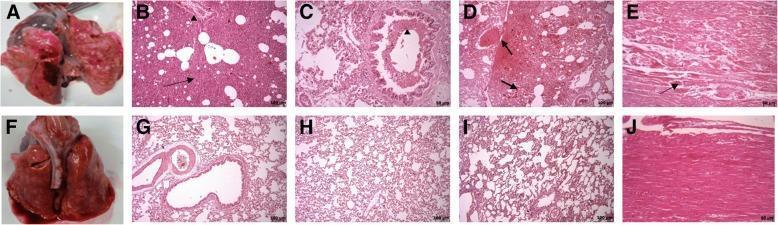
A comparison of macroscopic and histopathological findings in an animal model of acute respiratory distress syndrome (ARDS) across control (A-E) and treatment (F-J) groups. Macroscopic images A and F show excised lung tissue; the control lung (A) displays severe hyperemia, hemorrhage, and edema, while the treatment lung (F) shows minimal hyperemia. Light microscopy images (H&E stain) further illustrate pathological differences. In the control group, lung sections (B, C, D) reveal severe interstitial pneumonia, extensive inflammatory cell infiltration (arrowheads), and intra-alveolar hemorrhage (arrows). Cardiac section E shows localized myofibrillar necrosis (arrow). Conversely, the treatment group (G, H, I) demonstrates significant preservation of lung architecture with reduced cellular infiltration and approximately normal alveolar septa. The cardiac section (J) shows healthy myocardium without necrosis. This visual serves as a comparative study on the efficacy of therapeutic interventions, such as mesenchymal stem cells, in mitigating organ damage associated with inflammatory lung injury.
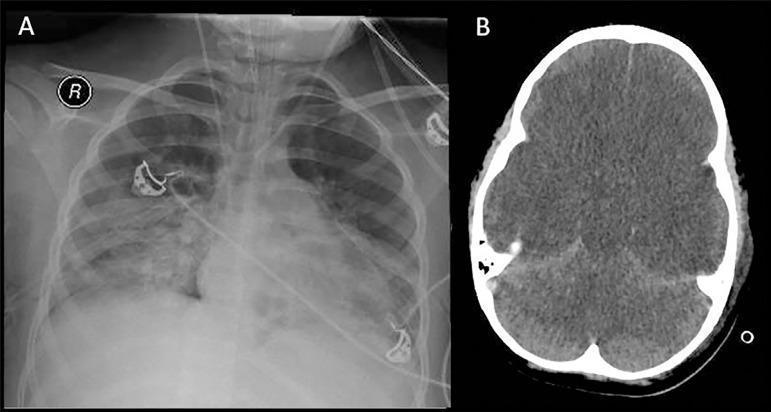
This composite clinical imaging figure illustrates critical multi-organ involvement in a pediatric patient with septic shock. Panel A is an anteroposterior chest radiograph showing diffuse, bilateral pulmonary infiltrates. The lung fields exhibit extensive hazy opacities that obscure normal vascular markings, consistent with Acute Respiratory Distress Syndrome (ARDS). Medical support devices, including an endotracheal tube and ECG leads, are visible. Panel B is an axial non-contrast brain computed tomography (CT) scan demonstrating severe, diffuse cerebral edema. Key diagnostic features include the loss of normal gray-white matter differentiation and significant effacement of the cerebral sulci and ventricular system. These findings indicate global hypoxic-ischemic injury and increased intracranial pressure. Together, the images provide a visual representation of the systemic complications of severe sepsis and hypoxemia, frequently encountered in critical care medicine and neurology when assessing for brain death.
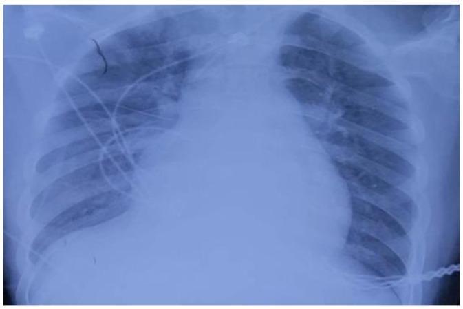
Anterior-posterior (AP) bedside chest radiograph demonstrating significant cardiopulmonary pathology. The cardiac silhouette is markedly enlarged, suggesting cardiomegaly or a pericardial effusion. The lung fields show extensive bilateral opacities, most prominent in the right lung, characterized by a ground-glass appearance and exudative changes consistent with acute pulmonary edema, diffuse alveolar disease, or pneumonia. There is an overall increase in bronchovascular markings and lung texture bilaterally. Several medical devices are visible, including ECG leads and what appears to be an endotracheal tube and central venous catheter, indicating intensive care monitoring. The costophrenic angles are partially obscured by overlying opacities. This image illustrates critical findings often associated with acute respiratory distress syndrome (ARDS) or cardiogenic shock in a clinical setting.
septic shock pathogenesis diagram mediators DIC organ failure
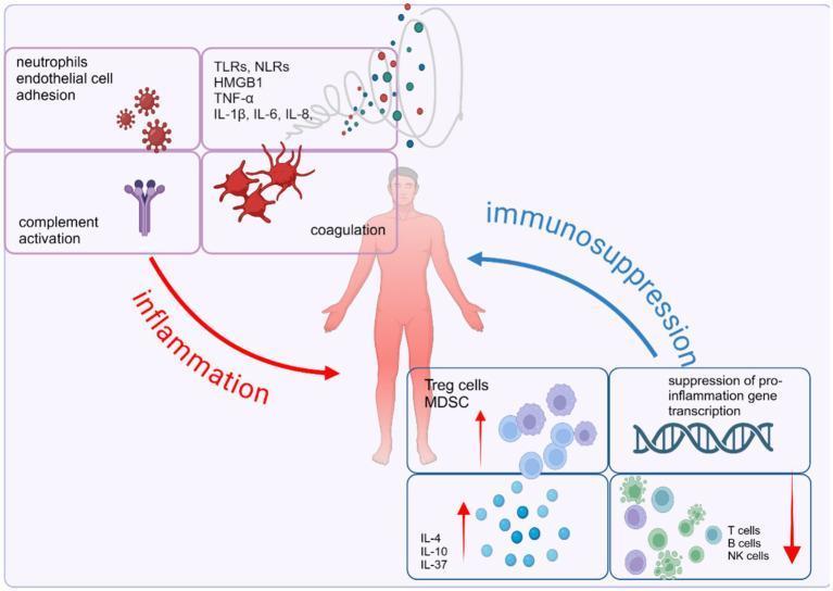
This pathophysiology diagram illustrates the dysregulated host immune response in sepsis, characterized by an imbalance between concurrent pro-inflammatory and anti-inflammatory pathways. Centered on a human figure, the left side (red 'inflammation' arrow) details the acute systemic inflammatory response syndrome (SIRS), featuring neutrophil-endothelial cell adhesion, complement activation, and coagulation cascades. A molecular cloud identifies key mediators including DAMPs/PAMPs (HMGB1, TLRs, NLRs) and pro-inflammatory cytokines (TNF-α, IL-1β, IL-6, IL-8). Conversely, the right side (blue 'immunosuppression' arrow) represents the compensatory anti-inflammatory response syndrome (CARS). This includes the expansion of suppressor cells (Treg cells, MDSCs) and anti-inflammatory cytokines (IL-4, IL-10, IL-37), the suppression of pro-inflammatory gene transcription, and the depletion or exhaustion of effector cells (T cells, B cells, and NK cells). The diagram serves as an educational tool for understanding the complex signaling pathways and cellular interactions that drive multi-organ dysfunction and immune homeostasis failure during septic shock.
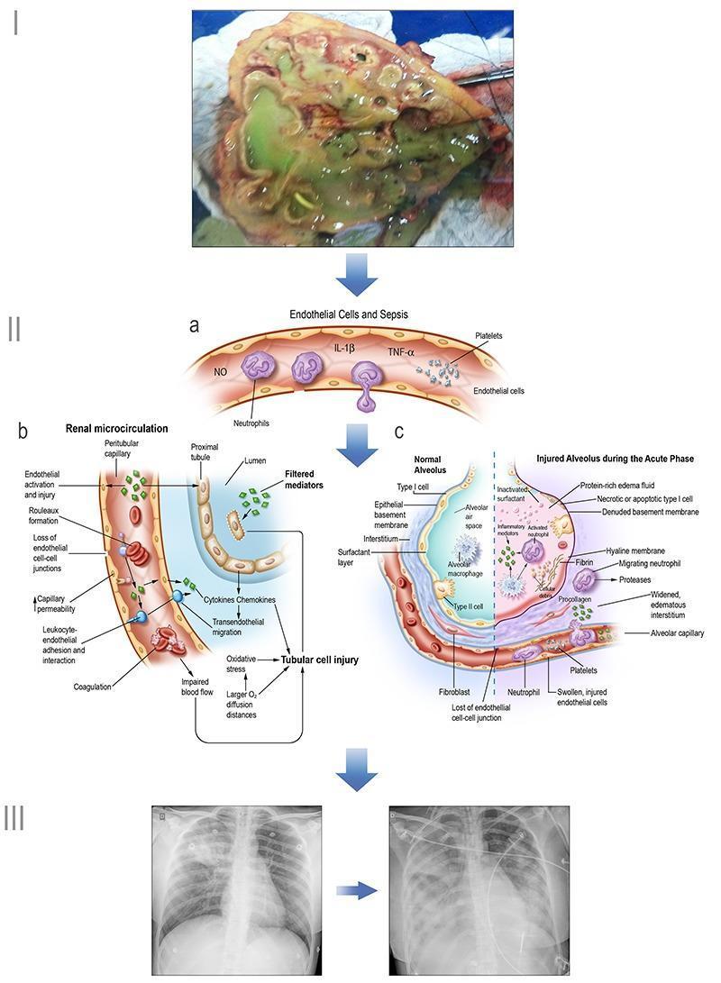
Educational infographic illustrating the pathogenesis of sepsis and multi-organ dysfunction. Section I shows a clinical photograph of a necrotic kidney (pyelonephritis). Section II contains three pathophysiology diagrams: (a) General endothelial changes in sepsis, depicting neutrophil extravasation, platelet activation, and release of inflammatory mediators like TNF-α, IL-1β, and NO; (b) Renal microcirculation injury, highlighting peritubular capillary changes (rouleaux formation, leukocyte adhesion, increased permeability) and subsequent tubular cell injury via oxidative stress and impaired blood flow; (c) Alveolar-capillary comparative diagram, contrasting a normal alveolus with one in the acute phase of ARDS, showing hyaline membrane formation, protein-rich edema, and Type I cell necrosis. Section III features diagnostic chest X-rays demonstrating progressive bilateral opacities consistent with acute respiratory distress syndrome. This visual summary correlates clinical findings, diagnostic imaging (radiography), and microscopic pathophysiology to explain systemic sepsis-induced organ failure.
SHOCK AND MULTIPLE ORGAN DYSFUNCTION SYNDROME (MODS)
From a Pathological Perspective
1. Definition of Shock
- Robbins, Cotran & Kumar - Pathologic Basis of Disease
2. Classification of Shock
| Type | Clinical Examples | Principal Pathogenic Mechanisms |
|---|---|---|
| Cardiogenic | Myocardial infarction, ventricular rupture, arrhythmia, cardiac tamponade, pulmonary embolism | Failure of myocardial pump - intrinsic damage, extrinsic compression, or outflow obstruction |
| Hypovolemic | Hemorrhage, vomiting, diarrhea, burns, trauma | Inadequate blood or plasma volume |
| Septic | Gram-positive/negative bacterial sepsis, fungal sepsis, superantigens (toxic shock syndrome) | Peripheral vasodilation and blood pooling; endothelial activation/injury; DIC; cytokine cascades |
| Neurogenic | Spinal cord injury, anesthesia | Acute vasodilation → hypotension |
| Anaphylactic | IgE-mediated hypersensitivity | Systemic vasodilation, increased vascular permeability |
3. Pathogenesis of Septic Shock (Detailed)
Step 1 - Microbial Activation of Innate Immunity
- Toll-like receptors (TLRs): Recognize PAMPs (pathogen-associated molecular patterns)
- G-protein-coupled receptors: Detect bacterial formyl peptides
- C-type lectin receptors (dectins): Recognize fungal cell wall components
Step 2 - Pro-Inflammatory and Counter-Inflammatory Responses
- SIRS (Systemic Inflammatory Response Syndrome): The proinflammatory surge causes vasodilation, vascular leakage, endothelial injury
- With time, counterregulatory CARS (Compensatory Anti-inflammatory Response Syndrome) kicks in, mediated by IL-10, TGF-β, expanded Treg cells, and MDSC (myeloid-derived suppressor cells)
- If CARS becomes excessive → immunosuppression → secondary infections → perpetuates organ failure
Step 3 - Endothelial Activation and Injury
- Vascular leakage: Inflammatory cytokines loosen endothelial tight junctions → protein-rich edema throughout the body → impedes nutrient delivery and waste removal
- Increased NO production: Activated endothelium produces NO and other vasoactive mediators (C3a, C5a, PAF) → smooth muscle relaxation → systemic hypotension
- Microvascular dysfunction: Loss of normal autoregulation; increased capillaries with intermittent/heterogeneous flow → oxygen supply-demand mismatch
Step 4 - Procoagulant State / DIC
- Proinflammatory cytokines increase tissue factor (TF) production by monocytes and endothelium
- Decrease endothelial anticoagulant factors: thrombomodulin, protein C, TFPI (tissue factor pathway inhibitor)
- Increase PAI-1 (plasminogen activator inhibitor) → dampens fibrinolysis
- Neutrophil Extracellular Traps (NETs): Stimulate both intrinsic and extrinsic coagulation pathways
- Vascular stasis + decreased washout of activated factors → systemic thrombin activation → fibrin-rich microthrombi in small vessels throughout organs
- In 50% of septic patients: full-blown DIC with consumption coagulopathy → paradoxical bleeding
Step 5 - Metabolic Abnormalities
- Insulin resistance and hyperglycemia: TNF, IL-1, glucagon, cortisol, catecholamines → gluconeogenesis; proinflammatory cytokines suppress insulin release and impair GLUT-4 expression
- Hyperglycemia → impairs neutrophil function, increases endothelial adhesion molecule expression
- Adrenal dysfunction: Initial surge in cortisol → followed by functional adrenocortical insufficiency; may develop frank adrenal necrosis from DIC (Waterhouse-Friderichsen syndrome)
- Lactic acidosis: Cellular hypoxia → aerobic respiration replaced by anaerobic glycolysis → lactate accumulation → metabolic acidosis → arteriolar dilation → more blood pooling
Step 6 - Organ Dysfunction
- High cytokine levels directly diminish myocardial contractility
- Increased vascular permeability → ARDS (shock lung)
- Ischemic renal tubular injury
- Gut mucosal barrier breakdown → bacterial translocation → perpetuates sepsis
4. Stages of Shock
Stage 1 - Nonprogressive (Compensated) Stage
- Reflex compensatory mechanisms activated; vital organ perfusion maintained
- Mechanisms: baroreceptor reflexes, catecholamine release, ADH release, RAAS activation, generalized sympathetic stimulation
- Net effects: tachycardia, peripheral vasoconstriction, renal fluid conservation
- Cutaneous vasoconstriction → cool, clammy, pale skin ("shocky" skin); septic shock early = warm, flushed skin
- Coronary and cerebral vessels relatively preserved (less sensitive to sympathetic stimulation) → blood shunted to heart and brain
Stage 2 - Progressive (Decompensated) Stage
- Compensatory mechanisms overwhelmed; widespread tissue hypoxia
- Aerobic respiration replaced by anaerobic glycolysis → excessive lactic acid production
- Metabolic lactic acidosis → blunts vasomotor response → arteriolar dilation → blood pools in microcirculation
- Peripheral pooling worsens cardiac output further
- Endothelial anoxic injury → DIC risk
- Vital organs begin to fail
Stage 3 - Irreversible Stage
- Widespread cellular injury beyond repair
- Lysosomal enzyme leakage further aggravates shock state
- Myocardial contractility worsens (increased NO synthesis)
- Ischemic bowel → intestinal bacterial translocation → superimposed bacteremia
- Renal failure from ischemic tubular injury
- Despite best therapeutic interventions → death
5. Morphological (Pathological) Changes in Shock
"The cellular and tissue effects of shock are essentially those of hypoxic injury and are caused by a combination of hypoperfusion and microvascular thrombosis."
- Robbins, Cotran & Kumar Pathologic Basis of Disease
A. Brain - Ischemic/Hypoxic Encephalopathy
- Gross: Cerebral edema, loss of gray-white matter differentiation
- Microscopic: Ischemic neuronal change (pyknotic shrunken neurons with eosinophilic cytoplasm, "red dead neurons"); selective vulnerability of CA1 hippocampal neurons, Purkinje cells
- Spectrum: Mild cognitive changes → coma → brain death
- Neurons and cardiomyocytes are the most susceptible cells and cannot regenerate - unlike other organs
B. Heart - Myocardial Depression
- Subendocardial hemorrhage and necrosis (from ischemia due to hypoperfusion)
- Contraction band necrosis: Hypercontraction of myofibrils with eosinophilic transverse bands; classic in catecholamine-driven injury (neurogenic shock, anaphylaxis, reperfusion injury)
- Diffuse petechial hemorrhages in epicardium/endocardium
- Myocardial function is directly suppressed by cytokines (TNF, IL-1, NO) in septic shock
C. Kidney - Acute Tubular Necrosis (ATN) / Acute Kidney Injury (AKI)
- Gross: Swollen pale kidneys; cortex pale, medulla congested/dark
- Microscopic:
- Proximal tubular epithelial cell injury: Flattening, loss of brush border, nuclear pyknosis, tubular dilation
- ATN (Acute Tubular Necrosis): Frank necrosis of tubular epithelial cells; most severe in straight portion (S3 segment) of proximal tubule and ascending thick limb of loop of Henle (areas of highest metabolic demand and lowest oxygen tension)
- Intraluminal granular casts (shed epithelial cells + Tamm-Horsfall protein)
- Pigmented casts in myoglobinuric/hemoglobinuric ATN
- Glomerular fibrin microthrombi - hallmark of DIC in shock (most visible here)
- Outcome: Oliguria → anuria → if tubular basement membrane intact → tubular regeneration within 10-14 days (reversible)
D. Lung - Diffuse Alveolar Damage (DAD) / ARDS ("Shock Lung")
- Lungs are relatively resistant to ischemia in pure hypovolemic shock
- Sepsis or trauma → "shock lung" → DAD (Diffuse Alveolar Damage) = pathological substrate of ARDS
| Phase | Timing | Pathology |
|---|---|---|
| Exudative (Acute) | Day 1-7 | Alveolar edema, fibrin exudates, hyaline membrane formation (acellular eosinophilic deposits lining alveoli), Type I pneumocyte necrosis, intra-alveolar hemorrhage, neutrophil infiltration |
| Proliferative | Day 7-21 | Type II pneumocyte hyperplasia (attempt at repair), fibroblast proliferation, interstitial thickening |
| Fibrotic | >21 days | Interstitial and intra-alveolar fibrosis; honeycombing in severe cases |
- Gross: Heavy ("beefy"), congested, consolidated lungs; non-collapsing on opening pleural cavity
- Microscopic (Acute): Hyaline membranes, alveolar wall thickening, Type I cell necrosis (compared to normal alveolus showing aerated spaces with thin walls)
E. Liver - Centrilobular Necrosis / Shock Hepatitis ("Ischemic Hepatitis")
- Centrilobular (zone 3) is most vulnerable - most distal from hepatic artery supply (relative hypoxia even normally)
- Gross: Nutmeg liver (centrilobular congestion and necrosis alternating with pale periportal areas)
- Microscopic: Centrilobular hepatocyte necrosis, sinusoidal congestion, fatty change; periportal hepatocytes (zone 1) relatively preserved
- Kupffer cell activation in sepsis → release of inflammatory mediators → systemic amplification of MODS
- Lab: Markedly elevated transaminases ("shock liver"), elevated bilirubin, prolonged PT
F. Gastrointestinal Tract - Mucosal Ischemia and Bacterial Translocation
- Splanchnic circulation is the first sacrificed in shock (vasoconstrictive homeostasis)
- Gross: Mucosal erosions, superficial ulcerations ("stress ulcers"), hemorrhagic gastroenteropathy
- Microscopic: Mucosal epithelial cell apoptosis and necrosis; villus tip ischemia (small intestine); loss of mucus layer
- Critical consequence: Disruption of mucosal barrier → bacterial translocation → gram-negative bacteria and endotoxin enter portal/systemic circulation → amplifies sepsis → perpetuates MODS
- Splanchnic hypoperfusion + reperfusion → activates neutrophils and macrophages → key initiator of MODS
G. Adrenal Glands - Lipid Depletion ± Necrosis
- Adrenal cortical cell lipid depletion: Reflects increased use of stored lipids for cortisol synthesis ("stress response") - seen in all forms of shock
- In overwhelming sepsis/DIC: Bilateral adrenal hemorrhage and necrosis (Waterhouse-Friderichsen syndrome) - classically associated with meningococcemia
- Gross: Hemorrhagic adrenals; dark red/brown
- Results in relative adrenal insufficiency → worsens hypotension
H. Pancreas
- Acinar cell injury from ischemia → activates pancreatic enzymes → contributes to SIRS
- Pancreatitis itself can trigger SIRS → MODS (a major initiating mechanism in surgical patients)
6. Multiple Organ Dysfunction Syndrome (MODS)
Definition
- Maingot's Abdominal Operations
Types
| Type | Description |
|---|---|
| Primary MODS | Organ dysfunction results directly from a well-defined insult (e.g., crush injury directly causing renal failure) |
| Secondary MODS | Organ dysfunction occurring as a consequence of the host response (sepsis, SIRS) rather than the initial injury itself |
| Transient MODS | Organ dysfunction resolving within 48 hours with supportive treatment |
| Persistent/Chronic Critical Illness (CCI) | Prolonged ICU course with persistent multi-organ dysfunction |
Pathogenesis of MODS - The "Two-Hit" Model
Key Pathogenic Mechanisms
-
Splanchnic and pulmonary microcirculation as epicenters:
- Splanchnic hypoperfusion → gut mucosal injury → bacterial translocation → portal delivery of endotoxin to liver → Kupffer cell activation → systemic inflammatory mediator release → distant organ injury
- Gut is the "motor of MODS"
-
Neutrophil priming and activation:
- Primed neutrophils sequester in lung (most common first organ), liver, and other capillary beds
- Release ROS, proteases, NETs → microvascular endothelial injury
-
Endothelial injury (pan-endotheliopathy):
- Glycocalyx degradation
- Tight junction disruption → edema
- Loss of anticoagulant properties → microthrombi
-
Mitochondrial dysfunction:
- Cytopathic hypoxia: cells cannot utilize delivered oxygen due to mitochondrial injury from ROS and NO → organ failure even in presence of adequate oxygen delivery
-
Ischemia-Reperfusion Injury:
- Reperfusion of ischemic organs paradoxically worsens injury via burst of ROS from xanthine oxidase → free radical cascade
-
Immunosuppression phase (CARS):
- Exhaustion of immune effectors → lymphocyte apoptosis, anergy
- Increased susceptibility to nosocomial infections → secondary sepsis → perpetuates MODS
Sequence of Organ Failure in MODS
- Lung (ARDS) - first and most frequent; usually within hours to days
- Kidney (AKI/ATN) - second
- Liver (ischemic hepatitis) - third
- GI tract - mucosal failure, stress ulcers
- Coagulation (DIC) - consumptive coagulopathy
- Cardiovascular - myocardial depression, refractory hypotension
- Brain - encephalopathy, last to fail clinically
- Maingot's Abdominal Operations, p. 1051
Scoring Systems for MODS
| Scoring System | Description |
|---|---|
| APACHE II/III | Acute Physiology and Chronic Health Evaluation; general physiologic severity |
| SOFA (Sequential Organ Failure Assessment) | Grades 6 organ systems (respiratory, coagulation, liver, cardiovascular, CNS, renal) 0-4; score >2 = organ dysfunction; best MODS-specific tool |
| MODS Score (Marshall) | Describes organ dysfunction as a clinical outcome descriptor |
| LODS (Logistic Organ Dysfunction) | Ordinal scoring of organ dysfunction |
| SAPS (Simplified Acute Physiology Score) | Simplified general severity scoring |
7. Summary - Organ Pathology at a Glance
| Organ | Gross Changes | Microscopic Changes | Key Feature |
|---|---|---|---|
| Brain | Edema, swelling | Ischemic neurons (shrunken, eosinophilic) | Red dead neurons; CA1 hippocampus vulnerable |
| Heart | Petechial hemorrhages | Contraction band necrosis, subendocardial necrosis | Catecholamine excess |
| Kidney | Swollen, pale cortex | ATN - tubular cell necrosis, casts; glomerular fibrin thrombi | Oliguria → anuria; reversible if BM intact |
| Lung | Heavy, consolidated ("shock lung") | DAD: hyaline membranes, Type I cell necrosis, alveolar edema | Exudative → proliferative → fibrotic |
| Liver | Nutmeg liver | Centrilobular (zone 3) necrosis, sinusoidal congestion | Kupffer cells amplify MODS |
| GI tract | Mucosal erosions, stress ulcers | Villus tip necrosis, mucosal loss | Bacterial translocation - "motor of MODS" |
| Adrenal | Lipid depletion ± hemorrhage | Cortical cell lipid depletion; Waterhouse-Friderichsen (DIC) | Relative adrenal insufficiency |
| Pancreas | Edema, necrosis | Acinar cell injury | Initiates SIRS if primary organ |
8. Special Note - DIC in Shock
- Triggering factors: Endotoxin, TNF, IL-1 → tissue factor expression → thrombin generation → fibrin microthrombi
- Consequences: Microvascular occlusion → ischemic organ injury + consumption of clotting factors/platelets → paradoxical hemorrhage
- Pathology: Fibrin thrombi most visible in glomerular capillaries, adrenal microvasculature, hepatic sinusoids
- DIC is the pathological link between septic shock and multi-organ ischemia
9. Clinical Correlation
| Parameter | Hypovolemic | Cardiogenic | Septic (Early) | Septic (Late) |
|---|---|---|---|---|
| Skin | Cold, clammy | Cold, clammy, mottled | Warm, flushed | Cold, mottled |
| HR | ↑ | ↑ | ↑ | ↑ |
| BP | ↓ | ↓ | Normal/↓ | ↓↓ |
| CO | ↓ | ↓↓ | ↑ | ↓ |
| SVR | ↑ | ↑ | ↓↓ | Variable |
| CVP/PCWP | ↓ | ↑ | ↓ | Variable |
- Young healthy patients with hypovolemic shock: >90% survival with appropriate management
- Septic or cardiogenic shock: Substantially poorer outcomes even with optimal care
- Robbins, Cotran & Kumar - Pathologic Basis of Disease, p. 137
- Robbins, Cotran & Kumar - Pathologic Basis of Disease (pp. 134-137) - definitions, types, pathogenesis, morphology, clinical features
- Robbins & Kumar - Basic Pathology (pp. 96-100) - septic shock mechanism, DIC, stages
- Maingot's Abdominal Operations (p. 1051-1064) - MODS definition, scoring, sequence of organ failure
- Mulholland & Greenfield's Surgery 7e - MODS pathogenesis, splanchnic circulation, CCI