
Liver cirrhosis
liver cirrhosis histology fibrosis nodules
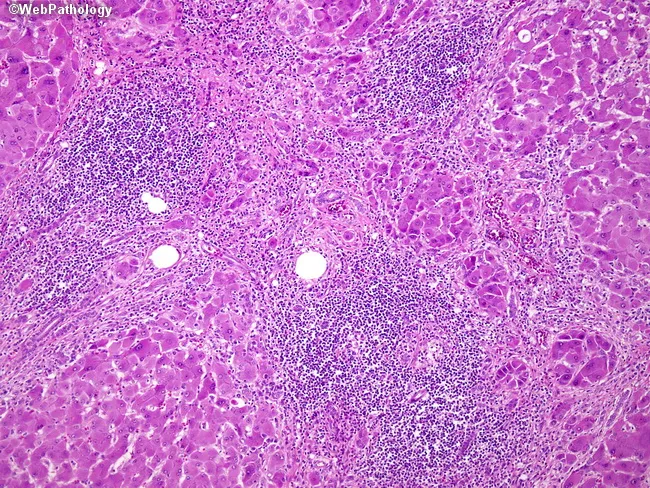
Imaging modality: Light microscopy of a liver biopsy stained with hematoxylin and eosin. The histologic specimen demonstrates established cirrhosis with nodular regeneration. The hepatic parenchyma is reorganized into regenerative nodules encircled by dense fibrous septa, reflecting bridging fibrosis. Nodules are predominantly micronodular (<3 mm) in classic early chronic hepatitis B–related cirrhosis, though macronodular (>3 mm) foci may be present as disease progresses. The nodules are composed of regenerating hepatocytes arranged in thickened cords with disrupted lobular architecture; surrounding scar tissue contains portal-type inflammatory infiltrates and dense mononuclear cell aggregates. A notable ductular reaction is evident, with proliferation of small bile ductules at the periphery of fibrous bands. The portal tracts show chronic inflammatory cells, including lymphocytes and plasma cells, and interface activity may be seen. Overall the image captures the hallmark features of cirrhosis: irregular nodularity, parenchymal extinction, and fibrotic remodeling driven by chronic inflammatory insult from hepatitis B infection. Clinically, this histology confirms cirrhosis, supports staging (nodular pattern and septal fibrosis), and informs prognosis and antiviral management. Potential differential diagnoses include alcoholic cirrhosis and nonalcoholic steatohepatitis with cirrhosis, which can mimic nodular architecture but differ in etiologies and associated features.
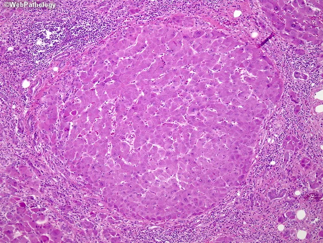
This liver biopsy histology, stained with Hematoxylin and Eosin, is presented at low power to emphasize cirrhotic nodularity produced by chronic hepatitis B infection. The hepatic parenchyma shows regenerative nodules separated by fibrous septa, creating a mosaic architecture typical of cirrhosis. Nodules are micronodular (<3 mm) and macronodular (>3 mm), reflecting different stages of fibrogenesis and nodular regeneration. Within the nodules, hepatocytes form plates and cords with preserved central venules in some regions, while surrounding fibrous tissue contains chronic inflammatory infiltrates and a ductular reaction indicating ongoing bile ductular proliferation. The interface between nodules and septa is irregular, with disruption of normal lobular architecture and portal tract remodeling. Staining highlights eosinophilic cytoplasm of regenerating hepatocytes, occasional binucleation, and prominent vascular channels within expanded fibrous stroma. Overall, the image supports a diagnosis of cirrhosis secondary to chronic hepatitis B, with implications for portal hypertension, hepatic insufficiency risk, and hepatocellular carcinoma surveillance. This histologic pattern informs fibrosis staging (e.g., advanced F4) and guides clinical management, including antiviral therapy optimization, risk stratification, transplant evaluation, and prognosis in HBV-related liver disease. Correlative clinical data, laboratory markers, and imaging are recommended to complete the assessment and monitor response to therapy over time.
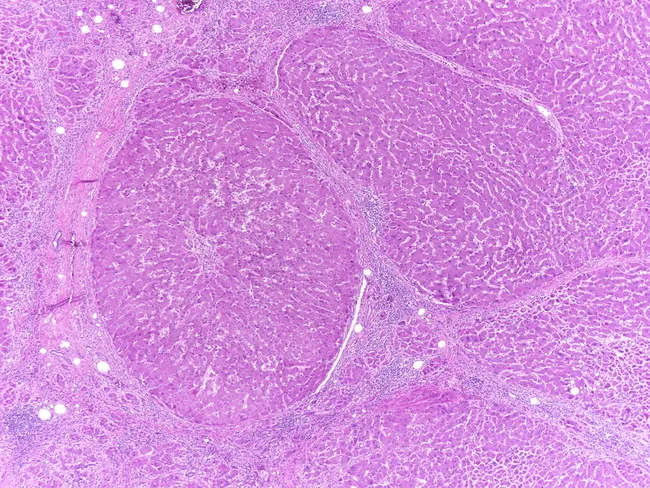
Histology image of cirrhotic liver tissue stained with hematoxylin and eosin (H&E). The hepatic architecture is markedly disrupted by broad fibrous septa bridging across regenerating nodules. The nodular parenchyma comprises thickened plates of hepatocytes organized into irregular lobules, with islands of regenerated hepatocytes separated by dense stromal bands. The nodules appear macronodular, with diameters exceeding the 3-millimeter threshold, corresponding to macronodular cirrhosis; features of micronodular cirrhosis, such as small regenerative nodules, are less evident here. Chronic inflammatory cell infiltrates are present within the fibrous septa and portal tracts, consistent with a long-standing inflammatory process. Vascular remodeling and porto-systemic shunting may be suggested by bridging fibrosis and nodular regeneration. In chronic hepatitis B infection, hepatocytes often show ballooning degeneration and occasional Councilman-like necrosis, though the hallmark is the regenerative nodularity and bridging fibrosis rather than overt tumor. Overall, this image demonstrates the morphological hallmarks of cirrhosis: altered lobular architecture, regenerating nodules, fibrous septa, and chronic inflammatory infiltration. Clinically, such histology correlates with portal hypertension risk, impaired synthetic function, and elevated hepatocellular carcinoma risk in chronic HBV carriers. This slide provides a robust reference for teaching nodularity classification and cirrhosis staging from a histopathologic perspective.
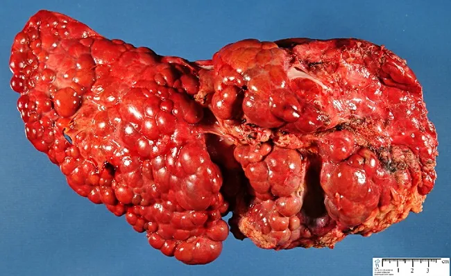
Gross pathology photograph of the liver showing advanced cirrhosis with coarse, nodular surface. The specimen demonstrates macronodular cirrhosis, in which regenerative nodules exceed 3 mm in diameter, a histologic and macroscopic hallmark of fibrosis and architectural disarray associated with chronic tyrosinemia type I. The context is hereditary infantile tyrosinemia due to fumarylacetoacetate hydrolase deficiency, a metabolic liver disease that progresses to cirrhotic remodeling and risk of hepatocellular carcinoma if untreated. The hepatic nodularity and firm consistency reflect extensive parenchymal scarring with bridging fibrous septa linking nodules. Color is a deep reddish-brown, with variable areas of congested or mottled parenchyma; surface nodularity may obscure normal lobular anatomy. This image provides a macroscopic correlate to the cellular and architectural changes described in histology, including regenerative nodules embedded in dense fibrous tissue. Clinically, such findings support severe liver disease in tyrosinemia and justify monitoring for portal hypertension and hepatocellular carcinoma risk. In educational and research contexts, this photograph is valuable for teaching gross hepatic morphology, cirrhotic evolution in metabolic disorders, and the distinction between macronodular vs micronodular cirrhosis. Potential uses include medical education, pathology slide correlation, and diagnostic radiology-pathology correlation studies. These features reinforce differential considerations for cirrhosis etiologies in pediatrics worldwide today.
Liver Cirrhosis
Definition
Etiology
| Category | Specific Causes |
|---|---|
| Major (>90% of cases) | Alcoholic liver disease, Non-alcoholic fatty liver disease (NAFLD/MASLD), Chronic hepatitis B |
| Other viral | Chronic hepatitis C |
| Cholestatic/Autoimmune | Primary biliary cirrhosis (PBC), Primary sclerosing cholangitis (PSC), Autoimmune hepatitis |
| Metabolic/Genetic | Hemochromatosis, Wilson disease, α1-antitrypsin deficiency, Glycogen storage diseases |
| Vascular/Outflow obstruction | Budd-Chiari syndrome, Veno-occlusive disease, Right-sided heart failure |
| Drugs and toxins | Methotrexate, amiodarone, others |
| Mechanical | Biliary atresia, Cystic fibrosis |
| Cryptogenic | No identifiable cause |
- Goldman-Cecil Medicine, Table 139-1, p. 1615
Pathogenesis
Hepatic Stellate Cell Activation (Central Mechanism)
-
Lose their vitamin A stores
-
Proliferate and develop prominent rough endoplasmic reticulum
-
Secrete extracellular matrix: collagen types I and III, sulfated proteoglycans, and glycoproteins
-
Transform into contractile myofibroblasts
-
Goldman-Cecil Medicine, p. 1614
Sinusoidal Changes
Reversibility
Morphology
Gross
- The liver surface shows irregular nodularity with broad fibrous scars separating bulging regenerative nodules
- Orange-tawny color, firm consistency
Microscopic
- Micronodular cirrhosis (<3 mm nodules): typical of early alcoholic or hemochromatosis-related cirrhosis
- Macronodular cirrhosis (>3 mm nodules): typical of post-viral cirrhosis; higher HCC risk
- Ductular reactions: proliferating small bile ductules at the periphery of fibrous bands, increasing with disease progression
Complications
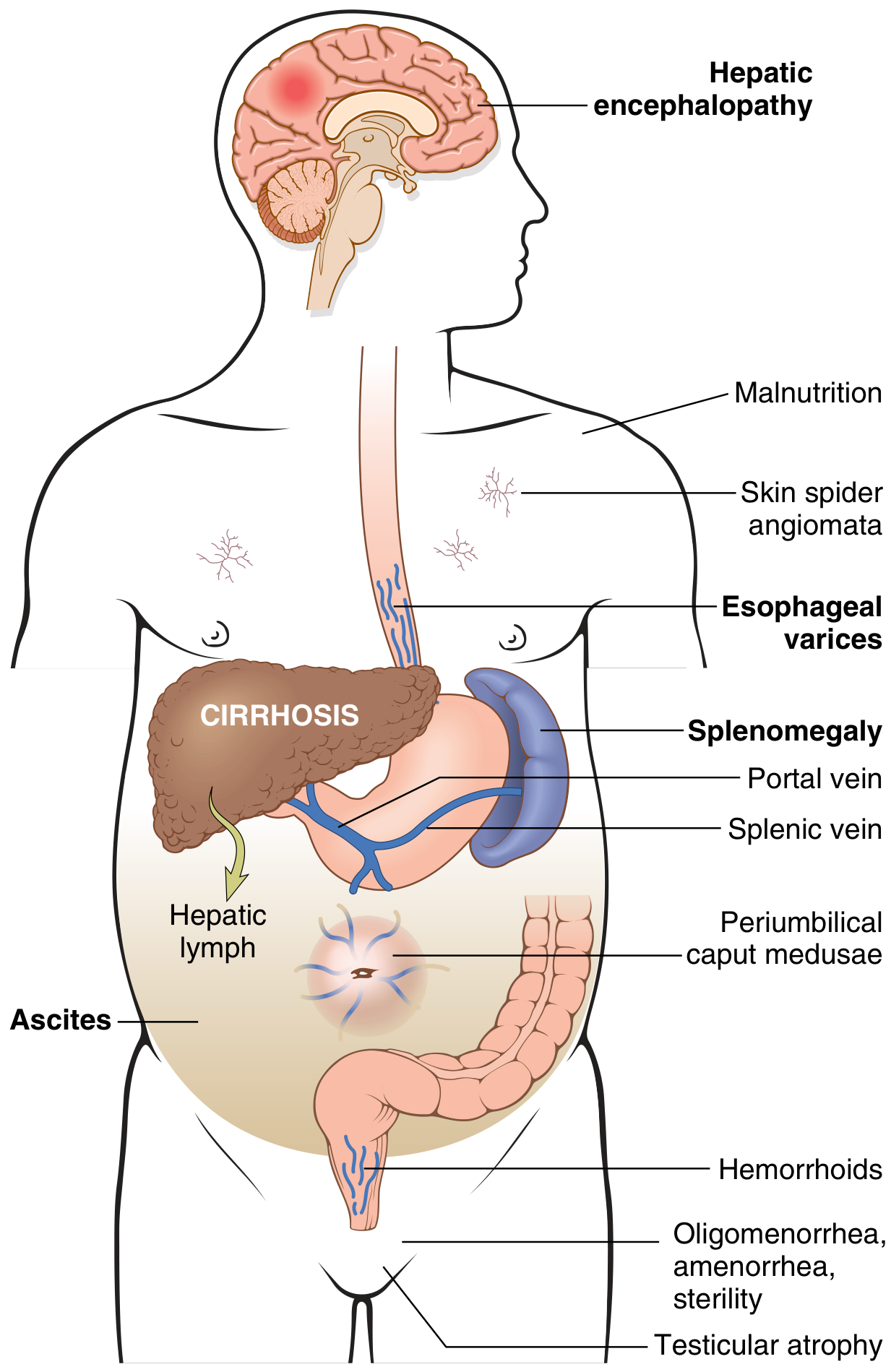
1. Portal Hypertension
- Increased sinusoidal resistance: (a) Fixed component - fibrosis and nodule compression; (b) Functional component - active vasoconstriction from reduced intrahepatic NO and enhanced vasoconstrictors
- Increased portal venous inflow: due to splanchnic arteriolar vasodilation (mediated by increased NO and other vasodilators in the splanchnic circulation)
2. Esophagogastric Varices
- Develop in ~40% of patients with advanced cirrhosis
- Occur as portosystemic collaterals wherever portal and systemic circulations share capillary beds
- Coronary vein reversal is an early event
- May cause massive, fatal hematemesis especially with concurrent coagulopathy
3. Ascites
- Peritoneal fluid accumulation; ~85% of cases are due to portal hypertension from cirrhosis
- Transudate: protein <3 g/dL; serum-to-ascites albumin gradient (SAAG) ≥1.1 g/dL
- Management: sodium restriction + diuretics (spironolactone, furosemide); refractory ascites (10% of patients) treated with large-volume paracentesis or TIPS
4. Spontaneous Bacterial Peritonitis (SBP)
- Infection of ascitic fluid without an apparent cause
- Serious, life-threatening complication requiring prompt antibiotic therapy
5. Hepatic Encephalopathy (HE)
- Results from both portal hypertension (portosystemic shunting of nitrogenous compounds) AND liver insufficiency
- Ranges from subtle cognitive changes to coma (grade I-IV)
6. Hepatorenal Syndrome (HRS)
- Functional renal failure in advanced cirrhosis
- Results from extreme splanchnic vasodilation leading to renal vasoconstriction
- Type 1 HRS: rapidly progressive (creatinine doubling in <2 weeks); poor prognosis
- Type 2 HRS: slower progression, associated with refractory ascites
7. Splenomegaly and Hypersplenism
- Congestive splenomegaly with splenic weight up to 1000 g
- Leads to thrombocytopenia and pancytopenia from splenic sequestration
8. Coagulopathy and Jaundice
- Impaired synthesis of clotting factors (I, II, V, VII, IX, X)
- Jaundice from failure to excrete bilirubin; chronic severe jaundice causes pruritus
9. Hyperestrogenemia (in males)
- Palmer erythema (local vasodilation)
- Spider angiomas
- Gynecomastia
- Testicular atrophy
- Oligomenorrhea/amenorrhea in women
10. Cirrhotic Cardiomyopathy
- High-output cardiac failure with decreased peripheral oxygen utilization
- End result of the hyperdynamic circulatory state
11. Hepatopulmonary Syndrome (HPS) vs. Portopulmonary Hypertension
- HPS: Pulmonary capillary dilation (up to 500 μm vs. normal 8 μm) → arterial hypoxemia (right-to-left shunt equivalent); hallmark is platypnea-orthodeoxia
- Portopulmonary HTN: Vasoconstrictive substances bypass hepatic metabolism → endothelial proliferation → irreversible pulmonary hypertension
12. Hepatocellular Carcinoma (HCC)
- Can develop at any stage of cirrhosis, not just decompensated
- Cirrhosis is the single greatest risk factor for HCC
- Requires 6-monthly ultrasound surveillance ± AFP
Clinical Stages and Natural History
| Stage | Characteristics | Median Survival |
|---|---|---|
| Compensated | Asymptomatic or mild symptoms; normal synthetic function; portal pressure below variceal threshold | >12 years |
| Decompensated | Ascites, variceal hemorrhage, encephalopathy, or jaundice | ~1.6 years |
Diagnosis and Assessment
Clinical Signs
- Jaundice, spider angiomas, palmar erythema
- Leukonychia, clubbing, Dupuytren's contracture
- Muscle wasting (sarcopenia), ascites, caput medusae
- Splenomegaly, gynecomastia, testicular atrophy, asterixis
Laboratory Findings
- Elevated bilirubin, AST, ALT, GGT, ALP
- Decreased albumin, prolonged PT/INR
- Thrombocytopenia (hypersplenism)
- Elevated creatinine in HRS
Imaging
- Ultrasound: nodular liver surface, splenomegaly, ascites, portal vein flow
- Elastography (FibroScan): non-invasive fibrosis staging
- CT/MRI: HCC surveillance, portal vein thrombosis, morphology
Liver Biopsy
- Gold standard for staging fibrosis (METAVIR: F0-F4; F4 = cirrhosis)
Prognostic Scoring
| Parameter | 1 point | 2 points | 3 points |
|---|---|---|---|
| Bilirubin (mg/dL) | <2 | 2-3 | >3 |
| Albumin (g/dL) | >3.5 | 2.8-3.5 | <2.8 |
| PT prolongation (s) | <4 | 4-6 | >6 |
| Ascites | None | Controlled | Refractory |
| Encephalopathy | None | Grade 1-2 | Grade 3-4 |
- Class A (5-6): well-compensated; Class B (7-9): significant compromise; Class C (10-15): decompensated
-
Incorporates: serum bilirubin, creatinine, INR
-
MELD-Na adds sodium for improved accuracy
-
Better validated for predicting short-term mortality and transplant prioritization than Child-Pugh
-
Harrison's Principles of Internal Medicine 22E, p. cited; Tietz Textbook of Laboratory Medicine, p. cited
Management
Treat the Underlying Cause
- Alcohol-related: strict abstinence (reversibility demonstrated histologically)
- Hepatitis C: Direct-acting antivirals (DAAs) - even patients with decompensated cirrhosis can be treated
- Hepatitis B: Antiviral therapy (tenofovir, entecavir)
- NAFLD/MASLD: Weight loss, metabolic syndrome management
- Autoimmune hepatitis: Corticosteroids, azathioprine
- Wilson disease / Hemochromatosis / PBC: Disease-specific therapies
Managing Complications
| Complication | Management |
|---|---|
| Ascites | Low-sodium diet + spironolactone ± furosemide; large-volume paracentesis for refractory cases; TIPS |
| Variceal hemorrhage | Acute: IV octreotide + endoscopic band ligation + antibiotics; Prevention: non-selective beta-blockers (propranolol/carvedilol) |
| Hepatic encephalopathy | Lactulose (reduces gut ammonia absorption); rifaximin; treat precipitating cause |
| SBP | IV cefotaxime/albumin infusion; long-term prophylaxis with norfloxacin |
| HRS | Terlipressin + albumin (Type 1); midodrine + octreotide; TIPS; bridge to transplant |
| HPS | Liver transplantation (reversal post-transplant); O₂ supplementation |
| HCC surveillance | Ultrasound ± AFP every 6 months |
Liver Transplantation
- Definitive treatment for end-stage cirrhosis
- Indications: MELD ≥15-18, decompensated cirrhosis, HCC within Milan criteria
- Evaluation requires multidisciplinary liver transplant team
Key Points Summary
- Cirrhosis = fibrosis + nodular regeneration - an irreversible (but potentially reversible with treatment) architectural disruption
- The central cellular mechanism is hepatic stellate cell activation
- Compensated vs. decompensated is the most prognostically important distinction
- Portal hypertension drives most major complications (varices, ascites, HE, SBP, HRS)
- MELD score is the standard for transplant prioritization; Child-Pugh for clinical staging
- HCC surveillance is mandatory in all cirrhotic patients regardless of etiology