
CDC full form
CDC stands for Centers for Disease Control and Prevention.
It is the national public health agency of the United States, headquartered in Atlanta, Georgia, and operating under the Department of Health and Human Services (HHS).
Cytopathic effect
The cytopathic effect (CPE) refers to structural changes in host cells caused by viral infection that are visible under light microscopy. It is a key concept in virology and diagnostic microbiology.
Searching sources...
Reading File
Finding Sources
Finding Sources
Reading File
Reading File
Now let me get more detail on the types of CPE and virus-specific patterns:
Reading File
Finding Sources
Reading File
Reading File
Let me fetch the CPE image to include it in the answer:
Loading Image
Here is a complete, textbook-sourced overview of the cytopathic effect (CPE):
Cytopathic Effect (CPE)
Definition
The cytopathic effect refers to the structural and morphological changes induced in host cells by viral infection, typically culminating in cell death. These changes are often characteristic of a particular virus, making CPE a useful tool in diagnostic virology.
"The cellular changes induced by the virus, which usually culminate in cell death, are often characteristic of a particular virus and are referred to as the cytopathic effect of the virus."
- Sherris & Ryan's Medical Microbiology, 8th Ed.
Mechanism
When a lytic virus infects a permissive cell (one that allows full viral replication), daughter viruses are produced and the cell undergoes CPE. Changes affect multiple cellular compartments:
| Compartment | Changes |
|---|---|
| Nucleus | Inclusion body formation, nuclear swelling, nucleolar changes, margination of chromatin |
| Cytoplasm | Inclusion bodies, vacuolization |
| Cell membrane | Cell rounding, loss of adherence to substrate, cell fusion (syncytia formation) |
| Final outcome | Cellular lysis / disintegration |
Types of CPE
1. Cell Rounding and Lysis
Cells detach from the monolayer, round up, and eventually lyse. Seen with enteroviruses (rapid, complete cell destruction).
2. Syncytia (Giant Cell Formation)
Multiple cells fuse into one large multinucleated giant cell. Seen with:
- RSV (respiratory syncytial virus) - the classic example
- HSV, VZV, HIV, Measles, Paramyxoviruses
3. Inclusion Body Formation
Viral factories appear as discrete structures within the nucleus or cytoplasm:
- Nuclear inclusions: HSV (Cowdry type A "owl eye" bodies), Adenovirus ("smudge cells"), CMV
- Cytoplasmic inclusions: Poxvirus, Rabies (Negri bodies), CMV
- Both: Measles
4. Cell Aggregation (Grape-like Clusters)
Cells enlarge, round up, and clump. Classic for Adenovirus - infected cells do not lyse but form characteristic grape-like clusters.
5. Cytoplasmic Vacuolization
Vacuoles appear in the cytoplasm of infected cells.
6. Hemadsorption
Not a CPE per se, but a related phenomenon - virus-encoded hemagglutinin (e.g., Influenza, Parainfluenza) incorporates into the plasma membrane, causing RBCs to adhere to infected cells. This may precede visible CPE.
Virus-Specific CPE Summary
| Virus | CPE in Cell Culture | Histologic Features |
|---|---|---|
| Enterovirus | Tear-shaped cells, rapid rounding + lysis | - |
| HSV | Focal areas of swollen, rounded cells ("sweeping globular") | Nuclear inclusions (Cowdry A); multinucleated syncytia; 3 Ms (multinucleation, margination of chromatin, molding of nuclei) |
| CMV | Focal plaques in human diploid fibroblasts (HDF) | "Owl eye" nuclear inclusion; also cytoplasmic inclusions; NOT multinucleated |
| Adenovirus | Grape-like clusters; cells don't lyse | "Smudge cells"; nuclear inclusions |
| RSV | Syncytial (fused) cells | Syncytia; no inclusions |
| Measles | Syncytia | Nuclear + cytoplasmic inclusions; Warthin-Finkeldey giant cells |
| Rabies | - | Cytoplasmic inclusions (Negri bodies) |
| HPV | - | Koilocytic atypia (perinuclear halo + atypical hyperchromatic nuclei) |
| EBV | No distinctive CPE | - |
CPE in Diagnostic Virology
- CPE is the basis of viral culture: a clinical sample is inoculated onto a cell monolayer and observed over 1-3 weeks for characteristic changes.
- The Shell vial assay speeds up detection - a coverslip of cells is centrifuged after inoculation, then stained with fluorescent antibodies at 1-2 days, detecting viral antigens before CPE becomes visible.
- The Tzanck smear (for HSV/VZV) is a direct application of CPE identification - multinucleated giant cells with nuclear inclusions confirm herpetic infection.
- In tissue biopsy, specific CPE permits identification of most DNA viruses; however, many viruses have overlapping features (e.g., HSV vs. VZV cannot be distinguished without clinical context).
Viral Infection Outcomes (CPE vs. No CPE)
| Outcome | Description |
|---|---|
| Lytic infection | Active virus production + CPE + cell death |
| Persistent/Chronic | Low-level virus production, little or no CPE |
| Latent | Viral genome persists in host cell, no production; reactivates later |
| Abortive | No progeny produced; cell may still die from early viral functions |
| Transformation | Virus induces unregulated cell growth (tumorigenesis) |
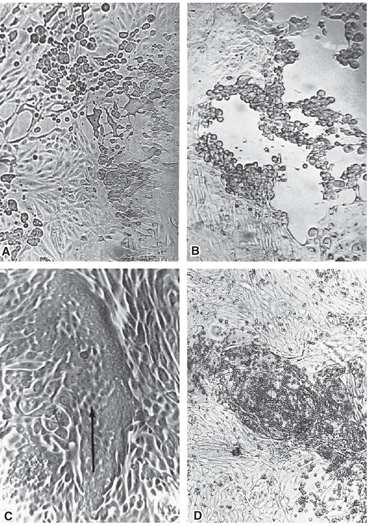
CPE in cell culture (60x, unfixed/unstained): A - Enterovirus (rapid rounding + destruction), B - Herpesvirus (focal swollen/rounded cells), C - Paramyxovirus (syncytia, arrow), D - Hemadsorption with RBCs adhering to infected cells.
- Jawetz, Melnick & Adelberg's Medical Microbiology, 28th Ed.
- Sherris & Ryan's Medical Microbiology, 8th Ed.
- Quick Compendium of Clinical Pathology, 5th Ed.
This is a shared conversation. Sign in to Orris to start your own chat.