
Tell me about the investigations.and management of AUB IN DIFFERENT AGE GROUPS
"abnormal uterine bleeding" AND management
PALM-COEIN classification AUB causes diagram
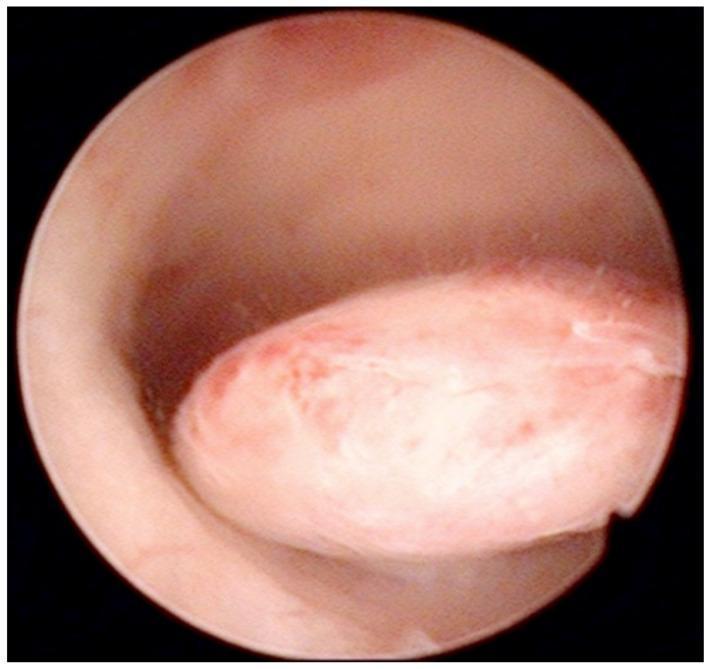
This diagnostic image is an intraoperative hysteroscopic photograph of the uterine cavity. The visual focus is a well-defined, oval-shaped pedunculated endometrial polyp protruding from the posterior uterine wall. The polyp demonstrates a smooth, glistening surface with a pale pink to white color, interspersed with focal areas of erythema and fine vertical striations across its body. Its morphology is slightly lobulated, and it is attached to the uterine wall by a distinct stalk. The surrounding endometrial tissue appears healthy, smooth, and pink, although there is a slight darkening near the base of the pedicle, likely representing minor blood accumulation. This clinical photograph illustrates a common structural cause of abnormal uterine bleeding (AUB), frequently categorized under the 'Polyp' component of the FIGO PALM-COEIN classification system. Such findings are critical in the diagnostic workup of perimenopausal and postmenopausal women to differentiate benign focal abnormalities from endometrial hyperplasia or malignancy.
This clinical photograph displays the volar (palmar) surface of a human hand, annotated to illustrate the Verdan classification system for flexor tendon injury zones. The image serves as an educational anatomical diagram for hand surgery and trauma management. The hand is divided into five primary zones and three specific thumb zones (T1–T3) using black demarcation lines. Zone 1 covers the distal segments of the fingers; Zone 2 (often called 'No Man's Land') extends from the distal palmar crease to the mid-portion of the middle phalanges; Zone 3 encompasses the central palm (lumbrical origin); Zone 4 corresponds to the carpal tunnel region; and Zone 5 includes the area proximal to the wrist crease (forearm). The thumb is separately classified into T1 (distal to the IP joint), T2 (between the MCP and IP joints), and T3 (thenar eminence). This visual guide is essential for medical students and surgical residents to understand the prognosis and surgical approach for flexor tendon repairs based on the anatomical site of injury.
Anatomical diagram and chart representing the skeletal structure of a human hand, specifically used for legal medicine and insurance assessment (AUB). The illustration shows the carpal bones, metacarpals, and phalanges of the five digits. It features specialized annotations representing disability values (Invaliditätswerte) for joint stiffening (ankylosis) in functionally favorable positions. The labels utilize a fractional format 'x/20 Fi' (Fingerwert), which refers to standardized assessment scales for hand function impairment. Labels '4/20 Fi', '6/20 Fi', and '8/20 Fi' are linked to specific joints of the index finger, while '10/20 Fi', '14/20 Fi', and '18/20 Fi' are associated with the thumb ray (pollex). This clinical algorithm diagram is designed for orthopedic surgeons and medical examiners to determine functional loss in medical-legal contexts regarding upper extremity injuries.
Abnormal Uterine Bleeding (AUB): Investigations and Management Across Age Groups
Definition and Classification (PALM-COEIN System)
| PALM (Structural) | COEIN (Non-structural) |
|---|---|
| Polyp | Coagulopathy |
| Adenomyosis | Ovulatory dysfunction |
| Leiomyoma | Endometrial |
| Malignancy & Hyperplasia | Iatrogenic |
| Not otherwise classified |
Note: Postmenopausal bleeding is not included in PALM-COEIN and is considered its own distinct diagnosis. - Sabiston Textbook of Surgery, p. 2795
Epidemiology
- AUB affects 10-30% of women of reproductive age
- ~20% of AUB is postmenarchal bleeding in adolescents (due to immature HPO axis)
- ~50% is perimenopausal bleeding related to incipient ovarian failure
- 75% of cases have no demonstrable organic cause and are anovulatory in origin
- Heavy menstrual bleeding is the presenting complaint in the majority - Goldman-Cecil Medicine, p. 2555
General Investigations (All Age Groups)
- Amount, duration, and pattern of blood loss
- Prospective charting of bleeding days
- Last menstrual period, obstetric history
- Medications (OCP, anticoagulants, tamoxifen, hormones)
- Family history of coagulopathy
- Pelvic exam: tenderness, uterine size/mass, cervical lesions
- Papanicolaou smear
- Signs of anemia
- Full blood count + platelet count
- Coagulation profile (PT, APTT)
- Von Willebrand disease screening (if heavy bleeding since menarche, family history, or multi-system bleeding signs)
- Thyroid function tests (TSH)
- Fasting blood glucose
- Serum prolactin
- Pregnancy test (mandatory in all reproductive-age women)
- Cervical cancer screening if not up to date
- STI screening
- Pelvic ultrasound (2D/3D, or saline sonohysterogram): first-line imaging to assess uterine cavity, endometrial thickness, and ovaries
- Hysteroscopy: gold standard for direct visualization; combined with endometrial biopsy has greater sensitivity and specificity than either alone
- All women ≥45 years with AUB (including intermenstrual bleeding)
- Women <45 years with: unopposed estrogen exposure (obesity, PCOS), persistent AUB refractory to medical management, elevated familial cancer risk
- Postmenopausal bleeding with endometrial thickness >4 mm
AUB by Age Group
1. Adolescents (Menarche to ~18 years)
Etiology
- Anovulation is physiologic for an average of 18 months after menarche while the hypothalamic-pituitary-ovarian axis matures
- Menorrhagia in adolescents is most commonly caused by anovulation
- Up to 24% of adolescents with menorrhagia may have an undiagnosed bleeding disorder (e.g., von Willebrand disease, ITP) - this is a critical and often missed diagnosis
- Primary amenorrhea causes: pregnancy, chromosomal abnormalities (Turner syndrome, Swyer syndrome), hypothalamic hypogonadism, congenital absence of uterus/cervix/vagina, structural anomalies (transverse vaginal septum, imperforate hymen)
Investigations
- Complete blood count
- Coagulation profile (PT, APTT)
- Von Willebrand disease screening (essential if heavy bleeding from menarche)
- Pelvic ultrasonography (to document pelvic organ presence if amenorrhea)
- Chromosome analysis if clinically indicated (primary amenorrhea workup)
- Pregnancy test
Note: Endometrial biopsy is rarely indicated in this age group unless there is prolonged unopposed estrogen exposure or suspicion of malignancy.
Management
- Anovulatory bleeding / menorrhagia: Hormonal contraception (combined OCP) for cycle control - first-line
- Coagulation disorders: treat the underlying bleeding disorder specifically (tranexamic acid, desmopressin for vWD)
- Acute heavy bleeding: hospital admission, IV conjugated estrogens or high-dose OCP
2. Reproductive-Age Women (~18-45 years)
Etiology
- Pregnancy complications (threatened/incomplete/missed miscarriage, ectopic pregnancy) - must always be excluded first
- Anovulatory disorders (PCOS, hyperprolactinemia, thyroid disease, Cushing's)
- Structural causes: uterine leiomyomas (most common benign gynecologic tumor, affecting up to 70% of women by age 50), endometrial polyps, adenomyosis
- Coagulopathy: ITP, von Willebrand disease
- Iatrogenic: OCP complications, IUD, anticoagulants
- Systemic disease: hepatic/renal failure, leukemia, endocrinopathies
- Menorrhagia: structural lesions, coagulopathy, hepatic/renal failure
- Polymenorrhea: luteal phase defect, short follicular phase
- Oligomenorrhea: prolonged follicular phase
- Intermenstrual bleeding: cervical pathology, IUD
Investigations
- Mandatory: Pregnancy test first
- FBC, coagulation studies, TFTs, prolactin, fasting glucose
- Pelvic ultrasound +/- saline sonohysterogram
- Endometrial biopsy: if age ≥45, or age <45 with risk factors (obesity, PCOS, persistent AUB, family history of endometrial cancer, tamoxifen use)
- Hysteroscopy: if focal lesion suspected, or biopsy insufficient
- Cervical cytology + STI swabs
Management
| Indication | Drug | Mechanism |
|---|---|---|
| Anovulatory (no pregnancy desired) | Combined OCP (q6h × 5-7 days for acute; cyclic for maintenance) | Induces shedding, cycle control |
| Anovulatory (pregnancy desired) | Ovulation induction (clomiphene) | Restores ovulation |
| Chronic anovulation | Cyclic progestin or OCP (≥4 withdrawal bleeds/year) | Prevents endometrial hyperplasia |
| Ovulatory AUB | NSAIDs, tranexamic acid, levonorgestrel IUD | Reduce prostaglandins, fibrinolysis |
| Acute profuse bleeding | IV conjugated estrogens 25 mg q4h (up to 3 doses) + simultaneous progestin | Rapid hemostasis |
| Fibroids | Elagolix 300 mg BD or relugolix 40 mg daily, UAE, or surgery | GnRH receptor antagonists |
| PCOS | OCP or progestins | Cycle regulation |
- Endometrial ablation: effective for persistent bleeding; not 100% effective (29% eventually require hysterectomy at 60 months in RCTs)
- Uterine artery embolization (UAE): for fibroids; ~31% ultimately require hysterectomy in RCTs
- Hysteroscopic polypectomy: for endometrial polyps
- Myomectomy: fibroid-preserving surgery
- Hysterectomy: reserved for failure of/intolerance to medical therapy; always requires endometrial sampling before proceeding; dilation and curettage alone is NOT an effective means of controlling bleeding
3. Perimenopausal Women (~45 years to menopause)
Etiology
- Most common: Anovulation due to declining ovarian follicle numbers and decreasing inhibin B levels
- Structural lesions (fibroids, polyps) remain common
- Increasing risk of endometrial hyperplasia and carcinoma
- Bleeding disorders
Investigations
- Endometrial biopsy is mandatory in all perimenopausal women with AUB to exclude endometrial hyperplasia/cancer
- Risk factors for endometrial cancer: nulliparity, diabetes, obesity
- Pelvic ultrasound (endometrial thickness)
- Hysteroscopy + biopsy if focal lesion suspected
- FBC, TFTs, coagulation studies
Management
- Non-smokers: Combined OCP for cycle control (most effective)
- Smokers (increased thrombotic risk - avoid estrogen): Cyclic progestin to provide regular withdrawal bleed
- Levonorgestrel IUD: excellent option for heavy menstrual bleeding + contraception
- If malignancy/hyperplasia excluded: same options as reproductive-age women
- Hysterectomy if medical therapy fails
4. Postmenopausal Women
Etiology (in order of frequency)
- Endometrial/vaginal atrophy (most common) - thinning due to estrogen deficiency
- Endometrial polyps
- Endometrial hyperplasia
- Endometrial carcinoma (10-20% of cases) - 90% of endometrial cancer patients present with postmenopausal bleeding
- Uterine fibroids/adenomyosis
- Medications: HRT, anticoagulants
- Cervical carcinoma
- Tamoxifen use: increases endometrial polyps, hyperplasia, cancer risk; doubles risk after 1-2 years, quadruples after 5 years (time-dependent, dose-independent)
Investigations
-
Pelvic ultrasound - assess endometrial stripe:
- Endometrial thickness ≤4 mm: endometrial cancer effectively excluded (biopsy may be avoided)
- Important caveat: the 4 mm cutoff has been shown to under-diagnose endometrial cancer in Black patients - clinical judgment required
- Endometrial thickness >4 mm: endometrial biopsy indicated
-
Office endometrial biopsy (Pipelle): excellent sensitivity; if cervical stenosis prevents access, surgical procedure may be indicated
- Focal lesion suspected on imaging
- Difficulty visualizing endometrium
- Persistent bleeding despite normal ultrasound
Management
- Atrophic vaginitis/endometrial atrophy: topical estrogen therapy
- Endometrial polyps: hysteroscopic polypectomy
- Endometrial hyperplasia without atypia: progestins (cyclic or continuous)
- Atypical hyperplasia/carcinoma: surgical staging (hysterectomy + BSO ± lymph node dissection)
- HRT-related bleeding: adjust or discontinue HRT
- Aromatase inhibitors (anastrozole, letrozole, exemestane): may reverse tamoxifen-induced endometrial abnormalities
Management of Acute Heavy Bleeding (Emergency)
- Admit to hospital
- IV access + IV fluid resuscitation
- Transfusion if anemia is profound
- IV conjugated estrogens: 25 mg IV every 4 hours for up to 3 doses until bleeding ceases; start simultaneous oral progestin (medroxyprogesterone acetate 5-10 mg for 10 days)
- If stable but profuse: oral OCP (1 pill q6h × 5-7 days) - bleeding should cease within 24 hours; warn patient about heavy withdrawal bleed 2-4 days after stopping
- If bleeding continues despite medical therapy: surgical curettage
- Hysterectomy in refractory cases
Summary Table: AUB by Age Group
| Age Group | Most Common Cause | Key Investigation | First-line Treatment |
|---|---|---|---|
| Adolescent | Anovulation (HPO axis immaturity), coagulopathy | FBC, coagulation, vWD screen | Combined OCP, treat coagulopathy |
| Reproductive age | Pregnancy complications, PCOS, structural (fibroids/polyps) | Pregnancy test, USS, +/- biopsy if risk factors | OCP, progestins, NSAIDs, LNG-IUD |
| Perimenopausal | Anovulation + structural lesions | Endometrial biopsy (mandatory), USS | OCP (non-smokers), cyclic progestin (smokers) |
| Postmenopausal | Atrophy (most common), carcinoma (10-20%) | Pelvic USS (endometrial stripe), endometrial biopsy | Treat cause; surgical staging if malignancy |
- Sabiston Textbook of Surgery, 21st ed. (PALM-COEIN, postmenopausal workup)
- Goldman-Cecil Medicine (epidemiology, anovulatory management)
- Textbook of Family Medicine, 9e (life-cycle approach, age-group management)
- Berek & Novak's Gynecology (surgical management, hysterectomy indications)
- Bailey and Love's Short Practice of Surgery, 28th ed. (structural/non-structural classification, tamoxifen)
- Recent meta-analysis: Comishen et al. 2025 - J Thromb Haemost - etiology and diagnosis of heavy menstrual bleeding across adolescent and adult patients (PMID 39617186)
GIVE ME CASES OF EACH AS IF I'M IN OPD AND let's see the investigation and treatment
endometrial polyp hysteroscopy uterine bleeding
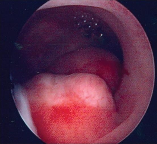
This clinical endoscopic image depicts a hysteroscopic view of the uterine cavity. The primary finding is a large, focal endometrial polyp characterized by a pale, whitish, and raised appearance that contrasts with the surrounding darker, vascularized endometrial tissue. The surface of the polyp shows irregular morphology and areas of focal reddish discoloration, suggestive of localized hemorrhage or hypervascularity. This diagnostic image demonstrates a common benign growth that can cause abnormal uterine bleeding (AUB) or postmenopausal bleeding (PMB). The visualization highlights the high sensitivity of hysteroscopy in detecting focal endometrial pathologies compared to transvaginal ultrasound, particularly in identifying the size, location, and vascular characteristics of endometrial polyps.
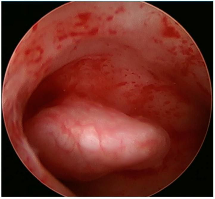
This clinical diagnostic image is a hysteroscopic view of the uterine cavity, showing an endometrial polyp. The image displays a smooth, rounded, and lobulated mass approximately 1.5 cm in size, situated on the right lateral wall of the uterus. The polyp exhibits a lighter, pinkish-white coloration compared to the surrounding pinkish-red uterine lining. Fine superficial blood vessels are visible on the surface of the polyp, which appears less vascular than the adjacent hyperemic endometrium. The surrounding uterine tissue shows evidence of mild spotting and irregular vascular patterns. This endoscopic finding is characteristic of a benign endometrial polyp, often investigated in cases of infertility or abnormal uterine bleeding. The image serves as an educational example of intrauterine pathology identified during a diagnostic hysteroscopy, illustrating the morphological differences between focal polyps and normal endometrial tissue.
uterine fibroid leiomyoma ultrasound sonogram
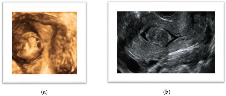
Educational diagnostic ultrasound images of a uterine leiomyoma (fibroid). Image (a) presents a 3D ultrasound in coronal view, demonstrating a solid, well-defined mass with a rounded and slightly lobulated morphology. The reconstruction reveals clear boundaries and a heterogeneous tissue density within the uterine environment. Image (b) shows a saline contrast sonohysterography (SHG) in a sagittal plane. This modality highlights a FIGO type 0 submucosal leiomyoma, appearing as a discrete, hypoechoic, rounded lesion entirely within the endometrial cavity. The instillation of saline provides a clear anechoic (dark) contrast around the lesion, delineating its relationship with the endometrial lining and myometrial junction. These images illustrate key diagnostic techniques for evaluating symptomatic uterine lesions, such as abnormal uterine bleeding or subfertility, and are essential for preoperative surgical planning.
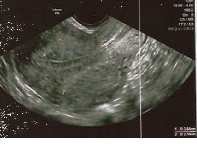
Diagnostic Image: This transvaginal ultrasound (TVUS) scan demonstrates a uterine leiomyoma (fibroid) following treatment with ulipristal acetate (UPA). The fibroid appears as a well-circumscribed, heterogeneous mass within the myometrium, exhibiting mixed echogenicity with interspersed hypoechoic areas and small anechoic cystic components, which may represent focal degeneration. The endometrium is visualized as a distinct, thin, echogenic linear structure adjacent to the mass, maintaining a regular proliferative appearance without significant distortion or displacement by the fibroid. Caliper markings are present on the image for size measurement. This clinical photograph is used in gynecology to monitor the response of uterine fibroids to medical management, focusing on volume reduction and the assessment of endometrial changes.
AUB in the OPD: Case-Based Approach Across Age Groups
🏥 CASE 1 - Adolescent (Age 14)
Presenting Complaint
- Menarche: 3 months ago
- Cycles: irregular, every 18-35 days
- Duration: 10-12 days per episode
- Clots: large clots passed
- Associated: pallor, fatigue, mild dizziness
- No sexual activity
- No prior medical history
- Family history: mother has "heavy periods," uncle has a bleeding disorder
What You're Thinking in OPD
- Physiologic anovulation (HPO axis immaturity - normal for 18 months post-menarche)
- Coagulopathy - Von Willebrand disease is present in up to 13-24% of adolescents with menorrhagia; the family history here is a red flag
- PCOS (less likely this early)
- Hypothyroidism
"The first described case of von Willebrand's disease was in a 13-year-old who died as a result of uncontrollable uterine bleeding." - Tintinalli's Emergency Medicine
OPD Examination
- Pallor of conjunctivae and nail beds
- No hirsutism, acne, acanthosis nigricans (argues against PCOS)
- No petechiae or ecchymoses (but does NOT rule out vWD)
- Pelvic exam: deferred (not sexually active, no indication)
- Abdominal exam: no masses
Investigations
| Test | Purpose | Expected Finding |
|---|---|---|
| FBC + reticulocyte count | Assess anemia | Hb low (may be 7-9 g/dL) |
| PT, APTT, platelet count | Coagulation screen | May show prolonged APTT in vWD type 1 (often normal!) |
| Von Willebrand Factor Ag, Activity (Ristocetin cofactor), Factor VIII | Specific vWD diagnosis | Reduced in vWD |
| TSH | Rule out hypothyroidism | Normal or low TSH |
| Blood group & crossmatch | If severe anemia | - |
| Urine or serum β-hCG | Mandatory - rule out pregnancy | Negative |
| Pelvic ultrasound | Rule out structural cause | Usually normal at this age |
Diagnosis
Management
- Iron supplementation (ferrous sulfate 200 mg TDS with vitamin C)
- If Hb < 7 g/dL or hemodynamically unstable: admit for IV iron or transfusion
- Combined OCP (30 µg ethinylestradiol + levonorgestrel): 1 tablet every 6 hours × 5-7 days → then taper; bleeding should stop within 24 hours
- Tranexamic acid (antifibrinolytic): reduces bleeding by 30-55%; can be used acutely and long-term
- Desmopressin (DDAVP): intranasal or IV; releases stored vWF from endothelial cells - specific for vWD type 1
- Combined OCP cyclically (first-line for ongoing cycle regulation)
- Levonorgestrel IUD: reduces heavy bleeding by up to 90%; FDA-approved for heavy menstrual bleeding; appropriate for adolescents (insertion under local anesthesia if needed)
- Continuous progestin (norethindrone, DMPA) if OCP contraindicated
🏥 CASE 2 - Reproductive Age (Age 28, PCOS)
Presenting Complaint
- LMP: 3 months ago
- Cycles: oligomenorrheic (4-5 cycles/year)
- Episodes: heavy, prolonged when they occur
- Associated: weight gain, hirsutism, acne
- Married 2 years, not conceived
- No contraception
What You're Thinking in OPD
OPD Examination
- BMI: 32 (obese)
- Hirsutism (Ferriman-Gallwey score)
- Acanthosis nigricans at nape of neck
- Acne
- Pelvic exam: uterus normal size, no masses
Investigations
| Test | Purpose |
|---|---|
| β-hCG (MANDATORY first) | Rule out pregnancy |
| FBC | Anemia from chronic blood loss |
| TSH, Prolactin | Rule out other causes of anovulation |
| LH:FSH ratio (LH elevated in PCOS), testosterone, DHEA-S | Hormonal profile |
| Fasting glucose + insulin, HOMA-IR | Insulin resistance (in PCOS) |
| Pelvic ultrasound (transvaginal) | Polycystic ovaries (≥12 follicles 2-9 mm, OR ovarian volume >10 mL); endometrial thickness |
| Endometrial biopsy (Pipelle) | Patient <45 but has risk factors: obesity + chronic anovulation + prolonged AUB - MANDATORY |
"Patients younger than 45 years old with unopposed estrogen exposure such as those with obesity and ovulatory dysfunction with persistent AUB should be biopsied." - Sabiston Textbook of Surgery, p. 2794
Diagnosis
Management
- Combined OCP (first-line): regulates cycles, opposes estrogen, treats hirsutism and acne
- Levonorgestrel IUD: excellent option - provides endometrial protection + heavy bleeding control
- Cyclic progestin (medroxyprogesterone acetate 10 mg/day × 10-14 days every 1-3 months): minimum 4 withdrawal bleeds per year to prevent endometrial hyperplasia
- Metformin: treats insulin resistance; may restore ovulation in PCOS
- Weight loss: restores ovulation in up to 50% of obese PCOS patients
- Ovulation induction: clomiphene citrate 50 mg days 2-6 (first-line)
- Letrozole: increasingly preferred in PCOS (better live birth rates)
- Metformin + clomiphene: if clomiphene-resistant
- High-dose progestins (medroxyprogesterone acetate 10-20 mg daily continuously × 3-6 months)
- Repeat biopsy in 3-6 months
🏥 CASE 3 - Reproductive Age (Age 35, Fibroids)
Presenting Complaint
- Cycles: regular, every 28 days
- Duration of heavy flow: 7-8 days (previously 4)
- Clots: large clots
- Associated: pelvic pressure, urinary frequency, constipation
- Parity: G2P2 (two previous normal deliveries)
- No oral contraceptive use
What You're Thinking in OPD
OPD Examination
- Pallor
- Abdomen: uterus palpable abdominally, irregular, firm, non-tender (14-16 week size)
- Bimanual: enlarged irregular uterus, no adnexal masses
Investigations
| Test | Finding Expected |
|---|---|
| FBC | Anemia (iron deficiency) |
| β-hCG | Negative |
| Pelvic ultrasound (TVS/TAS) | Multiple hypoechoic masses in myometrium; location classified by FIGO (Type 0-8); saline sonohysterogram if submucosal suspected |
| MRI pelvis | If ultrasound inadequate; maps fibroid number/location precisely before surgery |
| TFTs, Coagulation | Rule out contributory causes |
| Endometrial biopsy | Patient is 35 with structural lesion AND heavy bleeding - indicated to exclude hyperplasia/malignancy |
| Cervical smear | If overdue |
Diagnosis
Management
- GnRH receptor antagonists (oral):
- Elagolix 300 mg twice daily, OR
- Relugolix 40 mg daily
- Reduce uterine/fibroid volume; used for 6-12 months; side effects: hypoestrogenic symptoms, bone loss
- Levonorgestrel IUD: good for submucosal-free uterus; less effective if large/multiple submucosal fibroids
- Tranexamic acid (1 g TDS × 4 days/cycle): reduces flow by ~50% without affecting fertility
- NSAIDs (mefenamic acid 500 mg TDS): modest effect on flow; useful for dysmenorrhea
- Iron supplementation to correct anemia before surgery
| Fibroid Location | Procedure |
|---|---|
| Submucosal (Type 0, 1) | Hysteroscopic myomectomy (preserves fertility) |
| Intramural/subserosal (Types 2-7) | Laparoscopic or open myomectomy (fertility-sparing) |
| Any type, family complete | Uterine artery embolization (UAE): minimally invasive; 31% require hysterectomy long-term |
| Refractory, family complete | Hysterectomy (definitive) |
"Uterine artery embolization vs hysterectomy: 31% of those randomised to UAE ultimately underwent a hysterectomy." - Berek & Novak's Gynecology, p. 1411
🏥 CASE 4 - Perimenopausal (Age 48)
Presenting Complaint
- LMP: 3 weeks ago
- Cycles: irregular (18-60 day intervals) for 12 months
- Recently: one episode of prolonged heavy bleeding
- Associated: hot flushes, night sweats, mood changes
- BMI: 29 (overweight)
- No HRT
- Nulliparous (G0P0)
What You're Thinking in OPD
- Nulliparity ✅
- Overweight ✅
- Anovulatory cycles (unopposed estrogen) ✅
- Age >45 ✅
OPD Examination
- BP, BMI documented
- Thyroid: normal
- Pelvic exam: uterus slightly enlarged, no adnexal masses
- No vaginal lesions
Investigations
| Test | Purpose |
|---|---|
| β-hCG | Never forget - perimenopausal women can still ovulate |
| FBC | Anemia |
| TSH | Thyroid (common perimenopausal overlap) |
| Pelvic ultrasound (TVS) | Endometrial thickness, structural lesions; ET >4-5 mm suspicious |
| Endometrial biopsy (Pipelle) - MANDATORY | Exclude hyperplasia/carcinoma in ALL perimenopausal women with AUB |
| Hysteroscopy + D&C | If Pipelle fails/inadequate sample, focal lesion on USS, or persistent bleeding |
| FSH, LH, E2 | Confirm perimenopause transition if clinically needed |
| Coagulation profile | If suspected |
"Evaluation of a perimenopausal woman with abnormal bleeding should include an endometrial biopsy to exclude endometrial hyperplasia or cancer." - Textbook of Family Medicine, 9e
Diagnosis (scenarios based on biopsy result)
- Non-smoker: Combined OCP (low-dose 20 µg) - cycle regulation + contraception
- Smoker / VTE risk: Cyclic progestin (medroxyprogesterone acetate 10 mg × 14 days/month)
- Levonorgestrel IUD: excellent option - endometrial protection + heavy bleeding control
- Hot flushes: consider low-dose HRT or SSRIs if vasomotor symptoms significant
- Progestin therapy (continuous or cyclic)
- Repeat biopsy in 3-6 months
- LNG-IUD is superior to oral progestins for regression of hyperplasia
- Urgent referral to Gynaecological Oncology
- Hysterectomy + BSO + staging (standard treatment)
🏥 CASE 5 - Postmenopausal (Age 62)
Presenting Complaint
- Last menstrual period: 8 years ago
- Now: one episode of light fresh bleeding (2 days)
- No HRT currently
- Past history: hypertension, type 2 diabetes, BMI 34
- No abdominal pain, no fever
What You're Thinking in OPD
- Hypertension ✅
- Type 2 diabetes ✅
- Obesity (BMI 34) ✅
- Age >55 ✅
- This patient has the classic endometrial cancer profile
"Of patients diagnosed with endometrial cancer, 90% experience postmenopausal bleeding." - Sabiston Textbook of Surgery, p. 2795
"Between 10% and 20% of all postmenopausal bleeding is caused by malignancy." - Textbook of Family Medicine, 9e
OPD Examination
- Abdomen: no masses, no hepatomegaly
- Pelvic examination: atrophic vaginal mucosa (pale, dry - suggests atrophy), no vulval lesion, cervix appears normal
- Bimanual: uterus slightly bulky, no obvious adnexal mass
Investigations - URGENT PATHWAY
| Test | Finding/Action |
|---|---|
| Pelvic ultrasound (TVS) - FIRST-LINE | Measure endometrial stripe |
| → ET ≤ 4 mm | Endometrial cancer effectively excluded; likely atrophy |
| → ET > 4 mm | Endometrial biopsy MANDATORY |
| → Focal lesion / polyp | Biopsy regardless of ET thickness |
| Endometrial biopsy (Pipelle in OPD) | Histology: atrophy / polyp / hyperplasia / carcinoma |
| Hysteroscopy + directed biopsy | If Pipelle fails (cervical stenosis common post-menopause) OR persistent bleeding with normal USS OR focal lesion suspected |
| FBC, coagulation, LFTs, RFTs | Baseline, fitness for surgery if needed |
| CA125 | If adnexal mass or suspected ovarian involvement |
| Cervical cytology | Rule out cervical pathology |
| Colonoscopy | If colorectal source cannot be excluded |
Management Based on Findings
| Finding | Treatment |
|---|---|
| Endometrial/vaginal atrophy | Topical vaginal estrogen cream or pessary (low systemic absorption, safe) |
| Endometrial polyp | Hysteroscopic polypectomy (up to 5% malignant transformation) |
| Simple hyperplasia without atypia | Oral progestins or LNG-IUD; repeat biopsy in 3-6 months |
| Atypical hyperplasia | Hysterectomy + BSO (40% risk of coexisting carcinoma) |
| Endometrial carcinoma Stage I | TAH + BSO + pelvic ± para-aortic lymphadenectomy ± adjuvant radiotherapy |
| HRT-related bleeding | Adjust or discontinue HRT; re-evaluate |
| Tamoxifen-related | Investigate all bleeding; consider switching to aromatase inhibitor |
Quick OPD Summary Card
| Case 1 (14F) | Case 2 (28F) | Case 3 (35F) | Case 4 (48F) | Case 5 (62F) | |
|---|---|---|---|---|---|
| Dx | vWD / Anovulation | PCOS (AUB-O) | Fibroids (AUB-L) | Perimenopause ± hyperplasia | Atrophy vs Carcinoma |
| Must-do test | vWF screen | β-hCG + USS | USS + MRI | Endometrial biopsy | TVS + biopsy |
| First-line Rx | OCP + Tranexamic acid | OCP or cyclic progestin | LNG-IUD / Myomectomy | OCP or cyclic progestin | Topical E / Surgery |
| Red flag | Family Hx bleeding | Endometrial hyperplasia risk | Submucosal component | Cancer risk factors | ANY bleeding = urgent |