
Achilles tendon , whats the best evidence based Non Surgical interventional pain approach
Achilles tendinopathy non-surgical treatment interventional
Achilles tendinopathy best evidence non-surgical interventional treatment 2024 2025 guidelines
PMID: 36750789
Achilles tendinopathy PRP shockwave injection treatment
https://pmc.ncbi.nlm.nih.gov/articles/PMC12250464
Achilles tendinopathy ultrasound anatomy treatment diagram
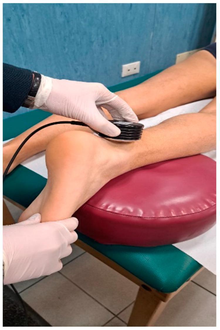
A clinical procedural photograph demonstrating the application of therapeutic ultrasound for mid-portion Achilles tendinopathy (AT). The image shows a patient in a prone or supine-modified position with the lower leg elevated on a maroon-colored bolster for comfort and anatomical access. A healthcare provider, wearing white examination gloves, is shown applying a black, circular ultrasound therapy probe to the posterior aspect of the ankle. The probe is positioned directly over the mid-portion of the Achilles tendon, approximately 2–6 cm proximal to the calcaneal insertion. The practitioner's second hand is seen stabilizing the heel to maintain the ankle in a neutral or slightly dorsiflexed position during the procedure. This modality is utilized in physical therapy and sports medicine to promote healing, increase local blood flow, and provide pain relief in patients with chronic tendinosis or degenerative tendon changes. The setting is a professional clinical environment featuring a padded treatment table and standard medical equipment.
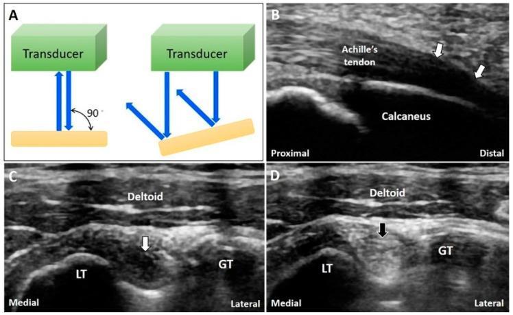
This educational composite focuses on the ultrasound artifact of anisotropy in musculoskeletal imaging. Panel A is a physics diagram illustrating that the ultrasound beam is reflected back to the transducer when incident at 90 degrees, but reflected away when the target is oblique, leading to signal loss. Panel B shows a long-axis ultrasound view of the Achilles tendon insertion on the calcaneus. White arrows indicate a hypoechoic (dark) area caused by anisotropy where the tendon fibers curve, which could be mistaken for tendinopathy or a tear. Panels C and D provide a short-axis comparison of the long head of the biceps tendon within the bicipital groove, situated between the lesser tubercle (LT) and greater tubercle (GT) under the deltoid muscle. In Panel C, a white arrow points to an anisotropic hypoechoic region within the tendon. Panel D demonstrates the resolution of the artifact (black arrow) after tilting the transducer to achieve a perpendicular angle of insonation, restoring normal hyperechoic fibrillar echogenicity. This figure teaches clinicians how to differentiate technical artifacts from true pathology during musculoskeletal ultrasound examinations.
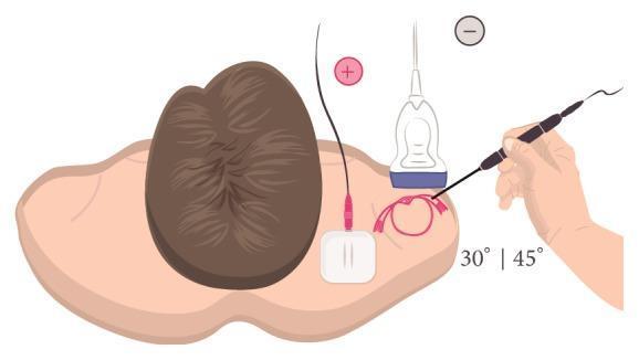
Educational illustration demonstrating the setup for Ultrasound (US)-guided percutaneous electrolysis for the treatment of shoulder tendinopathy, specifically targeting the supraspinatus tendon. The diagram depicts a patient in a supine position with the right shoulder area exposed. A linear ultrasound transducer is placed on the anterior shoulder for real-time visualization of the tendon. An acupuncture needle, functioning as the cathode (-), is inserted at a 30° to 45° angle to the skin surface, following a long-axis approach toward the target tissue under the probe. A surface electrode pad is placed over the upper trapezius muscle to serve as the anode (+), completing the galvanic current circuit. The illustration highlights the spatial relationship between the transducer, the needle insertion angle, and the electrical circuit configuration necessary for delivering therapeutic microtrauma to the affected tendon structure.
PMID: 39496560
Achilles tendon PRP injection platelet rich plasma procedure
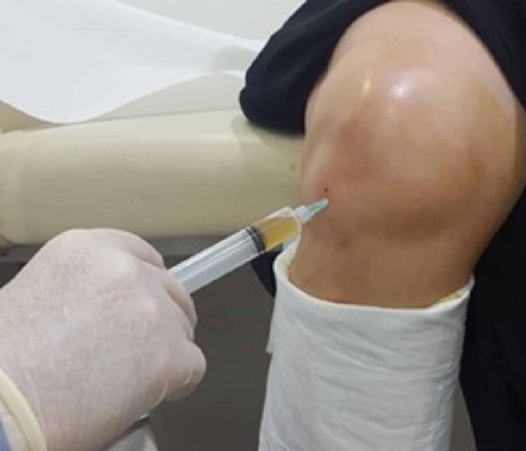
A clinical procedural photograph demonstrating an intra-articular injection of Platelet-Rich Plasma (PRP) into a patient's knee joint. A gloved hand is shown administering approximately 5cc of a translucent, straw-colored fluid via a syringe. The needle entry point is localized to the lateral aspect of the knee, positioned approximately 1 cm lateral to the patellar tendon and 1 cm inferior to the joint line. The anatomical landmarks of the patella and joint space are visible under the skin. The patient's lower leg is partially covered by a white protective cast or drape, and the procedure is being performed in a clinical setting on a treatment table. This image illustrates the standard clinical technique for regenerative therapy in the management of knee osteoarthritis.
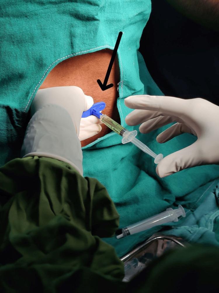
Clinical photograph demonstrating an epidural platelet-rich plasma (PRP) infiltration procedure in a patient with lumbar disc herniation. The image shows a sterile surgical field created by green fabric drapes. A clinician, wearing white sterile gloves, is performing the injection. A syringe containing a straw-colored, yellow fluid (PRP) is connected via a blue winged hub/connector to a needle already inserted into the skin of the lumbar region, as indicated by a black directional arrow. A second gloved hand is visible stabilizing the needle hub. In the foreground, an additional unused syringe rests on a metal instrument tray within the sterile field. The procedure illustrates a minimally invasive biological therapy for chronic low back pain and radiculopathy, serving as a teaching tool for interventional pain management and musculoskeletal regenerative medicine.
Achilles Tendinopathy: Best Evidence-Based Non-Surgical Interventional Pain Management
Quick Orientation: Two Distinct Entities
- Midportion (2-6 cm proximal to insertion) - most common, ~55-65% of cases
- Insertional (at the calcaneal attachment, including Haglund deformity variants) - responds differently, especially to loading exercises
Tier 1 - First-Line: Exercise-Based Loading (Strongest Evidence)
Eccentric Loading - Alfredson Protocol
- Classic protocol: 3 sets x 15 reps, twice daily, 12 weeks, on a decline board or step edge
- Works for midportion; modified (with load tolerance adjustment) for insertional
- Strong meta-analytic support; NNT ~3-4 for clinically meaningful pain improvement
- For insertional AT, purely eccentric loading (off a step edge moving into dorsiflexion) can aggravate the insertion - flat-surface eccentric or heavy slow resistance (HSR) is preferred
Heavy Slow Resistance (HSR) - Beyer Protocol
- Equivalent or superior to eccentric in patient adherence and outcomes (Beyer et al., RCT)
- 3x/week, progressive resistance, 12 weeks; concentric + eccentric combined
- Better tolerated by insertional AT and older/sedentary patients
Isometric Loading
- Recommended as an initial pain-relief strategy in irritable/high-pain presentations
- 5 x 45-second holds at 70% MVC; provides immediate analgesia (~1-2 weeks)
- Used as a "bridge" before progressing to isotonic loading
2024 CPG Key Point: Complete rest is NOT recommended. Continue activities within pain tolerance. Exercise loads should be "as high as tolerated." Night splints and passive modalities alone are NOT recommended.
Tier 2 - Adjunctive Interventional Therapies
1. Extracorporeal Shockwave Therapy (ESWT) - Best Adjunct Evidence
- Radial or focused ESWT, typically 3-5 sessions, 1 week apart
- Works via: collagen synthesis stimulation, upregulation of growth factors (TGF-beta, IGF-1), nociceptor hyperstimulation (hypoalgesia), neovascularization inhibition
- A 2021 double-blind RCT (JBJS) showed ESWT + eccentric exercise was superior to eccentric exercise alone for insertional AT
- Most effective for chronic (>3 months), recalcitrant cases
- Contraindications: overlying growth plate, coagulation disorders, pacemaker, pregnancy
2. High-Volume Image-Guided Injection (HVIGI)
- Typically: 10 mL 0.5% bupivacaine + 40 mg triamcinolone + 30-40 mL normal saline, delivered under ultrasound guidance into the paratenon/neovascular zone
- Mechanism: mechanical disruption of neovascularity (Doppler vessels associated with pain), strips the paratenon adhesions
- The 2024 systematic review (10 studies, n=460) showed significant reductions in pain, tendon thickness, and neovascularity - both short and long-term
- No adverse events reported despite corticosteroid use (notably, the low dose delivered into paratenon - NOT intra-tendinous - mitigates rupture risk)
- Best combined with a structured eccentric/HSR program post-injection
Important distinction: HVIGI deposits fluid in the peritendinous space, NOT into the tendon substance. This is mechanistically different from intratendinous corticosteroid injection (which carries rupture risk).
3. Platelet-Rich Plasma (PRP) Injection
- Mechanism: growth factor release (PDGF, VEGF, TGF-beta) promoting tenocyte proliferation
- The Kearney et al. 2021 RCT found no significant benefit over placebo for midportion AT
- 2024 AOPT guideline does NOT recommend PRP as first-line
- Meta-analyses (Nauwelaers 2021, Madhi 2020) show inconsistent results - some benefit in chronic mid-portion; little data for insertional
- May be considered in refractory cases after ESWT failure, but evidence remains weak
- Ultrasound guidance recommended to ensure peritendinous/intratendinous delivery
4. Prolotherapy (Hyperosmolar Dextrose)
- 20-25% dextrose solution injected into peritendinous tissue + tendon body
- Proposed mechanism: osmotic cellular injury triggers local growth factor release and healing cascade
- Small RCTs show reduction in pain and improved function vs. lidocaine control
- Cheaper than PRP; more readily available; low adverse event profile
5. Polidocanol Sclerotherapy (Ultrasound-guided)
- Targets neovascular ingrowth (associated with pain in tendinopathy)
- Polidocanol 5-10 mg/mL injected under Doppler guidance into the neovascular zone
- Originally from Ohberg & Alfredson; good short-term results in midportion AT
- Evidence remains limited; may be considered in Doppler-positive cases not responding to loading
What to AVOID
| Intervention | Reason |
|---|---|
| Intratendinous corticosteroid injection | High rupture risk; 2024 AOPT guideline recommends AGAINST |
| Complete rest / immobilization | Leads to tendon atrophy; 2024 CPG recommends against |
| Night splints as standalone | 2024 CPG: not recommended as primary treatment |
| Passive modalities alone (US, TENS, laser) | Insufficient standalone evidence |
Evidence Hierarchy Summary Table
| Intervention | Evidence Level | Best For | Grade |
|---|---|---|---|
| Eccentric / HSR loading | Meta-analyses, CPG 2024 | Midportion + insertional | A |
| ESWT + exercise | RCTs + SRs | Chronic / recalcitrant | A |
| HVIGI (peri-tendinous) | SR 2024 (n=460) | Midportion + insertional | B |
| PRP injection | Inconsistent RCTs | Refractory midportion only | C |
| Dextrose prolotherapy | Small RCTs | Chronic, refractory | C |
| Polidocanol sclerotherapy | Limited RCTs | Doppler+ midportion | C |
| Intratendinous steroid | Controlled trials | Contraindicated | D |
Suggested Clinical Pathway
1st → Progressive loading (eccentric/HSR) + education + activity modification [12 weeks]
         ↓ If inadequate response
2nd → Add ESWT (3-5 sessions) alongside continued loading
         ↓ If still refractory (>4-6 months total)
3rd → HVIGI under ultrasound guidance (with physiotherapy continuation)
         ↓ If still refractory
4th → Consider PRP (especially if Doppler positive) or prolotherapy
         ↓ If all fails
→ Surgical referral (tendon debridement ± FHL transfer for insertional)
Key References
- Chimenti et al. (2024) AOPT/JOSPT CPG - Midportion Achilles Tendinopathy CPG Revision 2024
- Ko VM et al. (2023) Network meta-analysis, BMC Musculoskelet Disord - PMID 36750789 - eccentric + soft tissue therapy ranked #1 for insertional AT short-term pain (SUCRA 84.8)
- Hassan R et al. (2024) High-volume injections SR, Br Med Bull - PMID 39496560 - HVI safe and effective, especially with corticosteroid in the paratenon
- Current Concepts in Nonoperative Management (PMC 2025) - comprehensive scoping review synthesizing all modalities
- Textbook of Family Medicine 9e - Alfredson eccentric program, corticosteroid risk, ESWT discussion
Images and Procedure protocol for HVIG
high volume injection Achilles tendinopathy protocol technique
high volume image guided injection Achilles tendon ultrasound guided peritendinous
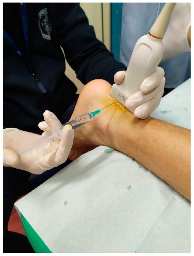
This clinical photograph demonstrates an ultrasound-guided high-volume image-guided injection (HVIGI) performed on the mid-portion of a patient's Achilles tendon. The patient is positioned prone with the ankle slightly elevated on a sterile disposable sheet. A clinician wearing protective gloves is using a high-frequency linear ultrasound transducer, applied with acoustic coupling gel, to visualize the tendon in real-time. Simultaneously, a needle attached to a large-capacity syringe containing clear saline or local anesthetic is inserted percutaneously into the paratendinous space. The procedure aims to mechanically strip the paratenon from the tendon and disrupt neo-vessels or nerves associated with chronic tendinopathy. This image serves as an educational reference for minimally invasive musculoskeletal procedures and ultrasound-guided interventional techniques in sports medicine and orthopedics for managing resistant Achilles tendinopathy.
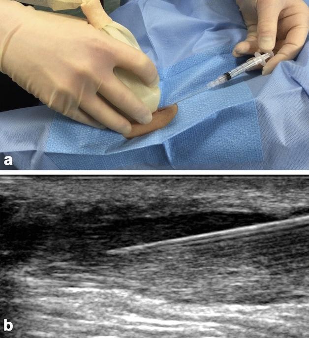
This composite educational image illustrates an ultrasound-guided stem cell injection for Achilles tendinosis. (a) A clinical photograph showing the sterile intraoperative setup where a practitioner, wearing sterile gloves, uses a high-frequency linear ultrasound probe (covered in a sterile sheath) to visualize the target area while inserting a syringe needle. The anatomical region is prepared with sterile blue drapes. (b) A corresponding longitudinal greyscale ultrasound image demonstrating the 'in-plane' needle technique. The needle appears as a distinct hyperechoic (bright) linear structure. It is positioned within a localized area of low reflectivity (hypoechoic) in the superficial portion of a thickened Achilles tendon, characteristic of focal tendinosis. The image captures the procedural moment of injection performed while withdrawing the needle distal-to-proximal through the disorganized fibrous tissue. This visual serves as a teaching tool for musculoskeletal interventional radiology, highlighting real-time imaging guidance to ensure precise delivery of biologics like bone marrow-derived mesenchymal stem cells (BMMSCs) into chronic tendon lesions.
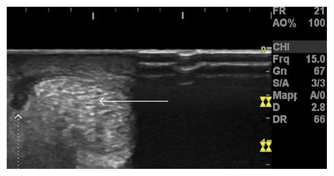
High-resolution cross-sectional (axial) ultrasound image of the Achilles tendon. The main body of the tendon (indicated by a solid white arrow) exhibits a heterogeneous echotexture with a mixture of hyperechoic and hypoechoic areas, representing its normal fibrous architecture under pathological stress. Laterally to the tendon body, a prominent hypoechoic, nearly anechoic region is visible (indicated by a dotted white arrow), which represents a peritendinous fluid collection. This finding is characteristic of acute paratendinopathy, where fluid accumulates between the tendon and its surrounding paratenon. The image serves as a clinical diagnostic example of musculoskeletal ultrasound used to differentiate between isolated tendinopathy and paratenon involvement in acute calf injuries.
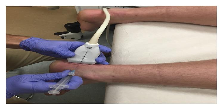
This clinical photograph demonstrates a sonographically guided injection procedure for chronic insertional Achilles calcific tendinosis. The patient is positioned prone with the lower leg and ankle resting on a padded examination table. A clinician wearing purple sterile gloves is shown performing a real-time ultrasound-guided intervention. One hand stabilizes a linear ultrasound probe placed in a short-axis orientation over the posterior calcaneal region to visualize the Achilles tendon insertion and associated enthesophytes. The other hand directs a 25-gauge needle attached to a syringe containing a lidocaine and dextrose mixture (prolotherapy). The needle is inserted at a shallow, medial-to-lateral trajectory, targeting the area anterior to the calcaneal enthesophyte. This procedural image illustrates the technique of ultrasound-guided fenestration and injection, emphasizing the coordinate placement of the imaging transducer and the needle to ensure precise delivery of the injectate into the pathology-affected tissues. The clinical focus is on musculoskeletal medicine, specifically the management of tendinopathy through minimally invasive regenerative techniques.
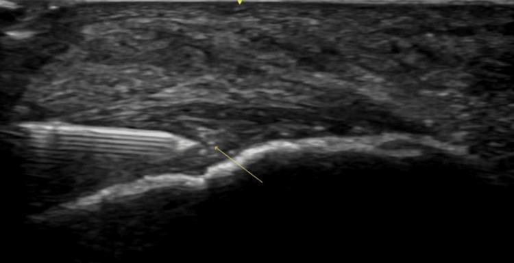
A diagnostic ultrasound image of the distal Achilles tendon in a long-axis view, illustrating an ultrasound-guided injection procedure. The Achilles tendon appears pathologically thickened with a heterogeneous, predominantly hypoechoic texture, consistent with insertional Achilles tendinopathy (IAT). Distinct anechoic voids and hyperechoic calcifications are visible near the tendon's insertion on the calcaneus. A hyperechoic needle is visualized using a long-axis, in-plane approach, with the tip (indicated by an arrow) positioned within the substance of the tendon. The procedure demonstrates a targeted injection (such as Botulinum toxin A or PRP) into the area of maximal tendinopathic change. The image serves as an educational example of musculoskeletal ultrasound intervention, showing real-time needle tracking and the characteristic sonographic features of chronic tendon degeneration.
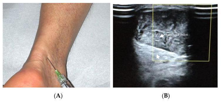
This composite educational figure illustrates the clinical and ultrasonographic aspects of a hyaluronic acid (HA) injection for Achilles tendinopathy. Panel A is a clinical photograph showing a dorsolateral approach to the Achilles tendon in the lower ankle region. A syringe with a 22-gauge needle and a green hub is shown at the entry point, demonstrating the trajectory required to reach the peritendinous space or mesotendon. Panel B is a corresponding ultrasound image (5–12 MHz linear probe) in the transverse plane. It depicts the internal anatomy of the Achilles tendon, which appears as a heterogeneous, ellipsoidal structure. Within the ultrasound's yellow focus box, the 22-gauge needle is clearly visible as a hyperechoic linear structure inserted at a 30-degree angle. Labels identify both the 'Achilles Tendon' and the 'needle' within the mesotendon. This content is designed for orthopedics and sports medicine education, demonstrating precise needle placement for viscosupplementation or regenerative therapy while avoiding intratendinous injection.
HVIGI high volume image guided injection Achilles tendinopathy procedure protocol technique needle placement 2024
https://pmc.ncbi.nlm.nih.gov/articles/PMC8264817
PMID: 34765229
High-Volume Image-Guided Injection (HVIGI) for Achilles Tendinopathy
Rationale & Mechanism
- In chronic AT, neovessels (detected on Power/Color Doppler) grow into the tendon from the anterior paratendinous space
- These vessels travel with sensory nerves (substance P-carrying nociceptors) - this is the source of pain
- High-volume fluid injection stretches, occludes, and tears these neovasculature bundles
- The accompanying nerve supply is destroyed by mechanical trauma and/or ischemia
- Result: significant pain reduction and improved Doppler-negative tendon appearance
Pre-Procedure Workup
| Step | Detail |
|---|---|
| Diagnostic US + Power Doppler | Confirm midportion AT, quantify neovascularity, exclude partial/full tear, identify injection target zone |
| VISA-A score | Baseline functional score (0-100; normal = 100) |
| Indication | Chronic AT (>3 months), failed structured exercise x 12 weeks |
| Imaging | Longitudinal + transverse B-mode; Power Doppler anterior to tendon |
Injectate Composition (Evidence-Based)
- 10 mL 0.5% bupivacaine hydrochloride (or 1% lidocaine)
- 20-40 mg methylprednisolone acetate (Depo-Medrol) OR 25 mg hydrocortisone acetate
- 40 mL normal saline (0.9% NaCl)
- Total volume: ~50 mL
- 10 mL 0.5% bupivacaine / 1% lidocaine
- 40 mL normal saline
- Total volume: 50 mL
- Slightly inferior short-term outcomes but equivalent at 24 weeks
Boesen et al. (2019): With-corticosteroid group showed +30.6 VISA-A at 6 weeks vs +13.8 without corticosteroid - significant early advantage; differences equalize by 24 weeks.
Step-by-Step Procedure Protocol
Position & Preparation
- High-frequency linear ultrasound probe (10-15 MHz)
- Sterile gel and probe cover
- 21-gauge needle (green)
- Extension tube/three-way stopcock
- Three 20 mL syringes (pre-filled: one with bupivacaine ± steroid, two with saline)
- Sterile drapes, gloves, chlorhexidine prep
Clinical Photo: HVIGI Procedure in Progress
Clinical Photo: Ultrasound-Guided Peritendinous Injection (Medial Approach)
Ultrasound Appearance: Paratendinous Fluid (Target Zone)
Step-by-Step Injection Protocol
- Disinfect skin with chlorhexidine or betadine
- Apply sterile US probe cover and sterile gel
- Inject 1-2 mL 1% lidocaine subcutaneously at the planned needle entry point
- Place probe transversely (short-axis) across the Achilles tendon
- Identify: tendon body, paratenon, Kager's fat pad anteriorly, sural nerve posterolaterally
- Insert 21G needle from the medial side - this avoids the sural nerve (which runs posterolateral)
- Use in-plane, long-axis needle technique relative to the probe
- Direct needle tip to the anterior paratendinous space between tendon and Kager's fat pad
- Confirm needle tip position on US before injecting - look for the hyperechoic needle tip in the peritendinous space (NOT inside the tendon substance)
- First: inject the bupivacaine ± steroid mixture (10 mL) - confirms needle position, achieves local anesthesia, begins paratenon elevation
- Connect extension tube to needle (keep needle in place)
- Inject the 40 mL saline in divided pushes via a 3-way stopcock
- Watch on US in real time: the fluid stripe should spread anteriorly along the tendon - you will see the paratenon stripping away
- The patient may feel pressure/fullness - this is normal
- Withdraw needle, apply pressure x 2 minutes
- Repeat Power Doppler scan - look for reduction in intratendinous neovascularity
- Cover with small sterile dressing
Post-Procedure Care Protocol
| Timeframe | Instructions |
|---|---|
| Day 0-2 | Rest, avoid loading. Paracetamol + ice PRN for post-injection ache |
| Day 3 | Begin isometric calf loading (5 x 45 sec holds) as pain allows |
| Week 1-2 | Progress to heavy slow resistance (HSR) program on flat surface |
| Week 3-12 | Full eccentric/HSR program with gradual return to activity |
| Follow-up | VISA-A score at 6 and 12 weeks; repeat US/Doppler at 6 weeks |
Critical point: HVIGI is NOT a standalone treatment. All evidence comes from patients who continued a structured exercise program post-injection. The injection reduces the pain barrier to allow loading to resume.
Key Anatomical Target: Kager's Fat Pad Interface
- NOT intratendinous (risk of rupture)
- NOT subcutaneous (no effect)
- The paratenon should visibly "lift off" the anterior tendon surface on US as fluid is injected
Contraindications
- Active local or systemic infection
- Allergy to bupivacaine / local anesthetic
- Coagulopathy / anticoagulant therapy (relative)
- Confirmed full-thickness tendon tear on imaging
- Known allergy to corticosteroid component (if used)
Evidence Snapshot
| Study | n | Outcome |
|---|---|---|
| Chan et al. 2008 (original) | 30 | Significant pain reduction, improved VISA-A |
| Boesen et al. 2019 | 28 | HVI + steroid: +30.6 VISA-A at 6wk; HVI alone: +13.8 |
| Wheeler et al. 2016 | 34 | 50 mL > 30 mL for pain improvement |
| Hassan SR 2024 (n=460, SR) | 460 | Significant reduction pain, tendon thickness, neovascularity; no adverse events |
Where is paratenon
Achilles tendon paratenon anatomy cross section layers diagram
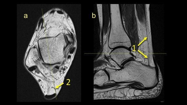
This diagnostic image consists of two MRI panels (a and b) illustrating the anatomy of the plantaris tendon and its relationship to the paratenon at the level of the ankle. Panel (a) is an axial T1-weighted image showing a transverse view of the ankle, where arrow 2 highlights the plantaris tendon inserting into the medial part of the paratenon, located superficially and posterior to the talus. Panel (b) is a sagittal cross-section where arrow 1 points to the plantaris tendon as it courses along the posterior aspect of the lower leg. The arrows in (b) demonstrate the longitudinal extension of the tendon; as it moves superiorly, the structure appears to fan out or thicken. The images provide anatomical detail regarding the distal lower leg, specifically the Kager's fat pad region and the fibrotic connections between the plantaris tendon, Achilles tendon paratenon, and local ligamentous structures. This comparison is used to visualize the plantaris tendon's morphology and its integration into the surrounding soft tissue layers for clinical and educational assessment of ankle anatomy.
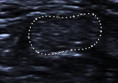
This transverse grayscale ultrasound image demonstrates the normal sonographic appearance of a human tendon, likely the Achilles tendon. The structure is outlined by a white dotted region of interest (ROI) markers, revealing an ovoid shape in cross-section. The tendon displays a characteristic fibrillary internal architecture, characterized by a uniform distribution of bright, hyperechoic dots and short linear echoes, which represent the parallel collagen fiber bundles imaged in the short axis. The borders are well-defined and smooth against the surrounding hypoechoic paratenon and subcutaneous tissues. The image serves as a baseline for understanding musculoskeletal sonography, specifically illustrating optimal perpendicular beam incidence to avoid anisotropy. In this orientation, the tendon appears correctly hyperechoic and organized, without the artifactual hypoechogenicity often mistaken for tendinopathy or partial-thickness tears.
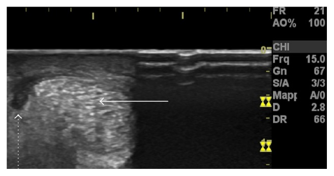
**Imaging Modality:** High-resolution musculoskeletal ultrasound (MSKUS). **Anatomical Region:** Achilles tendon, axial (transverse) cross-sectional view. **Observed Pathology:** Acute paratendinopathy with associated peritendinous fluid collection. **Visual Features:** * **Achilles Tendon:** The main body of the tendon (indicated by a solid white arrow) displays a characteristic speckled, hyperechoic fibrillar pattern in the transverse plane. * **Fluid Collection:** A distinct, well-defined hypoechoic (dark) crescentic area is visible at the lateral periphery of the tendon (indicated by a dotted arrow). * **Localization:** The hypoechoic region is situated between the tendon substance and the overlying paratenon, consistent with inflammatory exudate or fluid accumulation. * **Surrounding Tissue:** The superficial soft tissues and skin layers are visible superior to the tendon structure. **Clinical Significance:** This sonographic presentation is characteristic of acute paratendinopathy, where the primary inflammatory process involves the tendon sheath (paratenon) rather than the tendon transition itself. The presence of peritendinous fluid is a key diagnostic marker for differentiating acute paratenon inflammation from chronic tendinosis or mid-portion tears.
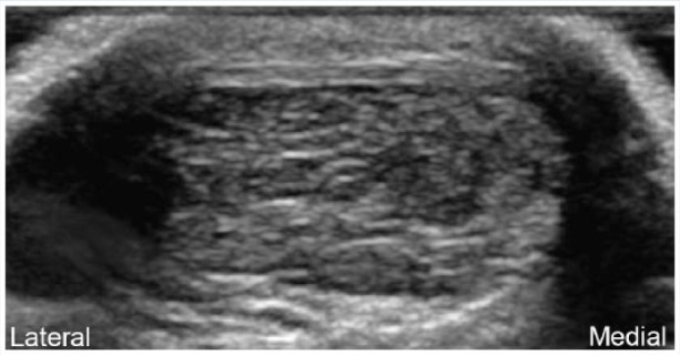
**Imaging Modality:** Musculoskeletal Ultrasound (grayscale) **Anatomical Region:** Posterior ankle, transverse view of the Achilles tendon. **Observations:** The image shows a transverse cross-section of the mid-portion Achilles tendon. Significant morphological changes are present, characterized by marked fusiform thickening and an overall increase in the cross-sectional area of the tendon. **Pathology/Features:** * **Echo-texture:** There is a loss of the normal organized fibrillar pattern. The tendon displays heterogeneous echogenicity with prominent, ill-defined hypoechoic (darker) areas. * **Distribution:** The hypoechoic changes are localized primarily to the central and medial aspects of the tendon body. This distribution corresponds to the anatomical territory of the soleus-derived fascicles. * **Borders:** The paratenon appears slightly thickened, though the anterior and posterior margins of the tendon remain identifiable despite the internal reorganization. **Diagnostic Context:** These sonographic findings are highly indicative of mid-portion Achilles tendinopathy (tendinosis). The specific localization to the medial and central fascicles suggests selective involvement of the soleus component of the Achilles complex. No discrete anechoic clefts suggestive of a full-thickness tear are visualized in this view.
Achilles tendon paratenon anatomy location layers diagram
Achilles tendon paratenon Kager fat pad sagittal MRI anatomy illustration
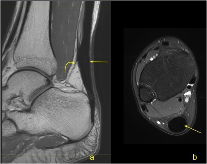
This diagnostic imaging set consists of two MRI views of the ankle and Achilles tendon in a 63-year-old patient following a partial rupture eight months prior. Image 'a' is a sagittal PD-weighted (proton density) MRI showing the Achilles tendon and Kager's fat pad. A straight yellow arrow indicates the area of tendon remodeling, characterized by persistent thickening and altered signal intensity despite clinical recovery. A curved yellow arrow points to peritendinous adhesions within Kager's fat pad, visible as linear low-signal bands. Image 'b' is an axial PD-weighted MRI with fat suppression. The straight yellow arrow highlights the thickened, hypointense Achilles tendon in cross-section. The use of fat suppression in this view increases the conspicuity of possible edema or fluid within the paratenon and adjacent soft tissues. These images illustrate the typical long-term morphological changes and postoperative sequelae, such as fibrosis and adhesions, following traumatic Achilles injury.
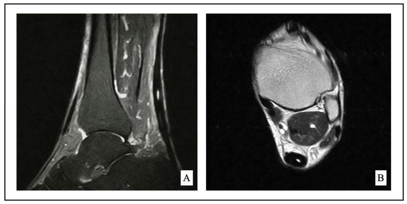
This diagnostic image consists of two MRI views of the ankle demonstrating midportion Achilles tendinopathy. Panel A is a sagittal fat-saturated MRI, which highlights fluid and edema as high-signal intensity. It reveals a thickened Achilles tendon with internal high-signal changes and disruption of the normal linear fibrillar pattern, characteristic of tendinosis and chronic inflammation. Adjacent structures including Kager's fat pad and the retrocalcaneal region show subtle reactive changes. Panel B is an axial T1-weighted MRI providing a cross-sectional view of the mid-tendon. This view demonstrates focal fusiform thickening (hypertrophy) and loss of the normal anterior concave or flat border of the Achilles tendon. The surrounding paratenon and soft tissues are visualized to assess for paratenonitis or fluid collections. These images are clinically significant for orthopedic and radiological evaluation of chronic Achilles tendon pathology, often used to guide management decisions between conservative physical therapy and surgical debridement.
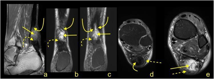
This set of multi-planar MRI images (sagittal, coronal, and axial) illustrates the status of an Achilles tendon four months after a rupture managed with conservative treatment. The modality includes PD-weighted (a), T2-weighted with fat suppression (b, c), and PD-weighted with fat suppression (d, e) sequences. Key diagnostic features include the clear separation of the proximal stump (indicated by curved solid arrows) and the distal stump (indicated by dashed curved arrows), both of which exhibit highly irregular, frayed outlines. Notably, there is a lack of visible fibrotic adhesions bridging the gap between the two stumps, signifying a failure of primary tendon healing or 'non-union'. The paratenon is clearly preserved but demonstrates pathological thickening (indicated by dashed straight arrows), suggesting chronic reactive changes. The surrounding Kager's fat pad and regional soft tissues are otherwise unremarkable. This case serves as an educational example of the long-term MRI appearance of unsuccessful conservative healing in total midportion Achilles tendon ruptures.
The Paratenon - Where It Is and What It Does
What Is It?
- An outer layer (epitenon) - thin, smooth fibrous membrane
- An inner layer - loose areolar connective tissue rich in mucopolysaccharides
- Together they form a gliding sleeve that allows the tendon to move 1.5-2 cm up and down relative to surrounding tissues during walking/running
Spatial Location - Layers from Skin to Bone (Posterior to Anterior)
SKIN
  ↓
Subcutaneous fat
  ↓
Paratenon - POSTERIOR surface (superficial/dorsal side of tendon)
  ↓
ACHILLES TENDON (the tendon body itself)
  ↓
Paratenon - ANTERIOR surface ← THIS IS THE HVIGI TARGET ZONE
  ↓
Kager's Fat Pad (triangular fat space)
  ↓
Calcaneus (bone)
"The paratenon does NOT extend around the tendon's deep (anterior) surface, where Kager's fat pad directly contacts the tendon itself."
MRI: Sagittal View - Paratenon and Kager's Fat Pad
MRI: Axial (Cross-Section) View - Where Paratenon Sits
Ultrasound: Paratenon as Fluid Collection
Why It Matters for HVIGI
- The anterior paratenon / Kager's fat pad interface is where Power Doppler shows neovascularization
- HVIGI needle is directed to this exact space - anterior to tendon, between tendon and fat pad
- The 50 mL of fluid mechanically strips the paratenon away from the tendon surface, tearing those neovasculature bundles
- Paratenon on the posterior/superficial side of the tendon is where inflammation and adhesions occur (paratenonitis) - felt as painful crepitus
Functions of the Paratenon
| Function | Detail |
|---|---|
| Gliding | Allows 1.5-2 cm of tendon excursion relative to surrounding tissue |
| Blood supply | Primary vascular conduit to the tendon (posterior tibial artery branches) |
| Proprioception | Contains sensory nerve endings |
| Healing | In rupture, paratenon contributes to repair cell migration |