
Topic pneumonia Q1. What is the most likely diagnosis? Q2.what are the differential diagnosis? Q3 . What are the treatment
pneumonia chest X-ray consolidation
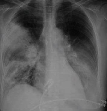
This diagnostic image is a posterior-anterior (PA) view chest X-ray demonstrating multifocal pneumonia with prominent consolidations. In the right lung, there is dense consolidation involving the upper lobe, characterized by increased radiopacity that obscures the regional vascular markings. Patchy, ill-defined airspace opacities are also visible extending into the right lower lobe. The left lung field reveals significant consolidation primarily localized within the lower lobe, partially obscuring the left hemidiaphragm silhouette. The cardiac silhouette appears normal in size, and the trachea is midline. The visual findings are consistent with severe pulmonary infection, such as atypical pneumonia (e.g., Legionella species). This clinical imaging serves as an educational example of multifocal bacterial consolidation and its radiographic distribution in the context of acute respiratory distress and sepsis.
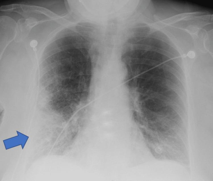
A frontal (anteroposterior) chest X-ray demonstrating clinical signs of pneumonia. A prominent blue arrow indicates a large area of increased opacity in the right lower lung field, suggestive of lobar consolidation or a dense pulmonary infiltrate. The opacity is heterogeneous with ill-defined borders, blending into the surrounding parenchyma. Beyond the focal area, there are diffuse bilateral reticulonodular interstitial markings, more pronounced in the mid and lower lung zones. The right costophrenic angle is relatively sharp, while the left shows mild blunting. Several medical devices are present: multiple radiopaque ECG leads/electrodes are positioned on the upper chest and shoulders, and a radiopaque enteric tube (nasogastric or feeding tube) is visible traversing the mediastinum toward the stomach. The image illustrates typical radiological findings of an infectious process in an acute clinical setting, highlighting focal consolidation against a background of generalized pulmonary congestion or inflammation.
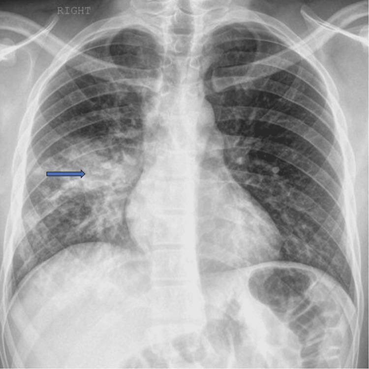
This diagnostic image is a posterior-anterior (PA) chest X-ray demonstrating common radiographic features of focal pneumonia. The primary finding is located in the right lung field, where patchy airspace opacities and an area of pulmonary consolidation are visible, highlighted by a blue arrow. The consolidation is characterized by increased density that obscures the underlying pulmonary vasculature, consistent with fluid or inflammatory exudate replacing air in the alveoli. The left lung field appears clear without similar focal opacities. The mediastinal contours, including the cardiac silhouette and trachea, are within normal limits. The costophrenic angles are sharp, suggesting the absence of significant pleural effusion. Bony structures, including the ribs, clavicles, and vertebrae, as well as the surrounding soft tissues, show no acute abnormalities. This image serves as a clinical example of localized infectious process, such as community-acquired pneumonia, in an adult patient.
"community-acquired pneumonia"[MeSH Terms] AND "treatment"[MeSH Terms]
Pneumonia: Diagnosis, Differentials & Treatment
Q1. Most Likely Diagnosis
- Fever (often high-grade, chills)
- Productive cough (purulent, blood-tinged, or rusty sputum)
- Dyspnea / chest pain (pleuritic in nature)
| Feature | Typical (Bacterial) Pneumonia | Atypical Pneumonia |
|---|---|---|
| Onset | Abrupt, rapid | Gradual/smoldering |
| Fever | High-grade (>38.5°C) | Low-grade |
| Cough | Productive, purulent | Dry, non-productive |
| WBC | >15,000 (neutrophilia) | Mildly elevated or normal |
| CXR | Lobar consolidation | Patchy bilateral infiltrates |
| Organisms | S. pneumoniae, H. influenzae, S. aureus | Mycoplasma, Chlamydia, Legionella, viruses |
- Decreased / bronchial breath sounds
- Dullness to percussion over affected lobe
- Egophony ("E to A" change)
- Increased tactile fremitus
- Chest X-ray (lobar opacity, infiltrates)
- CBC (leukocytosis with left shift)
- Sputum Gram stain + culture
- Blood cultures (in severe or hospitalized cases)
- Urine Legionella and pneumococcal antigens
- Pulse oximetry / ABG

Q2. Differential Diagnoses
Infectious Differentials
| Condition | Distinguishing Features |
|---|---|
| Pulmonary Tuberculosis | Chronic cough, night sweats, weight loss, upper lobe cavitation on CXR, AFB on sputum |
| Lung Abscess | Putrid sputum, fever, air-fluid level on CXR, often anaerobes/aspiration history |
| Viral Pneumonitis (influenza, COVID-19) | Bilateral ground-glass on CT, myalgia, viral prodrome |
| Fungal Pneumonia (Histoplasma, Blastomyces, PCP) | Immunocompromised host, endemic exposure; PCP in HIV patients |
| Atypical/Opportunistic | In HIV/AIDS: P. jirovecii, Cryptococcus, Coccidioides, atypical mycobacteria |
Non-Infectious Differentials
| Condition | Distinguishing Features |
|---|---|
| Pulmonary Embolism | Acute dyspnea, pleuritic chest pain, no fever initially, D-dimer elevated, V/Q mismatch |
| Congestive Heart Failure | Bilateral crackles, orthopnea, raised JVP, cardiomegaly, B-lines on lung ultrasound |
| Lung Cancer | Chronic cough, hemoptysis, weight loss, mass lesion on CXR/CT, no fever |
| ARDS / Chemical Pneumonitis | Aspiration history, bilateral opacities, hypoxia out of proportion |
| Pleural Effusion | Stony dullness, absent breath sounds, shifting dullness |
| Bronchitis | No consolidation on CXR, milder systemic symptoms |
Note: Blastomycotic pneumonia clinically and radiographically resembles community-acquired bacterial pneumonia, viral pneumonia, tuberculosis, and lung cancer - patients often receive multiple antibiotic courses before the correct diagnosis is made. - Harrison's Principles of Internal Medicine 22E
Q3. Treatment
Severity Assessment - PSI/PORT or CURB-65 Score
- Confusion
- Urea >7 mmol/L (BUN >19 mg/dL)
- Respiratory rate ≥30/min
- Blood pressure (systolic <90 or diastolic ≤60 mmHg)
- Age ≥65 years
| Score | Severity | Setting |
|---|---|---|
| 0-1 | Mild | Outpatient |
| 2 | Moderate | Consider hospitalization |
| 3-5 | Severe | Hospital / ICU |
Antibiotic Treatment by Setting
1. Outpatient CAP (No Comorbidities)
- Amoxicillin 1g TID × 5 days, OR
- Doxycycline 100 mg BD × 5 days (covers atypicals), OR
- Azithromycin 500 mg day 1, then 250 mg days 2-5 (macrolide)
2. Outpatient CAP (With Comorbidities: DM, COPD, heart/liver/renal disease, immunosuppression)
- Respiratory fluoroquinolone: Levofloxacin 750 mg OD or Moxifloxacin 400 mg OD × 5-7 days, OR
- Beta-lactam + macrolide: Amoxicillin-clavulanate + Azithromycin
"Although atypical pathogens commonly cause CAP, controlled trials show that beta-lactam antibiotics are as effective as macrolides and quinolones in most cases." - Textbook of Family Medicine 9e
3. Hospitalized CAP (Non-ICU)
- Beta-lactam + macrolide (e.g., IV Ceftriaxone 1-2 g OD + Azithromycin 500 mg OD), OR
- Respiratory fluoroquinolone monotherapy (Levofloxacin 750 mg OD)
4. Severe CAP (ICU)
- IV beta-lactam + IV macrolide (Ceftriaxone + Azithromycin), OR
- IV beta-lactam + fluoroquinolone
- Add anti-MRSA coverage (Vancomycin or Linezolid) if CA-MRSA suspected
- Add anti-Pseudomonal coverage (Piperacillin-tazobactam or Cefepime) if structural lung disease/recent hospitalization
5. Hospital-Acquired Pneumonia (HAP) / Ventilator-Acquired Pneumonia (VAP)
- Broader coverage targeting gram-negatives including Pseudomonas, Klebsiella, Serratia
- Piperacillin-tazobactam OR Cefepime OR Meropenem + anti-MRSA agent
Supportive Management
- Oxygen to maintain SpO2 ≥94% (≥88-92% in COPD)
- IV fluids for dehydration or sepsis
- Antipyretics/analgesics (paracetamol for fever and pleuritic pain)
- Bronchodilators if wheeze present
- Chest physiotherapy to aid sputum clearance
- DVT prophylaxis in hospitalized patients
- Nutrition support
Vaccination (Prevention)
- Influenza vaccine: annually for all age >50, chronic lung disease, diabetes, immunocompromised
- Pneumococcal vaccine (PCV13/PPSV23): all adults >65 and high-risk patients; revaccinate after 10 years if first dose given before age 65
In short notes
Pneumonia - Short Notes
Q1. Most Likely Diagnosis
- Fever + chills, productive cough (purulent/rusty sputum)
- Dyspnea, pleuritic chest pain
- Tachypnea, tachycardia
- Dullness to percussion
- Bronchial breath sounds + egophony
- Increased tactile fremitus
Q2. Differential Diagnoses
| Condition | Key Clue |
|---|---|
| Pulmonary TB | Night sweats, weight loss, upper lobe cavitation |
| Lung Abscess | Putrid sputum, air-fluid level on CXR |
| Pulmonary Embolism | No fever, elevated D-dimer, risk factors |
| Heart Failure | Bilateral crackles, orthopnea, raised JVP |
| Lung Cancer | Chronic cough, mass lesion, no fever |
| Viral Pneumonitis | Bilateral ground-glass, viral prodrome |
| PCP (HIV patient) | Immunocompromised, dry cough, bilateral infiltrates |
| Bronchitis | No consolidation on CXR, milder illness |
Q3. Treatment
Step 1 - Assess Severity (CURB-65)
- Score 0-1 → Outpatient | Score 2 → Hospital | Score 3+ → ICU
Step 2 - Antibiotics
- Amoxicillin 1g TDS × 5 days, OR Doxycycline 100mg BD, OR Azithromycin
- Levofloxacin 750mg OD, OR Amoxicillin-clavulanate + Azithromycin
- IV Ceftriaxone + Azithromycin, OR Levofloxacin monotherapy
- IV Beta-lactam + IV Macrolide ± add Vancomycin (if MRSA) or Piperacillin-tazobactam (if Pseudomonas risk)
Step 3 - Supportive
- O2 (SpO2 ≥94%)
- IV fluids
- Paracetamol for fever/pain
- DVT prophylaxis
- Chest physiotherapy
Prevention
- Annual influenza vaccine
- Pneumococcal vaccine (PCV13/PPSV23) for age >65 and high-risk groups