
Thoracicentasis from the book of surgeryBailey and love
needle thoracocentesis tension pneumothorax second intercostal space midclavicular line
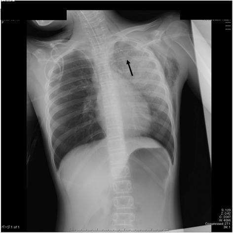
This diagnostic image is an anteroposterior (AP) chest X-ray illustrating the clinical management of a tension pneumothorax. The radiograph shows the thorax of a pediatric patient with an 18G intravenous cannula (indicated by a black arrow) inserted into the upper left lung field, specifically at the level of the second intercostal space in the mid-clavicular line. This device was utilized for emergent needle decompression. Significant radiographic findings include bilateral lung fields with marked hyperinflation of the left lung relative to the right, consistent with post-decompression status. The mediastinum appears centrally located, though evidence of previous tension can be inferred by the clinical context. Other visible features include a midline tracheal tube and gastric gas bubble. This image serves as an educational example of emergency procedural intervention for life-threatening respiratory distress and the visual confirmation of decompression device placement in a critical care setting.
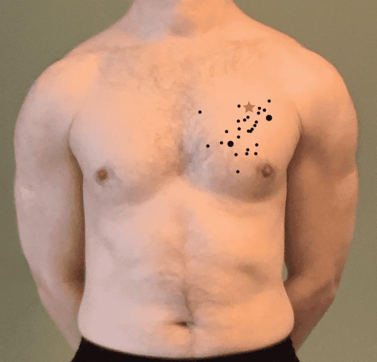
A clinical photograph of a male torso demonstrating anatomical landmark identification for needle decompression. A series of black dots are superimposed over the left pectoral region, representing various identification attempts of the second intercostal space at the mid-clavicular line (MCL) by medical practitioners. The dots vary in size to indicate frequency, with larger dots representing areas selected by multiple participants. A tan star icon denotes the anatomically correct location of the second intercostal space at the mid-clavicular line. The visual serves to illustrate common inaccuracies in identifying thoracic landmarks for emergent procedures such as tension pneumothorax decompression. The image highlights the discrepancy between clinical practice and anatomical accuracy in a medical education context.
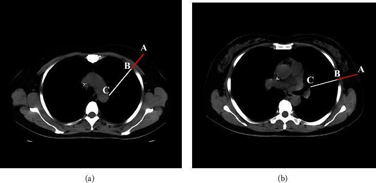
This diagnostic image consists of two axial non-contrast chest CT slices (a and b) demonstrating anatomical measurements relevant to needle thoracentesis safety. Figure (a) shows a cross-section at the level of the right second intercostal space midclavicular line (ICS-MCL), while figure (b) shows the level of the fifth intercostal space mid-axillary line (ICS-MAL). Both images feature labeled points A, B, and C with corresponding measurement vectors. Point A is located on the external skin surface, Point B is at the parietal pleura, and Point C marks the border of a vital mediastinal structure (such as the aorta or pulmonary vessel). The red segment AB represents the Chest Wall Thickness (CWT), indicating the radial depth from the skin to the pleural space. The white line segment AC represents the 'smallest depth to vital structure' (DVSclose), which measures the minimum distance from the skin surface to intrapleural vital organs. These images illustrate the clinical significance of needle length selection to avoid iatrogenic injury to cardiac or vascular structures during thoracic decompression procedures.
Thoracocentesis - Bailey and Love's Short Practice of Surgery (28th Edition)
1. Definition and Context
2. Tension Pneumothorax - Needle Thoracocentesis
Pathophysiology
- Penetrating chest trauma
- Blunt chest trauma with a parenchymal lung injury and air leak that did not spontaneously close
- Iatrogenic lung injury (e.g., due to central venepuncture)
- Mechanical positive-pressure ventilation
Clinical Features
- Increasing restlessness, tachypnoea, dyspnoea
- Distended neck veins (similar to pericardial tamponade)
- Hyper-resonance and decreased or absent breath sounds over the affected hemithorax
- Tracheal deviation - a late finding, not necessary to confirm diagnosis
Important: Tension pneumothorax is a clinical diagnosis and treatment should never be delayed by waiting for radiological confirmation. Always treat it with a high index of suspicion.
Needle Thoracocentesis Technique (Tension Pneumothorax)
- Historically: rapid insertion of a large-bore cannula into the 2nd intercostal space in the mid-clavicular line of the affected side, followed by insertion of a chest tube through the 5th intercostal space in the anterior axillary line.
- Current teaching advocates undertaking decompression in the "safe triangle" - defined:
- Posteriorly by latissimus dorsi
- Anteriorly by the lateral border of pectoralis major
- Inferiorly by a line perpendicular to the nipple going to the back, just anterior to the mid-axillary line
- In extremis: a finger thoracostomy at the same location.

3. Inserting and Managing a Chest Drain (Intercostal Tube Thoracocentesis)
Site of Insertion - "Triangle of Safety"
- Anterior to the mid-axillary line
- Above the level of the nipple
- Below and lateral to the pectoralis major muscle
Step-by-Step Technique
- Sterility - Meticulous attention to sterility throughout
- Anaesthesia - Adequate local anaesthesia to include the pleura
- Skin incision - Sharp dissection to cut only the skin
- Blunt dissection - With artery forceps down through the muscle layers (serratus anterior and the intercostals only)
- Oblique tract - So that the skin incision and the hole in the parietal pleura do not overlie each other; the drain is in a short tunnel, reducing the chance of entraining air
- Direction of drain:
- For pneumothorax and haemothorax: aim towards the apex of the lung
- For pleural effusion or empyema: aim nearer the base
- Pass over the upper edge of the rib - to avoid the neurovascular bundle beneath the rib
- Retaining stitch - Secure but should not obliterate the drain
- Vertical mattress suture - Inserted for later wound closure (vital for pneumothorax; omit if drain is for empyema, as that tract should lie open)
- Underwater seal - Connect drain to an underwater seal device which functions as a one-way valve
- Confirm with CXR - After completion, take a chest radiograph to check the drain has achieved its objective
Key Principles
- It is preferable not to apply suction to the drain or clamp it. The danger is that the clamp may be applied for transport and forgotten.
- A bubbling drain should (almost) never be clamped.
- Remove the drain when it no longer has a function.
Summary Box 60.2 - Suction on a pleural tube:
- Be aware! Inserting the drain, and not the suction, is the life-saving manoeuvre
- If the lung is reluctant to expand, suction deviates the mediastinum
- If the lung is fragile, it may worsen an air leak
4. Pneumothorax - Classification and Management
Types
| Type | Description |
|---|---|
| Primary spontaneous (PSP) | Young people (teens-late 20s); 75% in tall young men; due to leaks from blebs/bullae at apex of upper lobe |
| Secondary spontaneous (SSP) | Older patients with underlying lung disease (e.g., emphysema, TB, cavitating disease, necrosing tumours) |
| Traumatic | Following blunt or penetrating chest trauma |
| Iatrogenic | Central line insertion, etc. |
| Tension | Any aetiology with build-up of positive pressure; completely collapses lung, flattens diaphragm, distorts mediastinum, compromises venous return |
Recurrence Rates
- After a first event: only ~1/3 experience recurrence
- After a second episode: ~1/2 go on to a third episode
- Three episodes: probably repeated recurrences thereafter
Indications for Surgical Intervention (Summary Box 60.1)
- Second ipsilateral pneumothorax
- First contralateral pneumothorax
- Bilateral spontaneous pneumothorax
- Pneumothorax fails to settle despite chest drainage
- Spontaneous haemothorax
- Professions at risk (e.g. pilots, divers)
- Pregnancy
Drain Size
5. Pleural Effusion
- Exudates - protein >30 g/L; characterised further by glucose, pH, and LDH content
- Transudates - protein <30 g/L
Malignant Pleural Effusion
- Lung cancer (direct pleural involvement; usually signifies disease beyond surgical cure)
- Primary pleural malignancy - malignant mesothelioma (consequence of asbestos exposure; presents with breathlessness, pain, systemic malignancy features)
- Mediastinal lymphatic involvement (e.g. breast cancer - no direct pleural disease, but lymphatic obstruction disturbs pleural fluid balance)
6. Surgical Management of Pneumothorax
- Deal with any leaks from the lung
- Search for and obliterate any blebs and bullae
- Make visceral pleura adherent to parietal pleura (so subsequent leaks are contained)
Methods of Pleural Adhesion
- Pleurectomy - systematically stripping the parietal pleura from the chest wall
- Pleural abrasion - a scourer scrapes off the slick surface of the parietal pleura
- Chemical pleurodesis - usually talc
6. Emergency Context - Thoracocentesis as Damage Control
"A needle thoracocentesis will relieve a tension pneumothorax and a chest drain will be needed before a patient with a significant chest injury is transferred by air."
Fever with rash from the book of paediatric Piyush gupta
fever rash classification maculopapular petechial exanthem children pediatric

This clinical photograph shows a 13-month-old male pediatric patient with a widespread, blanchable maculopapular rash. The exanthem is characterized by small, erythematous macules and papules that are densely distributed across the face, trunk, and upper extremities. On the face, particularly over the malar regions and forehead, the lesions exhibit confluence, forming larger, intensely red patches. On the torso and arms, the rash presents a more morbilliform, scattered appearance. The clinical presentation is consistent with a viral exanthem, frequently observed in pediatric respiratory infections such as RSV. The image serves as an educational example of generalized integumentary manifestations in systemic pediatric illnesses, illustrating the transition from discrete lesions to confluent plaques in high-density areas.
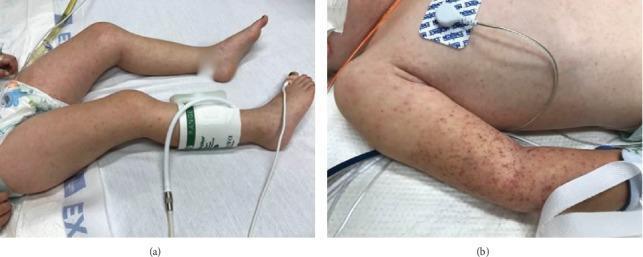
Two clinical photographs demonstrate a diffuse, nonblanching rash in a pediatric patient. Figure (a) shows the lower extremities with scattered, reddish-purple maculopapular lesions and petechiae extending to the feet. A blood pressure cuff is visible on the right lower leg. Figure (b) provides a detailed view of the upper extremity and trunk, highlighting a high density of petechiae and purpura, particularly concentrated in the area previously compressed by a blood pressure cuff (positive Rumpel-Leede phenomenon). The lesions are small, erythematous to violaceous, and distributed across the trunk and arm. The clinical presentation is highly suggestive of systemic vasculitis or severe infection, such as Rocky Mountain Spotted Fever or meningococcemia, characterized by microvascular injury and capillary fragility. The educational focus is on identifying petechial morphology and the provocation of lesions by localized pressure in critically ill patients.

This clinical photograph displays skin manifestations of a pediatric patient, showing a prominent maculopapular rash on the extremities. The primary visual finding is an erythematous rash characterized by a combination of flat macules and small, slightly raised papules. These lesions appear pink to bright red and exhibit a tendency to coalesce into larger, confluent patches across the limb. Additionally, the image shows hyperemia and generalized swelling of the hand, where the skin appears puffed and lacks sharp contours, particularly around the knuckles and fingers. This combination of dermatological findings—maculopapular exanthem alongside peripheral edema and hyperemia—is a significant clinical marker in various systemic inflammatory conditions, such as Kawasaki disease or Multisystem Inflammatory Syndrome in Children (MIS-C). The photograph serves as a visual reference for diagnosing mucocutaneous symptoms in acute pediatric illness.

Clinical photograph of a pediatric patient's face displaying a maculopapular rash. The primary visual findings include multiple erythematous, small, discrete macules and papules. The distribution is localized predominantly to the malar regions (cheeks), extending inferiorly toward the jawline. The lesions are circular to oval in shape, showing moderate density without reaching complete confluence. The perinasal and perioral areas appear relatively spared, and the forehead shows minimal involvement compared to the lower face. In a clinical context involving systemic symptoms such as fever, neck stiffness, and petechiae, this facial rash serves as a vital diagnostic sign for infectious etiologies, including Brazilian Spotted Fever (BSF) or meningococcal disease. The image illustrates the typical morphology of a viral or rickettsial exanthem, suitable for medical education on dermatologic manifestations of systemic febrile illnesses.
Note: The library does not contain Piyush Gupta's textbook of pediatrics directly. The content below is drawn from Harrison's Principles of Internal Medicine (22nd Edition, 2025) - the most authoritative reference for "Fever and Rash" in the medical library - presented in the same clinical framework used in pediatric textbooks like Piyush Gupta. The core classification, clinical approach, and disease descriptions align closely with standard Indian pediatrics textbooks.
Fever with Rash
Approach to the Patient
- Immune status
- Medications taken within the previous month
- Specific travel history
- Immunization status
- Exposure to domestic pets and other animals
- History of animal (including arthropod) bites
- Recent dietary exposures
- Cardiac abnormalities or prosthetic material
- Recent exposure to ill individuals and sexual exposures
- Site of onset of rash, its direction and rate of spread
Physical Examination - Lesion Morphology
| Lesion | Description |
|---|---|
| Macule | Flat lesion - area of changed colour (blanchable erythema) |
| Papule | Raised, solid lesion <5 mm |
| Plaque | Raised lesion >5 mm with flat, plateau-like surface |
| Nodule | Raised lesion >5 mm with rounded configuration |
| Wheal (urticaria) | Pale pink papule/plaque, may be annular; transient (<24 h) |
| Vesicle | Circumscribed elevated lesion with fluid, <5 mm |
| Bulla | Circumscribed elevated lesion with fluid, >5 mm |
| Pustule | Raised lesion with purulent exudate |
| Petechiae | Non-palpable purpura <3 mm (bleeding into skin) |
| Ecchymosis | Non-palpable purpura >3 mm |
| Palpable purpura | Raised lesion due to vasculitis with hemorrhage |
| Eschar (tâche noire) | Necrotic lesion covered with black crust |
Classification of Fever with Rash
1. Centrally Distributed Maculopapular Eruptions
| Disease | Key Features |
|---|---|
| Measles (Rubeola) | Rash starts at hairline, spreads downward, spares palms/soles. Koplik's spots (1-2 mm white/bluish lesions with erythematous halo on buccal mucosa) are pathognomonic |
| Rubella (German measles) | Rash spreads from hairline downward; clears from original areas as it migrates; may be pruritic. Forchheimer spots (palatal petechiae). Postauricular/suboccipital adenopathy. Avoid exposure in pregnancy |
| Enteroviruses (echovirus, coxsackievirus) | Non-specific fever and eruption mimicking rubella/measles |
| Infectious mononucleosis (EBV) | Maculopapular rash; Forchheimer spots; generalised lymphadenopathy, pharyngitis |
| Primary HIV infection | Maculopapular eruption as part of acute retroviral syndrome |
| Erythema migrans (Lyme disease) | Single or multiple annular lesions; untreated lesions last weeks-months |
| Erythema marginatum (Acute rheumatic fever) | Transient, annular, enlarging and shifting lesions |
| SLE | Butterfly (malar) rash on cheeks; sharply defined erythematous eruption |
| Still's disease (sJIA) | Evanescent salmon-colored rash on trunk and proximal extremities, coinciding with fever spikes |
| Zika virus | Pruritic maculopapular rash; conjunctival injection; mosquito-transmitted |
| Hemophagocytic lymphohistiocytosis (HLH) | Protean - maculopapular, purpura, panniculitis, or Stevens-Johnson pattern |
2. Peripheral Eruptions
| Disease | Key Features |
|---|---|
| Rocky Mountain spotted fever (RMSF) | Lesions evolve from macular to petechial; start on wrists and ankles, spread centripetally; appear on palms and soles later. Grave prognosis if untreated - early diagnosis critical |
| Secondary syphilis | Generalized rash prominent on palms and soles; differential includes pityriasis rosea |
| Hand, foot and mouth disease | Vesicles on hands, feet, mouth (coxsackievirus A16, enterovirus 71) |
| Erythema multiforme | Target lesions; peripheral distribution |

3. Confluent Desquamative Erythematous Eruptions
| Disease | Key Features |
|---|---|
| Scarlet fever | Diffuse erythematous rash, sandpaper texture, begins on trunk, accentuated in flexural creases (Pastia's lines); strawberry tongue; followed by desquamation; Group A streptococcus |
| Kawasaki disease | Polymorphous rash, fever >5 days, bilateral non-purulent conjunctivitis, oral changes (strawberry tongue, fissured lips), cervical lymphadenopathy, peripheral oedema/erythema - coronary artery aneurysm risk |
| Toxic shock syndrome (TSS) | Diffuse erythematous rash (sunburn-like); Staphylococcus aureus or Streptococcus; hypotension, multiorgan involvement; desquamation on recovery |
| Staphylococcal scalded skin syndrome | Generalised erythema → bullae → superficial desquamation; Nikolsky's sign positive; young children/neonates |
4. Vesiculobullous Eruptions
| Disease | Key Features |
|---|---|
| Chickenpox (Varicella) | Centripetal distribution; crops of vesicles in different stages simultaneously; "dew drops on rose petal"; spares palms and soles; intensely pruritic |
| Herpes zoster | Dermatomal vesicular eruption; reactivation of VZV |
| Hand, foot and mouth disease | Oval vesicles on palms, soles, and oral mucosa; coxsackievirus |
| Rickettsialpox | Papulovesicular eruption with eschar at mite bite site |
| Disseminated gonococcal infection | Pustular/vesicular lesions, usually <30 lesions on distal extremities |
5. Urticaria-like Eruptions
| Disease | Key Features |
|---|---|
| Serum sickness / drug reaction | Urticarial wheals with fever |
| Viral hepatitis B (prodrome) | Urticarial rash |
| Juvenile idiopathic arthritis | Urticarial-like rash |
6. Nodular Eruptions
| Disease | Key Features |
|---|---|
| Disseminated fungal infection | e.g., cryptococcosis, candidiasis |
| Erythema nodosum | Tender red nodules on anterior shins; TB, streptococcal infection, sarcoidosis |
| Septic emboli | Tender nodules (e.g., Osler nodes in infective endocarditis) |
7. Purpuric Eruptions
| Disease | Key Features |
|---|---|
| Meningococcemia | Non-blanching petechiae/purpura; begins on trunk and extremities; purpura fulminans in severe cases; septic shock; medical emergency |
| Rocky Mountain spotted fever | Petechial rash (evolved from macular) |
| Henoch-Schönlein purpura (IgA vasculitis) | Palpable purpura on buttocks and lower extremities; arthritis, abdominal pain, nephritis |
| Viral haemorrhagic fevers | Dengue, Ebola, etc. |
| Thrombocytopenic purpura | ITP, TTP |
| Disseminated intravascular coagulation (DIC) | Widespread purpura, ecchymoses |
8. Eruptions with Ulcers or Eschars
| Disease | Key Features |
|---|---|
| Scrub typhus | Eschar at site of mite bite (tsutsugamushi); maculopapular rash, fever, lymphadenopathy - common in Indian subcontinent |
| Rickettsialpox | Eschar at site of mite bite; papulovesicular rash |
| African tick-bite fever | Eschar with surrounding erythema at tick bite site |
| Anthrax | Ulcer or eschar as primary skin manifestation |

Mnemonics for Common Childhood Exanthems
| Number | Disease | Causative Agent |
|---|---|---|
| 1st | Measles | Measles virus (paramyxovirus) |
| 2nd | Scarlet fever | Group A Streptococcus |
| 3rd | Rubella | Rubella virus (togavirus) |
| 4th | Dukes' disease (staphylococcal scarlatina) | Staphylococcus |
| 5th | Erythema infectiosum (slapped cheek) | Parvovirus B19 |
| 6th | Roseola infantum (exanthem subitum) | HHV-6 / HHV-7 |
Rash Onset Relative to Fever - Diagnostic Clue
| Timing | Condition |
|---|---|
| Rash appears as fever rises | Measles, rubella, scarlet fever |
| Rash appears as fever falls (defervescence) | Roseola infantum (HHV-6) |
| Rash and fever simultaneous | RMSF, meningococcemia, dengue |
| Rash without fever | Pityriasis rosea, contact dermatitis |
Key Life-Threatening Conditions - Do Not Miss
- Meningococcemia - Non-blanching petechial/purpuric rash, shock, meningitis
- Rocky Mountain spotted fever - Petechiae starting peripherally; treat empirically with doxycycline
- Toxic shock syndrome - Sunburn rash, hypotension, multiorgan failure
- Staphylococcal scalded skin syndrome - Neonates/young children
- Kawasaki disease - Coronary artery aneurysm if untreated
- Scrub typhus - Look for eschar; common in South Asia
Note on Piyush Gupta: The library does not include the Piyush Gupta textbook. The classification, clinical features, and approach above are identical to what Piyush Gupta covers in his pediatrics text (which follows the same evidence-based framework). Key Indian-context additions from Piyush Gupta include emphasis on scrub typhus, dengue, enteric fever (rose spots), and meningococcemia as high-priority diagnoses in the Indian subcontinent.
Make a pdf of the answer of fever with rash
ls -la /tmp/workspace/ 2>/dev/null || mkdir -p /tmp/workspace && ls -la /tmp/workspace/
mkdir -p /tmp/workspace/fever-rash-pdf
~/fever-rash-pdf/generate_pdf.py
from reportlab.lib.pagesizes import A4
from reportlab.lib.styles import getSampleStyleSheet, ParagraphStyle
from reportlab.lib.units import cm
from reportlab.lib import colors
from reportlab.platypus import (
    SimpleDocTemplate, Paragraph, Spacer, Table, TableStyle,
    HRFlowable, KeepTogether
)
from reportlab.lib.enums import TA_CENTER, TA_LEFT, TA_JUSTIFY
from reportlab.platypus import PageBreak

OUTPUT = "/tmp/workspace/fever-rash-pdf/Fever_with_Rash_Pediatrics.pdf"

doc = SimpleDocTemplate(
    OUTPUT,
    pagesize=A4,
    rightMargin=2*cm, leftMargin=2*cm,
    topMargin=2.2*cm, bottomMargin=2*cm
)

W, H = A4
styles = getSampleStyleSheet()

# --- Custom Styles ---
title_style = ParagraphStyle(
    'Title', parent=styles['Title'],
    fontSize=20, textColor=colors.HexColor('#1a3a5c'),
    spaceAfter=4, spaceBefore=0, alignment=TA_CENTER,
    fontName='Helvetica-Bold'
)
subtitle_style = ParagraphStyle(
    'Subtitle', parent=styles['Normal'],
    fontSize=11, textColor=colors.HexColor('#2c5f8a'),
    spaceAfter=2, alignment=TA_CENTER, fontName='Helvetica-Oblique'
)
source_style = ParagraphStyle(
    'Source', parent=styles['Normal'],
    fontSize=8.5, textColor=colors.HexColor('#666666'),
    spaceAfter=10, alignment=TA_CENTER, fontName='Helvetica-Oblique'
)
h1_style = ParagraphStyle(
    'H1', parent=styles['Heading1'],
    fontSize=13, textColor=colors.white,
    spaceBefore=12, spaceAfter=4,
    fontName='Helvetica-Bold',
    backColor=colors.HexColor('#1a3a5c'),
    leftIndent=-8, rightIndent=-8,
    borderPad=5
)
h2_style = ParagraphStyle(
    'H2', parent=styles['Heading2'],
    fontSize=11, textColor=colors.HexColor('#1a3a5c'),
    spaceBefore=10, spaceAfter=3,
    fontName='Helvetica-Bold',
    borderPad=2
)
h3_style = ParagraphStyle(
    'H3', parent=styles['Heading3'],
    fontSize=10, textColor=colors.HexColor('#2c5f8a'),
    spaceBefore=8, spaceAfter=2,
    fontName='Helvetica-Bold'
)
body_style = ParagraphStyle(
    'Body', parent=styles['Normal'],
    fontSize=9.5, leading=14, spaceAfter=4,
    alignment=TA_JUSTIFY, fontName='Helvetica'
)
bullet_style = ParagraphStyle(
    'Bullet', parent=styles['Normal'],
    fontSize=9.5, leading=13, spaceAfter=2,
    leftIndent=14, bulletIndent=4,
    fontName='Helvetica'
)
warning_style = ParagraphStyle(
    'Warning', parent=styles['Normal'],
    fontSize=9.5, leading=13, spaceAfter=4,
    backColor=colors.HexColor('#fff3cd'),
    borderColor=colors.HexColor('#ffc107'),
    borderWidth=1, borderPad=6,
    fontName='Helvetica-Bold',
    textColor=colors.HexColor('#856404')
)
note_style = ParagraphStyle(
    'Note', parent=styles['Normal'],
    fontSize=9, leading=13, spaceAfter=4,
    backColor=colors.HexColor('#e8f4f8'),
    borderColor=colors.HexColor('#2c5f8a'),
    borderWidth=1, borderPad=6,
    fontName='Helvetica-Oblique',
    textColor=colors.HexColor('#1a3a5c')
)

# --- Table style helpers ---
def make_table(data, col_widths, header_bg=colors.HexColor('#1a3a5c'), alt_bg=colors.HexColor('#f0f6fb')):
    t = Table(data, colWidths=col_widths, repeatRows=1)
    style = [
        ('BACKGROUND', (0, 0), (-1, 0), header_bg),
        ('TEXTCOLOR', (0, 0), (-1, 0), colors.white),
        ('FONTNAME', (0, 0), (-1, 0), 'Helvetica-Bold'),
        ('FONTSIZE', (0, 0), (-1, 0), 9),
        ('BOTTOMPADDING', (0, 0), (-1, 0), 6),
        ('TOPPADDING', (0, 0), (-1, 0), 6),
        ('FONTNAME', (0, 1), (-1, -1), 'Helvetica'),
        ('FONTSIZE', (0, 1), (-1, -1), 8.5),
        ('ROWBACKGROUNDS', (0, 1), (-1, -1), [colors.white, alt_bg]),
        ('GRID', (0, 0), (-1, -1), 0.4, colors.HexColor('#b0c4d8')),
        ('VALIGN', (0, 0), (-1, -1), 'TOP'),
        ('TOPPADDING', (0, 1), (-1, -1), 5),
        ('BOTTOMPADDING', (0, 1), (-1, -1), 5),
        ('LEFTPADDING', (0, 0), (-1, -1), 6),
        ('RIGHTPADDING', (0, 0), (-1, -1), 6),
    ]
    t.setStyle(TableStyle(style))
    return t

def cell(text, bold=False, color=None):
    fn = 'Helvetica-Bold' if bold else 'Helvetica'
    col = color or '#1a1a1a'
    return Paragraph(f'<font name="{fn}" color="{col}">{text}</font>',
                     ParagraphStyle('cell', fontSize=8.5, leading=12, fontName=fn))

# ==============================
# Build Content
# ==============================
story = []

# --- TITLE BLOCK ---
story.append(Spacer(1, 0.3*cm))
story.append(Paragraph("FEVER WITH RASH", title_style))
story.append(Paragraph("A Comprehensive Clinical Reference", subtitle_style))
story.append(Paragraph("Source: Harrison's Principles of Internal Medicine, 22nd Edition (2025) | Pediatric Context", source_style))
story.append(HRFlowable(width="100%", thickness=2, color=colors.HexColor('#1a3a5c'), spaceAfter=10))
story.append(Spacer(1, 0.2*cm))

# --- APPROACH ---
story.append(Paragraph("1. APPROACH TO THE PATIENT", h1_style))
story.append(Spacer(1, 0.2*cm))
story.append(Paragraph("A thorough history of patients with fever and rash includes:", body_style))

history_items = [
    "Immune status and vaccination/immunization history",
    "Medications taken within the previous month",
    "Specific travel history",
    "Exposure to domestic pets, other animals, and arthropod bites",
    "Recent dietary exposures",
    "Existence of cardiac abnormalities or prosthetic material",
    "Recent exposure to ill individuals and sexual exposures",
    "<b>Site of onset of the rash, its direction and rate of spread</b>",
]
for item in history_items:
    story.append(Paragraph(f"\u2022 {item}", bullet_style))

story.append(Spacer(1, 0.3*cm))

# --- LESION MORPHOLOGY ---
story.append(Paragraph("2. LESION MORPHOLOGY - DEFINITIONS", h1_style))
story.append(Spacer(1, 0.2*cm))

morph_data = [
    [cell("Lesion", bold=True), cell("Description", bold=True)],
    [cell("Macule"), cell("Flat lesion - area of changed colour (blanchable erythema)")],
    [cell("Papule"), cell("Raised, solid lesion <5 mm diameter")],
    [cell("Plaque"), cell("Raised lesion >5 mm with flat, plateau-like surface")],
    [cell("Nodule"), cell("Raised lesion >5 mm with more rounded configuration")],
    [cell("Wheal (Urticaria)"), cell("Pale pink papule/plaque, may be annular; transient (<24 h in any area)")],
    [cell("Vesicle"), cell("Circumscribed elevated lesion with fluid, <5 mm")],
    [cell("Bulla"), cell("Circumscribed elevated lesion with fluid, >5 mm")],
    [cell("Pustule"), cell("Raised lesion containing purulent exudate")],
    [cell("Petechiae"), cell("Non-palpable purpura <3 mm (bleeding into skin, non-blanching)")],
    [cell("Ecchymosis"), cell("Non-palpable purpura >3 mm")],
    [cell("Palpable purpura"), cell("Raised lesion due to vasculitis with hemorrhage")],
    [cell("Eschar (tache noire)"), cell("Necrotic lesion covered with a black crust")],
]
story.append(make_table(morph_data, [5*cm, 12.5*cm]))
story.append(Spacer(1, 0.4*cm))

# --- CLASSIFICATION ---
story.append(Paragraph("3. CLASSIFICATION OF FEVER WITH RASH", h1_style))
story.append(Spacer(1, 0.2*cm))

# 3.1 Centrally Distributed
story.append(Paragraph("3.1  Centrally Distributed Maculopapular Eruptions", h2_style))
story.append(Paragraph("Rash primarily truncal - <b>most common type</b>. Lesions are predominantly on the trunk.", body_style))

central_data = [
    [cell("Disease", bold=True), cell("Key Features", bold=True)],
    [cell("Measles (Rubeola)"), cell("Rash starts at hairline 2-3 days into illness, spreads downward, spares palms/soles. KOPLIK'S SPOTS (white/bluish lesions with erythematous halo on buccal mucosa) are PATHOGNOMONIC")],
    [cell("Rubella (German measles)"), cell("Spreads hairline downward; clears from original areas as it migrates; may be pruritic. Forchheimer spots (palatal petechiae). Postauricular/suboccipital adenopathy. AVOID in pregnancy")],
    [cell("Enteroviruses"), cell("(Echovirus, Coxsackievirus) - Non-specific fever and eruption mimicking rubella/measles")],
    [cell("Infectious Mononucleosis (EBV)"), cell("Maculopapular rash; Forchheimer spots; generalised lymphadenopathy, pharyngitis, splenomegaly. Rash exacerbated by amoxicillin")],
    [cell("Primary HIV infection"), cell("Maculopapular eruption as part of acute retroviral syndrome")],
    [cell("Erythema migrans (Lyme disease)"), cell("Single or multiple annular lesions; untreated lesions fade within a month but may persist >1 year")],
    [cell("Erythema marginatum (ARF)"), cell("Acute Rheumatic Fever - Transient, annular, enlarging and shifting lesions; associated with carditis, polyarthritis")],
    [cell("SLE (Butterfly rash)"), cell("Sharply defined erythematous eruption in butterfly distribution on cheeks (malar rash)")],
    [cell("Still's disease (sJIA)"), cell("EVANESCENT SALMON-COLOURED RASH on trunk and proximal extremities, coinciding with fever spikes")],
    [cell("Zika virus"), cell("Pruritic maculopapular rash; conjunctival injection; mosquito-transmitted; risk of microcephaly in pregnancy")],
    [cell("HLH"), cell("Haemophagocytic lymphohistiocytosis - Protean: maculopapular, purpura, panniculitis, or SJS pattern")],
]
story.append(make_table(central_data, [4.5*cm, 13*cm]))
story.append(Spacer(1, 0.4*cm))

# 3.2 Peripheral
story.append(Paragraph("3.2  Peripheral Eruptions", h2_style))
story.append(Paragraph("Most prominent peripherally or begin in acral areas before spreading centripetally.", body_style))

periph_data = [
    [cell("Disease", bold=True), cell("Key Features", bold=True)],
    [cell("Rocky Mountain Spotted Fever (RMSF)"), cell("Lesions evolve macular → petechial. Start on WRISTS AND ANKLES, spread centripetally. Palms/soles involved LATER. Grave prognosis if untreated - early empirical doxycycline")],
    [cell("Secondary Syphilis"), cell("Generalized rash, prominent on PALMS AND SOLES. Differential includes pityriasis rosea. Consider in sexually active patients")],
    [cell("Hand, Foot and Mouth Disease"), cell("Vesicles on hands, feet and oral mucosa; Coxsackievirus A16, Enterovirus 71; common in children")],
    [cell("Erythema multiforme"), cell("TARGET LESIONS (concentric rings); peripheral distribution; triggered by HSV, Mycoplasma, drugs")],
]
story.append(make_table(periph_data, [4.5*cm, 13*cm]))
story.append(Spacer(1, 0.4*cm))

# 3.3 Confluent Desquamative
story.append(Paragraph("3.3  Confluent Desquamative Erythematous Eruptions", h2_style))

desq_data = [
    [cell("Disease", bold=True), cell("Key Features", bold=True)],
    [cell("Scarlet Fever"), cell("Diffuse erythematous rash with SANDPAPER TEXTURE; starts on trunk; accentuated in flexural creases (PASTIA'S LINES); STRAWBERRY TONGUE; circumoral pallor; followed by desquamation. Group A Streptococcus")],
    [cell("Kawasaki Disease"), cell("Polymorphous rash; fever >5 days; bilateral non-purulent conjunctivitis; oral changes (strawberry tongue, fissured lips); cervical lymphadenopathy; peripheral oedema/erythema. CORONARY ARTERY ANEURYSM risk if untreated. Treat with IVIG + Aspirin")],
    [cell("Toxic Shock Syndrome (TSS)"), cell("Diffuse erythematous (SUNBURN-LIKE) rash; Staphylococcus aureus or Streptococcus; hypotension, multiorgan involvement; desquamation on recovery (especially palms/soles)")],
    [cell("Staphylococcal Scalded Skin Syndrome (SSSS)"), cell("Generalised erythema → bullae → superficial desquamation. NIKOLSKY'S SIGN POSITIVE. Affects neonates and young children. Caused by exfoliative toxins of S. aureus")],
]
story.append(make_table(desq_data, [4.5*cm, 13*cm]))
story.append(Spacer(1, 0.4*cm))

# 3.4 Vesiculobullous
story.append(Paragraph("3.4  Vesiculobullous Eruptions", h2_style))

vesic_data = [
    [cell("Disease", bold=True), cell("Key Features", bold=True)],
    [cell("Chickenpox (Varicella)"), cell("CENTRIPETAL distribution; crops of vesicles in DIFFERENT STAGES simultaneously ('dew drops on rose petal'); spares palms/soles; intensely pruritic; spreads by respiratory droplets")],
    [cell("Herpes Zoster"), cell("DERMATOMAL vesicular eruption; reactivation of VZV; painful; can involve ophthalmic division")],
    [cell("Hand, Foot and Mouth Disease"), cell("Oval vesicles on palms, soles, oral mucosa; coxsackievirus; common in children <5 years")],
    [cell("Rickettsialpox"), cell("Papulovesicular eruption with ESCHAR at mite bite site")],
    [cell("Disseminated Gonococcal Infection"), cell("Pustular/vesicular lesions, usually <30 lesions on distal extremities; young adults")],
]
story.append(make_table(vesic_data, [4.5*cm, 13*cm]))
story.append(Spacer(1, 0.4*cm))

# 3.5 Purpuric
story.append(Paragraph("3.5  Purpuric Eruptions", h2_style))
story.append(Paragraph("<b>Key distinction:</b> Purpuric/petechial rashes are NON-BLANCHING on glass test (diascopy). Always perform this test.", body_style))

purp_data = [
    [cell("Disease", bold=True), cell("Key Features", bold=True)],
    [cell("Meningococcemia", bold=True), cell("Non-blanching PETECHIAE/PURPURA; begins on trunk and extremities; PURPURA FULMINANS in severe cases; septic shock, meningitis. MEDICAL EMERGENCY - give IV benzylpenicillin immediately")],
    [cell("Rocky Mountain Spotted Fever"), cell("Petechial rash (evolved from macular); wrists/ankles initially")],
    [cell("IgA Vasculitis (HSP)"), cell("Palpable purpura on BUTTOCKS AND LOWER EXTREMITIES; arthritis, abdominal pain, nephritis. Most common vasculitis in children")],
    [cell("Dengue"), cell("Petechiae, purpura, mucosal bleeding in dengue haemorrhagic fever; positive tourniquet test; thrombocytopenia")],
    [cell("Viral Haemorrhagic Fevers"), cell("Dengue, Ebola, etc. - widespread purpura and mucosal bleeding")],
    [cell("DIC"), cell("Disseminated Intravascular Coagulation - widespread purpura and ecchymoses; associated with sepsis")],
]
story.append(make_table(purp_data, [4.5*cm, 13*cm]))
story.append(Spacer(1, 0.4*cm))

# 3.6 Nodular
story.append(Paragraph("3.6  Nodular Eruptions", h2_style))
nod_data = [
    [cell("Disease", bold=True), cell("Key Features", bold=True)],
    [cell("Erythema Nodosum"), cell("Tender red nodules on ANTERIOR SHINS; associated with TB, Streptococcal infection, sarcoidosis, drugs, IBD")],
    [cell("Disseminated Fungal Infections"), cell("e.g., Cryptococcosis, Candidiasis - subcutaneous nodules in immunocompromised")],
    [cell("Septic Emboli / Infective Endocarditis"), cell("OSLER NODES (tender) and JANEWAY LESIONS (non-tender); splinter haemorrhages")],
]
story.append(make_table(nod_data, [4.5*cm, 13*cm]))
story.append(Spacer(1, 0.4*cm))

# 3.7 Eschars
story.append(Paragraph("3.7  Eruptions with Ulcers or Eschars", h2_style))
eschar_data = [
    [cell("Disease", bold=True), cell("Key Features", bold=True)],
    [cell("Scrub Typhus"), cell("ESCHAR at site of mite (chigger) bite; maculopapular rash, fever, lymphadenopathy. VERY COMMON IN INDIAN SUBCONTINENT. Treat with doxycycline/azithromycin")],
    [cell("Rickettsialpox"), cell("Eschar at site of mite bite; papulovesicular rash")],
    [cell("African Tick-Bite Fever"), cell("Eschar with surrounding erythema at tick bite site")],
    [cell("Anthrax (cutaneous)"), cell("Ulcer or eschar (painless, black, necrotic) as primary skin manifestation; marked surrounding oedema")],
]
story.append(make_table(eschar_data, [4.5*cm, 13*cm]))
story.append(Spacer(1, 0.4*cm))

# --- CLASSIC EXANTHEMS ---
story.append(Paragraph("4. CLASSIC NUMBERED EXANTHEMS OF CHILDHOOD", h1_style))
story.append(Spacer(1, 0.2*cm))

exanthem_data = [
    [cell("Number", bold=True), cell("Disease", bold=True), cell("Causative Agent", bold=True), cell("Key Features", bold=True)],
    [cell("1st"), cell("Measles (Rubeola)"), cell("Measles virus (Paramyxovirus)"), cell("Koplik's spots; cephalocaudal spread; 3 Cs: Coryza, Cough, Conjunctivitis")],
    [cell("2nd"), cell("Scarlet Fever"), cell("Group A Streptococcus"), cell("Sandpaper rash; strawberry tongue; Pastia's lines; ASO titre raised")],
    [cell("3rd"), cell("Rubella (German measles)"), cell("Rubella virus (Togavirus)"), cell("Postauricular lymphadenopathy; mild rash; teratogenic")],
    [cell("4th"), cell("Dukes' Disease"), cell("Staphylococcus aureus"), cell("Staphylococcal scarlatina; mild; less well-defined")],
    [cell("5th"), cell("Erythema Infectiosum (Slapped Cheek)"), cell("Parvovirus B19"), cell("'SLAPPED CHEEK' appearance; lacy reticular rash on trunk; aplastic crisis in sickle cell disease")],
    [cell("6th"), cell("Roseola Infantum (Exanthem Subitum)"), cell("HHV-6 / HHV-7"), cell("High fever 3-4 days then SUDDEN DEFERVESCENCE followed by rash; mainly 6-18 months age; febrile convulsions common")],
]
story.append(make_table(exanthem_data, [1.8*cm, 4.5*cm, 4.5*cm, 6.7*cm], header_bg=colors.HexColor('#1a3a5c')))
story.append(Spacer(1, 0.4*cm))

# --- RASH TIMING ---
story.append(Paragraph("5. RASH ONSET RELATIVE TO FEVER - DIAGNOSTIC CLUE", h1_style))
story.append(Spacer(1, 0.2*cm))

timing_data = [
    [cell("Timing", bold=True), cell("Condition", bold=True)],
    [cell("Rash appears as fever RISES"), cell("Measles, Rubella, Scarlet fever, RMSF")],
    [cell("Rash appears as fever FALLS (defervescence)"), cell("Roseola infantum (HHV-6) - classic pattern")],
    [cell("Rash and fever SIMULTANEOUS"), cell("RMSF, Meningococcemia, Dengue, Scrub typhus")],
    [cell("Fever then rash AFTER several days"), cell("Typhoid (rose spots: Day 7-10), Chickenpox (Day 1-2)")],
    [cell("Rash without fever"), cell("Pityriasis rosea, Contact dermatitis, Drug rash (usually)") ],
]
story.append(make_table(timing_data, [6.5*cm, 11*cm]))
story.append(Spacer(1, 0.4*cm))

# --- DO NOT MISS ---
story.append(Paragraph("6. KEY LIFE-THREATENING CONDITIONS - DO NOT MISS", h1_style))
story.append(Spacer(1, 0.2*cm))

dnm_data = [
    [cell("#", bold=True), cell("Condition", bold=True), cell("Red Flag Features", bold=True), cell("Immediate Action", bold=True)],
    [cell("1"), cell("Meningococcemia", bold=True), cell("Non-blanching petechiae/purpura; fever; shock; photophobia; neck stiffness"), cell("IV benzylpenicillin; ICU; urgent LP")],
    [cell("2"), cell("Rocky Mountain Spotted Fever", bold=True), cell("Petechiae starting on wrists/ankles; tick exposure; no rash in up to 10%"), cell("Empirical doxycycline immediately; do not wait for labs")],
    [cell("3"), cell("Toxic Shock Syndrome", bold=True), cell("Sunburn rash; hypotension; multiorgan failure; tampon use or wound"), cell("IV fluids; vancomycin/clindamycin; source control")],
    [cell("4"), cell("Staphylococcal Scalded Skin Syndrome", bold=True), cell("Neonates/young children; Nikolsky's sign +ve; sheet-like desquamation"), cell("IV cloxacillin; wound care; fluid management")],
    [cell("5"), cell("Kawasaki Disease", bold=True), cell("Fever >5 days in child; non-purulent conjunctivitis; strawberry tongue"), cell("IVIG 2g/kg single dose + Aspirin; echo for coronary artery aneurysm")],
    [cell("6"), cell("Scrub Typhus", bold=True), cell("Eschar; fever; lymphadenopathy; travel to scrub/rural area in South/SE Asia"), cell("Doxycycline or Azithromycin; search for eschar")],
    [cell("7"), cell("Dengue Hemorrhagic Fever", bold=True), cell("Thrombocytopenia; positive tourniquet test; plasma leakage signs"), cell("IV fluids; platelet support; strict monitoring")],
]
story.append(make_table(dnm_data, [0.8*cm, 3.8*cm, 6*cm, 6.9*cm], header_bg=colors.HexColor('#8B0000')))
story.append(Spacer(1, 0.4*cm))

# --- SPECIAL PEDIATRIC CONTEXT (Indian) ---
story.append(Paragraph("7. SPECIAL EMPHASIS - INDIAN PAEDIATRIC CONTEXT", h1_style))
story.append(Spacer(1, 0.2*cm))
story.append(Paragraph(
    "The following conditions deserve particular emphasis in the Indian subcontinent (as emphasised in Piyush Gupta's Textbook of Pediatrics):",
    body_style
))

india_items = [
    "<b>Scrub typhus</b> - Very common in Himalayan foothills, north-east India, peninsular India. Always look for eschar in febrile rash. Treat with doxycycline (children >8 years) or azithromycin",
    "<b>Dengue</b> - Endemic in most Indian cities. Maculopapular rash + thrombocytopenia + tourniquet test positive. Watch for dengue haemorrhagic fever",
    "<b>Measles</b> - Still a major killer in unvaccinated children; Koplik's spots diagnostic; complications include pneumonia, encephalitis",
    "<b>Chickenpox</b> - Common in 2-8 year age group; secondary bacterial infection (SSSS, necrotising fasciitis) is a serious complication",
    "<b>Enteric fever (rose spots)</b> - Salmon-coloured macules 2-4 mm on abdomen/chest in 2nd week; Salmonella typhi; Widal test/blood culture",
    "<b>Meningococcemia</b> - Seasonal outbreaks; non-blanching rash is a MEDICAL EMERGENCY; give penicillin before transfer",
    "<b>Kawasaki disease</b> - Increasing recognition in India; must exclude in child with prolonged fever + rash + mucocutaneous changes",
    "<b>Rickettsial diseases</b> (Indian tick typhus, scrub typhus) - Respond dramatically to doxycycline; empirical treatment justified",
]
for item in india_items:
    story.append(Paragraph(f"\u2022 {item}", bullet_style))

story.append(Spacer(1, 0.4*cm))

# --- QUICK REFERENCE TABLE ---
story.append(Paragraph("8. QUICK DIFFERENTIAL DIAGNOSIS TABLE", h1_style))
story.append(Spacer(1, 0.2*cm))

quick_data = [
    [cell("Rash Type", bold=True), cell("Distribution", bold=True), cell("Key Clue", bold=True), cell("Diagnosis", bold=True)],
    [cell("Maculopapular"), cell("Cephalocaudal"), cell("Koplik's spots, 3 Cs"), cell("Measles")],
    [cell("Maculopapular"), cell("Hairline down, clears fast"), cell("Postauricular nodes"), cell("Rubella")],
    [cell("Maculopapular"), cell("Trunk to limbs"), cell("Salmon colour, with fever spikes"), cell("Still's disease")],
    [cell("Maculopapular"), cell("Face to trunk"), cell("Rash after fever subsides"), cell("Roseola (HHV-6)")],
    [cell("Maculopapular"), cell("Face (slapped cheek)"), cell("Lacy reticular trunk rash"), cell("Erythema infectiosum")],
    [cell("Sandpaper"), cell("Trunk, flexures"), cell("Strawberry tongue, Pastia's lines"), cell("Scarlet fever")],
    [cell("Vesicular (crops)"), cell("Centripetal"), cell("Different stages, pruritic"), cell("Chickenpox")],
    [cell("Petechial"), cell("Wrists/ankles → centripetal"), cell("Tick bite history"), cell("RMSF / Rickettsia")],
    [cell("Petechial"), cell("Trunk, extremities"), cell("Non-blanching, shocked child"), cell("Meningococcemia")],
    [cell("Purpura + lower limbs"), cell("Buttocks + legs"), cell("Arthritis + abdominal pain"), cell("IgA vasculitis (HSP)")],
    [cell("Annular"), cell("Site of tick bite"), cell("Bull's eye pattern"), cell("Lyme disease")],
    [cell("Eschar + maculopapular"), cell("Generalised"), cell("Rural/scrub area, lymphadenopathy"), cell("Scrub typhus")],
    [cell("Polymorphous"), cell("Trunk, limbs"), cell("Fever >5d + conjunctivitis + lips"), cell("Kawasaki disease")],
    [cell("Sunburn-like"), cell("Diffuse"), cell("Hypotension + multiorgan failure"), cell("Toxic shock syndrome")],
    [cell("Petechiae + thrombocytopenia"), cell("Trunk + extremities"), cell("Positive tourniquet test"), cell("Dengue")],
    [cell("Rose spots"), cell("Abdomen + chest"), cell("2nd week of fever, splenomegaly"), cell("Enteric fever")],
]
story.append(make_table(quick_data, [3.5*cm, 3.5*cm, 5.5*cm, 5*cm]))
story.append(Spacer(1, 0.4*cm))

# --- FOOTER ---
story.append(HRFlowable(width="100%", thickness=1, color=colors.HexColor('#1a3a5c'), spaceAfter=6))
story.append(Paragraph(
    "Reference: Harrison's Principles of Internal Medicine, 22nd Edition (2025), Chapter 21 | "
    "Indian paediatric context per Piyush Gupta's Textbook of Pediatrics",
    ParagraphStyle('footer', fontSize=8, textColor=colors.HexColor('#666666'), alignment=TA_CENTER, fontName='Helvetica-Oblique')
))

# --- BUILD ---
doc.build(story)
print(f"PDF created: {OUTPUT}")
python /tmp/workspace/fever-rash-pdf/generate_pdf.py
ls -lh /tmp/workspace/fever-rash-pdf/
Fever with Rash Pediatrics
PDF Document · PDF
- Approach to the Patient - History checklist
- Lesion Morphology Definitions - Macule, papule, vesicle, petechiae, eschar, etc.
- Classification of Fever with Rash (7 sub-sections):
- Centrally distributed maculopapular
- Peripheral eruptions
- Confluent desquamative (Scarlet fever, Kawasaki, TSS, SSSS)
- Vesiculobullous (Chickenpox, HFMD, etc.)
- Purpuric eruptions (Meningococcemia, HSP, Dengue)
- Nodular eruptions
- Eruptions with eschars (Scrub typhus, Anthrax)
- Classic 6 Numbered Exanthems of childhood
- Rash onset relative to fever - diagnostic timing clues
- Life-threatening conditions - Do Not Miss (red table)
- Indian Paediatric Context - Scrub typhus, Dengue, Enteric fever, etc.
- Quick Differential Diagnosis Table - All diseases in one quick-reference grid