
Non Tuberculos mycobacterium ( mycobactriods abcess)
Mycobacterium abscessus treatment outcomes
Mycobacterium abscessus nontuberculous mycobacteria NTM lung infection chest CT
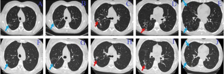
This Comparison Chart displays ten axial chest CT scan slices from a patient with a pulmonary Mycobacterium abscessus infection, illustrating disease progression and treatment response. Panels A-E (top row) represent pre-treatment baseline imaging, while panels F-J (bottom row) show the same anatomical levels following the initial phase of medical therapy. The pre-treatment scans (A-E) demonstrate multiple active pathologies: large thick-walled pulmonary cavities in the right lung (indicated by red arrows in panels C and D) and extensive bilateral 'tree-in-bud' opacities (indicated by blue arrows), which represent centrilobular nodules and branching linear structures characteristic of small-airway impaction and endobronchial spread of infection. In the post-treatment scans (F-J), there is a visible radiological improvement characterized by a significant reduction in the size and wall thickness of the cavities (red arrows in H and I) and a marked decrease in the density and distribution of the tree-in-bud lesions (blue arrows). The image serves as a clinical illustration of antibiotic efficacy in treating nontuberculous mycobacteria (NTM) lung disease, specifically highlighting the resolution of bronchogenic spread and cavitary lesions.
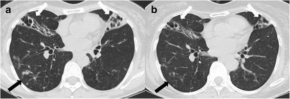
This figure presents two transverse chest computed tomography (CT) scans (axial view) illustrating the progression and treatment response of nontuberculous mycobacterial (NTM) lung disease. Scan (a) shows the initial presentation, characterized by bilateral bronchiectasis and pulmonary consolidations, specifically prominent in the right middle lobe and the lingular division of the left upper lobe (white arrows). Multiple 'tree-in-bud' opacities (black arrow) are visible in the peripheral lung zones, signifying active bronchiolitis and infectious small airway involvement. Scan (b) displays the same anatomical level after 12 months of antibiotic therapy, demonstrating a clear interval reduction in the density and extent of the consolidations (white arrows) and a significant decrease in the tree-in-bud appearances (black arrow). The images illustrate the classic radiographic features of the nodular bronchiectatic form of Mycobacterium abscessus infection and its typical response to targeted pharmacological intervention.
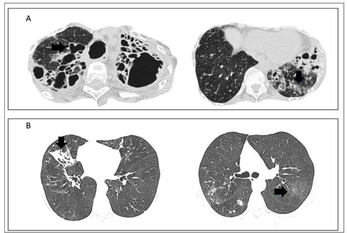
Diagnostic Image: This figure presents multiple axial high-resolution computed tomography (CT) scans of the chest from two different clinical cases of non-tuberculous mycobacteria lung disease (NTM-LD). Panel A (Top): Displays axial slices of an immunocompromised patient with Mycobacterium asiaticum infection. Notable findings include severe, extensive varicose bronchiectasis characterized by significant bronchial dilation and bronchial wall (parietal) thickening, highlighted by black arrows. Also visible are areas of centrilobular opacities with ground-glass attenuation, indicating small-airway involvement. Panel B (Bottom): Shows axial slices from a patient with Mycobacterium avium-intracellulare (MAC) complex. The left slice demonstrates focal consolidation and associated bronchiectasis localized to the right middle lobe. The right slice highlights ground-glass centrilobular opacities in the lingula and lower lobes (black arrows). Educational Significance: These images demonstrate typical radiological patterns of NTM-LD, including the 'nodular bronchiectatic' form which often involves the middle lobe and lingula, and the more severe cavitary/varicose form seen in immunocompromised states.
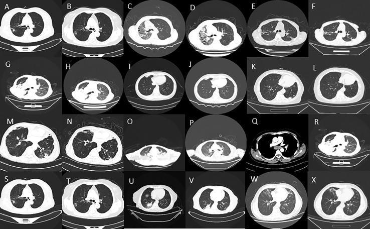
This diagnostic image consists of a 24-panel mosaic (labeled A–X) of axial chest CT scans demonstrating various pulmonary manifestations of Non-Tuberculous Mycobacteria (NTM) infections. The collection illustrates a spectrum of pathological patterns including small pulmonary nodules (prominent in A, B, S, T), patchy ground-glass opacities (E, F, M, W), and dense lobar or segmental consolidation (C, D). Some sections (G, H) show evidence of reduced lung volume and structural distortion, while others (I, J, K, L, R, V) demonstrate post-treatment intervals with relative clearing of parenchymal disease and more distinct bronchovascular markings. The images provide a comparison of disease morphology across different NTM species, such as Mycobacterium avium complex and M. abscessus. Educational focus is placed on recognizing the radiologic hallmarks of chronic mycobacterial lung disease, monitoring treatment response, and identifying the heterogeneity of infectious presentations in different patients.
Nontuberculous Mycobacteria (NTM) - Focus on Mycobacterium abscessus
Overview of NTM
- Pre-existing pulmonary disease (bronchiectasis, COPD, cystic fibrosis)
- Immunocompromise (HIV/AIDS, organ transplant, biologics)
- Nodular/bronchiectatic disease in otherwise healthy hosts (classic Lady Windermere syndrome - non-smoking white women >60 years old)
Classification: Runyon System
| Group | Growth Rate | Examples |
|---|---|---|
| I - Photochromogens | Slow | M. kansasii, M. marinum |
| II - Scotochromogens | Slow | M. gordonae, M. scrofulaceum |
| III - Nonchromogens | Slow | M. avium complex (MAC), M. xenopi |
| IV - Rapid Growers | Fast (<7 days) | M. abscessus, M. fortuitum, M. chelonae |
Mycobacterium abscessus - Key Facts
Subspecies
- M. abscessus subsp. abscessus - harbors erm(41) gene → inducible macrolide resistance
- M. abscessus subsp. massiliense - truncated/nonfunctional erm(41) → macrolide susceptible, better treatment outcomes
- M. abscessus subsp. *bolletii
Epidemiology
- 3rd most common NTM pathogen in the United States
- Endemic in southeastern states (Texas to Florida)
- Environmental reservoir: tap water is the major source
- Healthcare outbreaks linked to: dental unit water lines, polyethylene ear tubes, contaminated surgical equipment, cosmetic procedures (tattooing, acupuncture, pedicures)
Clinical Presentations
| Site | Details |
|---|---|
| Pulmonary | Most common; often in nonsmoking white women >60 without underlying lung disease, OR in cystic fibrosis patients |
| Skin/Soft Tissue/Bone | After accidental trauma, surgery, cosmetic procedures, tattoos, injections |
| Lymphadenitis | Less common than MAC |
| Catheter-associated | IV line infections, especially post-surgery |
| Otitis media | Associated with polyethylene ear tubes |
| Disseminated | Immunocompromised hosts (HIV, transplant) |
Diagnosis
- Significant clinical manifestations AND/OR radiographic evidence of progressive disease, PLUS:
- Reproducible positive sputum cultures (≥2 positive cultures), OR
- A single positive bronchoscopy culture
- Tree-in-bud opacities
- Bronchiectasis (often middle lobe / lingula - "Lady Windermere" pattern)
- Cavitary lesions (in fibrocavitary form)
- Nodular infiltrates
- RGM grow on culture media in < 7 days (subcultures)
- AFB smear positive (weakly acid-fast)
- Definitive ID by: PCR, 16S rRNA gene sequencing, MALDI-TOF
- Susceptibility testing is mandatory - routine anti-TB drugs are ineffective
Antimicrobial Susceptibility
- Macrolides (clarithromycin, azithromycin) - BUT see resistance caveat below
- Amikacin
- Imipenem / cefoxitin
- Tigecycline
- Linezolid
- Clofazimine
- Trimethoprim-sulfamethoxazole
- Ciprofloxacin (variable)
Critical Resistance Mechanism: erm(41) Gene
Treatment
General Principles
- Always seek infectious disease consultation
- Treatment is prolonged, complex, and multi-drug
- Susceptibility testing must guide therapy
- Overall culture conversion rates are low (~35%) for pulmonary disease
Skin, Soft Tissue, and Bone Infections
- Minimum 4 months (skin/soft tissue); ≥6 months for bone
- Regimen: Macrolide (clarithromycin or azithromycin) + parenteral agent (amikacin, cefoxitin, or imipenem)
Pulmonary Infection
- Regimen of at least 3 active drugs based on susceptibility testing:
- Amikacin + imipenem/cefoxitin + macrolide (if susceptible) ± linezolid ± tigecycline
- Recent meta-analysis (PMID: 41420504): overall therapeutic efficiency ~35%; amikacin, imipenem, linezolid, and/or tigecycline incorporation → improved outcomes; macrolide resistance → worse outcomes
- Surgical resection should be considered in patients with good lung reserve and localized infection
Treatment of RGM (Fortuitum / Chelonae) by Red Book
Recent Evidence (2025)
-
Treatment outcomes in M. abscessus pulmonary disease (PMID: 41420504, 2025, Ann Acad Med Singapore) - confirms ~35% culture conversion; amikacin/imipenem/linezolid/tigecycline associated with improved outcomes.
-
Tetracyclines for NTM (focus on RGM) (PMID: 41193247, 2025, Eur Respir Rev) - reviews role of doxycycline/tigecycline in rapidly growing NTM.
-
NTM after cosmetic procedures (PMID: 39395754, 2024, Int J Infect Dis) - systematic review of RGM infections (predominantly M. abscessus) following cosmetic surgeries, injections, and tattoos.
Key Comparison: M. abscessus vs Other Common NTM
| Feature | M. abscessus | MAC (M. avium) | M. kansasii |
|---|---|---|---|
| Growth | Rapid (Runyon IV) | Slow (Runyon III) | Slow (Runyon I) |
| Main disease | Lung, skin/soft tissue | Lung, disseminated (HIV) | Lung |
| First-line | Amikacin + imipenem ± macrolide | Clarithromycin + ethambutol + rifampin | Rifampin + ethambutol + isoniazid |
| Macrolide resistance | Common (erm41) | Acquired (point mutation) | Usually susceptible |
| Treatment success | ~35% (pulmonary) | ~60% | >80% |
| Surgical role | Important | Sometimes | Rare |
Prevention / Infection Control
- Standard precautions for hospitalized patients
- Avoid tap water contamination of central lines, dental equipment, and surgical wounds
- Use filtered (not tap) water for heater-cooler devices in cardiac surgery
- Decontamination protocols for dental unit water lines
- Report suspected heater-cooler device-associated infections to FDA MedWatch