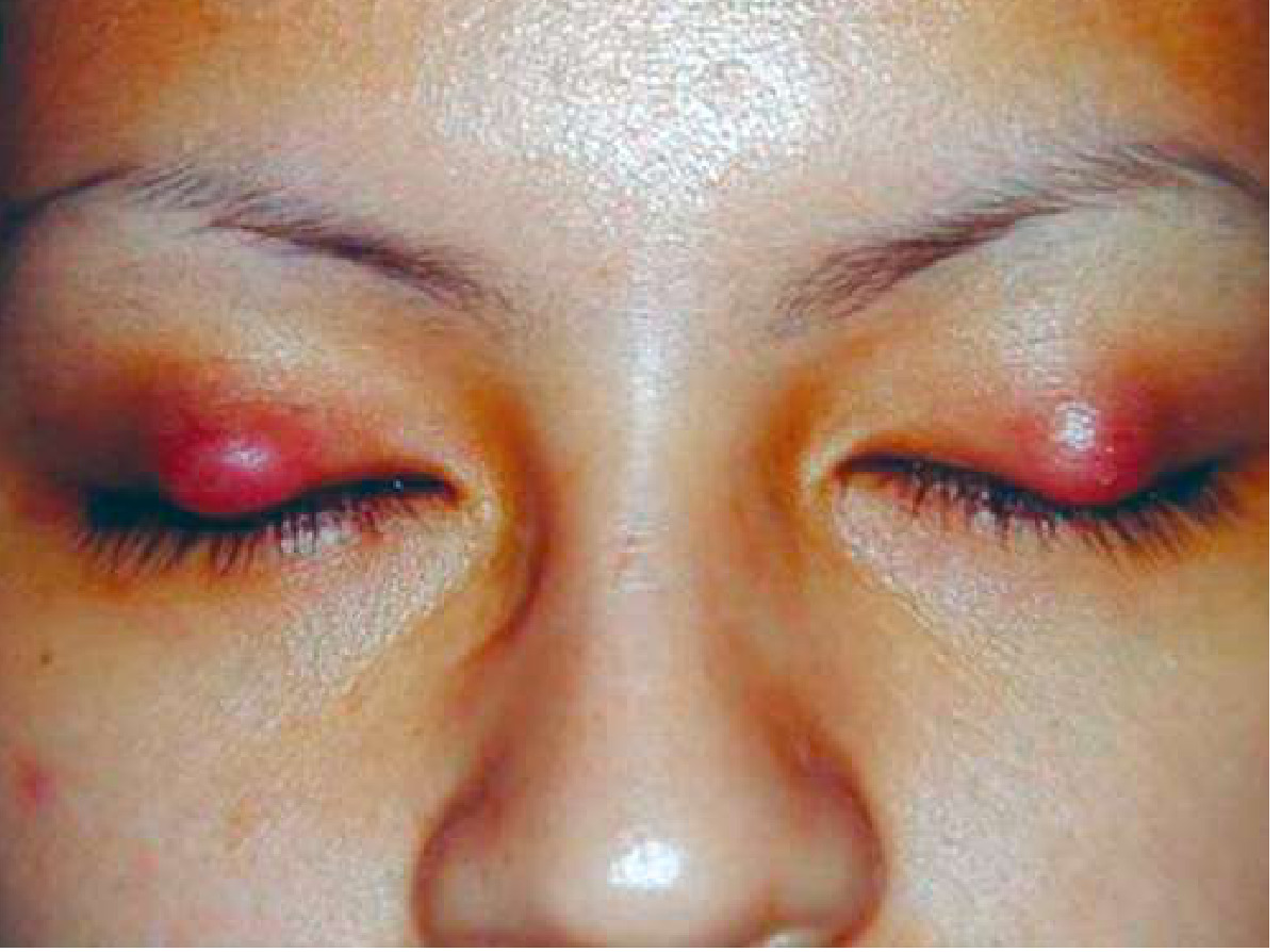
Itching on upper eye lid margin on right eye . Acute in onset what could be the reason and treatment
chalazion upper eyelid large swelling meibomian gland
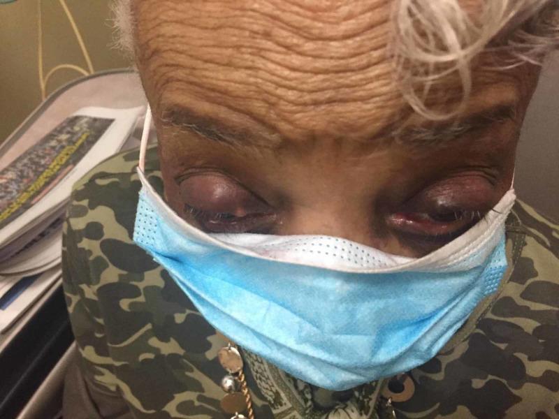
This clinical photograph shows the periocular region of an elderly patient with a darker complexion, displaying bilateral upper eyelid swelling consistent with chalazia. Both upper eyelids exhibit focal, nodular lesions with overlying erythema and a purplish discoloration, suggesting secondary inflammation. The lesion on the right upper eyelid is significantly larger and more protuberant than the one on the left, causing more pronounced distention of the palpebral tissue. The surrounding forehead skin shows deep rhythmic rhytids (wrinkles), indicative of the patient's age. The lower face is partially covered by a light blue surgical mask. This image illustrates the typical clinical presentation of chronic meibomian gland obstruction (chalazion) when occurring bilaterally with asymmetric progression. It serves as a pedagogical tool for ophthalmological and primary care physical examination findings related to eyelid pathologies.
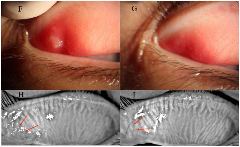
A multi-panel medical comparison showing a 32-year-old female patient with an upper eyelid chalazion before and after Optimal Pulse Technology (OPT) treatment. Panels F and G are clinical photographs of the everted upper palpebral conjunctiva. Panel F (baseline) demonstrates a raised, circumscribed chalazion with significant surrounding palpebral conjunctival hyperemia. Panel G (post-treatment) shows complete regression of the cyst and resolution of the hyperemia. Panels H and I provide infrared meibography of the same eyelid. Panel H shows significant meibomian gland dropout and architectural distortion at the site of the original chalazion (marked with red arrows). Panel I (post-treatment) displays a comparative increase in meibomian gland density and improved definition of glandular structures in the previously affected area. This comparison illustrates the efficacy of non-invasive light therapy in reducing eyelid inflammation and promoting meibomian gland recovery.
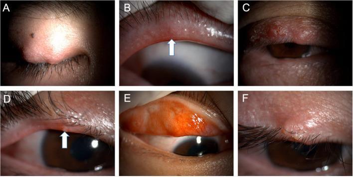
A composite of six clinical photographs (A-F) illustrating varying clinical presentations of recurrent or multiple chalaziosis on the eyelids. Panel A, C, and F show external views of localized, rounded, firm nodules within the tarsal plate of the upper eyelids, demonstrating typical chalazion morphology. Panel B (arrow) highlights telangiectasia and eyelid margin neovascularization, indicative of chronic blepharitis or meibomian gland dysfunction (MGD). Panel D (arrow) illustrates a lipid suppository or 'pouting' at a meibomian gland orifice, representing obstructed lipid secretions. Panel E shows an everted upper eyelid revealing significant palpebral conjunctival hyperemia and tarsal inflammation. Collectively, these images demonstrate the spectrum of meibomian gland-related eyelid pathology, including active granulomatous inflammation (chalazion), secondary vascular changes, and signs of underlying MGD, which are critical in the diagnosis of recurrent chalaziosis in ophthalmology and dermatology.
chalazion treatment management
Clinical Assessment
Most Likely Diagnoses
1. Inflamed Chalazion (Primary Diagnosis - Most Likely)
- The lesion is typically a firm, rubbery, painless-to-mildly tender nodule
- It may wax and wane, and can have a liquefied center mimicking an abscess
- Associated erythema and edema can develop, especially when inflamed
- Size, location (upper lid, centrally over tarsal plate), and appearance in your images are all consistent
2. Internal Hordeolum (Acute Bacterial Stye)
- Acute onset tender, tense swelling
- Associated eyelid cellulitis (erythema, edema, warmth)
- A point of fluctuance indicating pus collection
- Usually caused by Staphylococcus aureus
Distinction
| Feature | Chalazion | Internal Hordeolum |
|---|---|---|
| Onset | Gradual (days-weeks) | Acute (hours-days) |
| Pain | Mild/painless | Tender/painful |
| Texture | Rubbery, cystic | Tense, fluctuant |
| Pus | No (lipid granuloma) | Yes (bacterial abscess) |
| Cellulitis | Mild at most | Often prominent |
| Resolution | Weeks-months | Spontaneous in 5-7 days |
Treatment
Step 1: Conservative Management (First-line)
- Warm compresses: Apply a clean warm damp cloth to the closed eyelid for 10-15 minutes, 4 times daily. This softens blocked meibomian secretions and promotes drainage
- Eyelid scrubs: Nightly cleansing of the lid margin at the base of eyelashes with diluted baby shampoo (1:1 with water) or commercial lid scrub pads
- Do NOT squeeze or puncture the lesion - this risks spreading infection deeper
Step 2: Topical Antibiotics (if infection suspected)
- Erythromycin ointment applied to the lid margin after cleansing
- Alternatively: tobramycin, bacitracin, or fluoroquinolone drops (moxifloxacin, gatifloxacin)
- - Goldman-Cecil Medicine; Textbook of Family Medicine 9e
Step 3: Systemic Antibiotics (if cellulitis is present)
- Oral antibiotics (e.g., doxycycline 100 mg twice daily, or amoxicillin-clavulanate) if there is preseptal cellulitis - spreading redness/warmth to surrounding lid skin
- Oral doxycycline also helps with meibomian gland function in recurrent cases
Step 4: Intralesional Steroid Injection (if persisting >4-6 weeks)
- Triamcinolone acetonide 40 mg/mL, 0.2-0.4 mL injected via a 30-gauge needle through the conjunctival surface under topical anesthesia (tetracaine drops)
- Note: Carries risk of skin hypopigmentation, especially in darker skin tones
- - Pfenninger & Fowler's Procedures for Primary Care
Step 5: Surgical Incision & Curettage (if unresponsive to medical therapy)
- Performed by an ophthalmologist under local anesthesia
- Indicated when the chalazion is: unresponsive to 4-6 weeks of medical treatment, substantially large (as in this case), causing visual problems, or patient-requested
- Done via the conjunctival (inner) surface to avoid visible skin scarring
- CPT codes: 67800 (single), 67801 (multiple same lid), 67700 (I&D of hordeolum abscess)
Red Flags - Seek Urgent Ophthalmology Review If:
- Vision blurring or double vision
- Pain on eye movement (suggests orbital involvement)
- Redness/swelling spreading to cheek or forehead (preseptal/orbital cellulitis)
- Proptosis (eyeball pushed forward)
- Fever or systemic illness
- Recurrence in the same spot - may indicate sebaceous cell carcinoma (a recurrent chalazion in the identical location must be biopsied)
Important Note