
Explain about pneumonia it's x-ray findings with pictures diagnosis and treatment in detail
Reading File
Now let me search the medical library for comprehensive pneumonia content in parallel.
Finding Sources
Finding Sources
Searching the Web
pneumonia chest x-ray findings consolidation diagnosis treatment 2024
Now let me read the key sections from the textbooks and fetch the X-ray images in parallel.
Reading File
Reading File
Reading File
Reading File
Searching PubMed
community-acquired pneumonia diagnosis treatment guidelines
Now let me get the detailed CAP section from Goldman-Cecil and the main pneumonia treatment content, plus fetch key X-ray images.
Finding Sources
Loading Image
Loading Image
Now let me get the main CAP chapter from Goldman-Cecil and the Murray & Nadel pneumonia sections:
Finding Sources
Reading File
Loading Image
Now let me get the main comprehensive CAP content from Goldman-Cecil and Fishman's:
Reading File
Reading File
Loading Image
Let me now get the Goldman-Cecil detailed CAP chapter content and the Harriet Lane x-ray image, along with more radiology-focused content:
Reading File
Reading File
Finding Sources
Let me now fetch more X-ray images and the key diagnostic/treatment content from Goldman-Cecil:
Reading File
Loading Image
Finding Sources
Let me read the Harrison's CAP section and fetch a couple more important X-ray images:
Reading File
Finding Sources
Excellent - I found the Grainger & Allison radiology textbook with key content. Let me read it and fetch the X-ray image from there:
Reading File
Reading File
Now let me fetch the key radiology images from Grainger & Allison:
Loading Image
Loading Image
Reading File
I have comprehensive material. Let me now compile the full detailed answer with all the images, textbook content, and treatment algorithms.
Pneumonia: Complete Overview
1. Definition and Epidemiology
Pneumonia is an inflammation of the lung parenchyma, most often infectious, characterized by fever, cough, sputum production, pleuritic chest pain, and a pulmonary infiltrate on imaging. It is the most common infectious cause of hospitalization and death in the United States, accounting for about 7 annual health care visits per 1000 young adults but rising to 96 visits per 1000 adults aged 85 and older. Overall, ~7 million or more CAP cases occur annually in the US, and along with influenza, it remains among the top 10 leading causes of death. The mortality rate in outpatients is usually <5%, but ranges from 12 to 40% in hospitalized patients.
- Goldman-Cecil Medicine, p. 992; Harrison's Principles of Internal Medicine 22E, p. 1069
2. Classification
| Type | Setting | Common Pathogens |
|---|---|---|
| Community-Acquired Pneumonia (CAP) | Onset outside hospital or within 48h of admission | S. pneumoniae, H. influenzae, M. pneumoniae, Legionella, viruses |
| Hospital-Acquired (HAP) | >48h after admission | P. aeruginosa, MRSA, gram-negative bacilli |
| Ventilator-Associated (VAP) | After intubation | MDR organisms |
| Aspiration Pneumonia | Impaired gag reflex, alcohol, neurological disease | Anaerobes, gram-negatives |
| Immunocompromised | HIV, transplant, steroids | Pneumocystis jirovecii (PCP), fungi, CMV, unusual bacteria |
3. Etiology by Age/Patient Population
| Population | Likely Pathogens |
|---|---|
| Neonates | Group B streptococci, gram-negative bacilli |
| Infants 3-19 weeks | Chlamydia trachomatis |
| Children <5 years | RSV, parainfluenza (viral predominates - up to 90%) |
| Children >5 years | M. pneumoniae, C. pneumoniae, S. pneumoniae |
| Adults (outpatient, no comorbidities) | S. pneumoniae, M. pneumoniae, C. pneumoniae, H. influenzae, respiratory viruses |
| Adults with cardiopulmonary disease | All above + DRSP, enteric gram-negatives, possibly anaerobes |
| Severe CAP (ICU) | S. pneumoniae, Legionella, S. aureus, enteric gram-negatives, P. aeruginosa |
| Alcoholism | K. pneumoniae, oral anaerobes, S. pneumoniae, M. tuberculosis |
| COPD/smoking | H. influenzae, P. aeruginosa, M. catarrhalis |
- Fishman's Pulmonary Diseases and Disorders, p. 2202; Harrison's, Table 131-2
4. Pathophysiology
The most common route of infection is microaspiration of oropharyngeal organisms. Pneumonia impairs oxygenation through two key mechanisms: increased shunting and increased perfusion to low V/Q ratio units. In mild to moderate disease (PaO₂ ~74 mmHg), shunt increases to ~7.5% and low V/Q regions to ~4.2%. In severe disease requiring mechanical ventilation, shunt can exceed 22% and low V/Q regions >10%. With lobar pneumonia, the inflammatory exudate begins in distal air spaces adjacent to the visceral pleura and spreads via collateral routes (pores of Kohn) to produce uniform homogeneous opacification.
5. Clinical Features
Symptoms:
- Fever, chills, rigors
- Productive cough (purulent, blood-streaked, or "rusty" sputum in S. pneumoniae)
- Pleuritic chest pain
- Dyspnea, tachypnea
- Fatigue, malaise
Signs:
- Tachycardia, tachypnea, fever (may be absent in elderly)
- Dullness to percussion (consolidation)
- Increased tactile fremitus
- Bronchial breathing, crepitations (crackles) on auscultation
- Egophony ("E" sounds like "A")
- Decreased breath sounds (effusion)
- Confusion in elderly (may be the only sign)
Atypical presentation (Mycoplasma, Legionella, Chlamydia): gradual onset, dry cough, extrapulmonary features (headache, myalgia, GI symptoms, rash), chest radiograph often worse than clinical picture.
6. Chest X-Ray Findings with Pictures
The chest radiograph is the primary imaging tool for diagnosing and classifying pneumonia. Three main radiographic patterns are recognized:
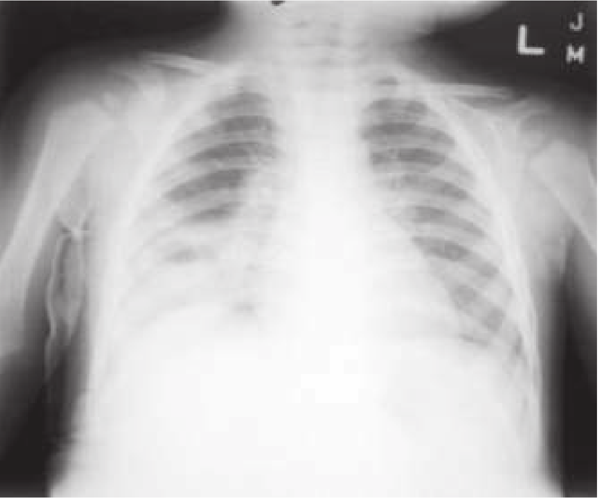
6A. Lobar / Segmental Pneumonia
The exudate fills alveoli uniformly, producing a homogeneous opacity that respects lobar or segmental boundaries. Air bronchograms (air-filled bronchi seen against a background of opacified lung) are a hallmark feature. Caused most commonly by S. pneumoniae, but also Klebsiella, Legionella, and H. influenzae.
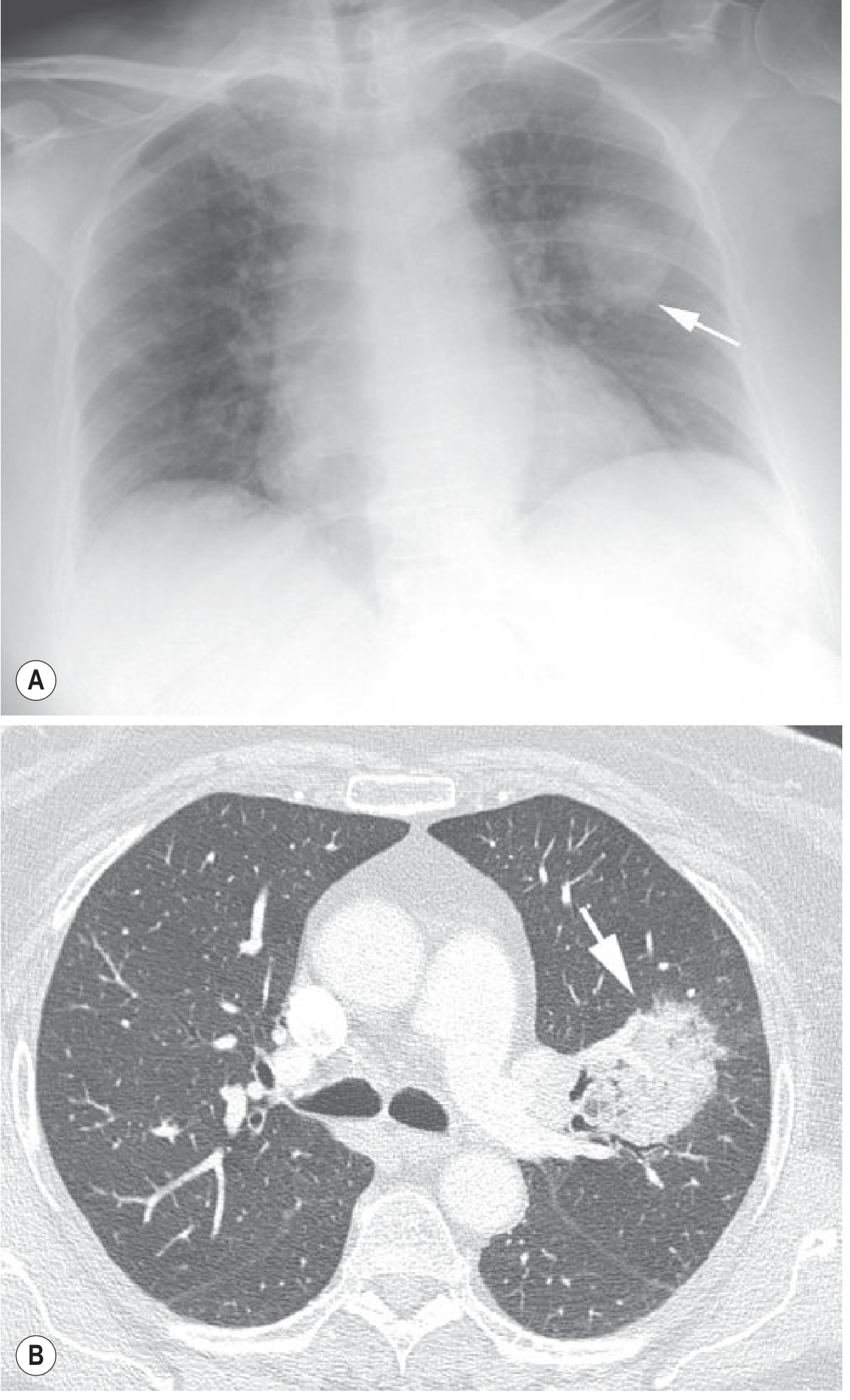
6B. Round Pneumonia
A discrete, well-marginated spherical consolidation that can mimic a pulmonary mass. Resolves with antibiotics.
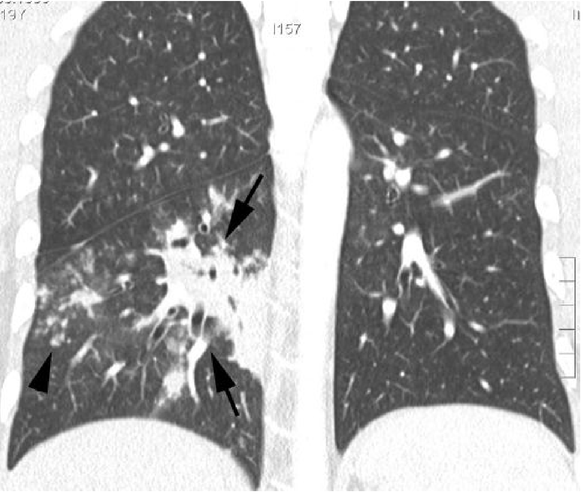
6C. Bronchopneumonia (Lobular Pneumonia)
Histologically peribronchiolar inflammation. Radiographically shows patchy, multifocal consolidations with poorly defined margins, predominantly in lower lobes. Air bronchogram is usually absent. Centrilobular ill-defined nodules and "tree-in-bud" opacities on HRCT. Caused by S. aureus, H. influenzae, P. aeruginosa, and anaerobes.
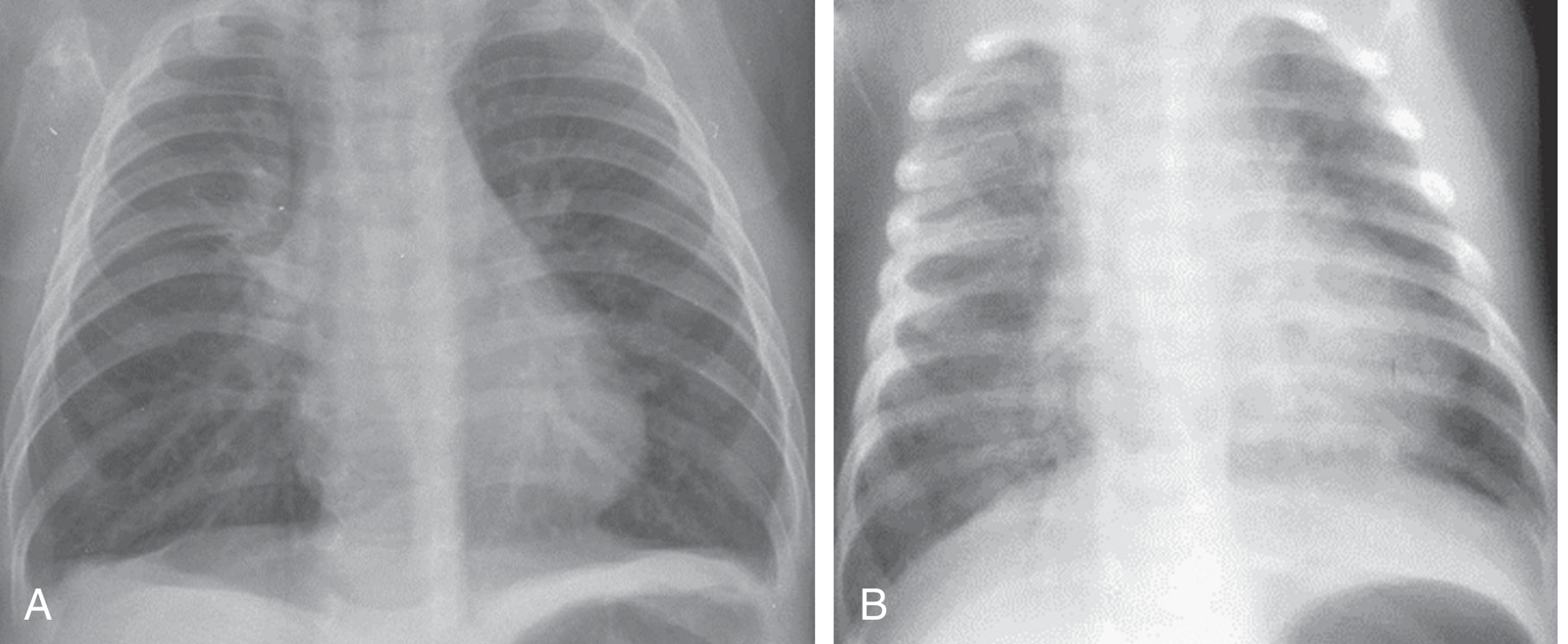
6D. RSV Bronchiolitis and Pneumonia (Before and After Treatment)
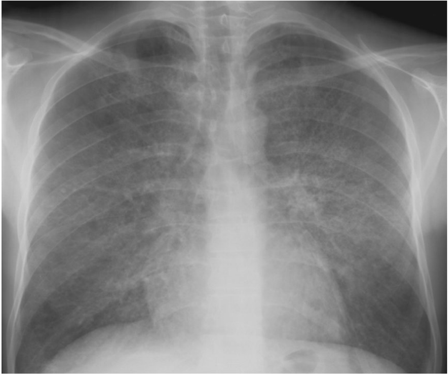
6E. Bilateral Diffuse Infiltrates (Severe/Interstitial Pneumonia)
Summary of CXR Findings
| Finding | Description | Association |
|---|---|---|
| Lobar/segmental consolidation | Homogeneous opacity, lobe-shaped | S. pneumoniae, K. pneumoniae |
| Air bronchogram | Air-filled bronchi visible within opacity | Lobar consolidation |
| Patchy infiltrates | Multifocal poorly-defined opacities | Bronchopneumonia |
| Interstitial pattern | Bilateral diffuse small opacities | Atypical/viral, Mycoplasma, PCP |
| Pleural effusion | Blunting of costophrenic angle | Parapneumonic effusion, empyema |
| Cavitation | Air-fluid level within opacity | S. aureus, K. pneumoniae, anaerobes, TB |
| Bulging fissure sign | Lobar expansion with bulging fissure | K. pneumoniae (classic) |
| Normal CXR | Up to 10% of confirmed PCP | Pneumocystis pneumonia |
7. Diagnosis
7A. Clinical Diagnosis
Pneumonia is diagnosed by clinical signs + abnormal chest radiograph showing pulmonary infiltrates. No single test is diagnostic; a combination of history, examination, and investigations is used.
7B. Laboratory Investigations
| Test | Finding/Purpose |
|---|---|
| CBC | Leukocytosis (bacterial); normal or low WBC (viral/atypical) |
| CRP / Procalcitonin | Elevated in bacterial; helps guide antibiotic stewardship |
| Blood cultures | Before antibiotics in hospitalized patients; positive in ~10-15% |
| Sputum Gram stain & culture | Guides narrowing of therapy |
| Urinary antigen | Legionella (sensitivity ~74-80%), pneumococcal (sensitivity ~70-80%) |
| Serology | Mycoplasma, Chlamydia (cold agglutinins, titres) |
| Respiratory multiplex PCR | Viral panel (influenza, RSV, COVID-19, adenovirus) |
| HIV test | If PCP or atypical pneumonia suspected |
| ABG / SpO₂ | Severity assessment, hypoxemia |
| BUN, creatinine, electrolytes | Part of CURB-65 and severity scoring |
| LFTs | Especially for Legionella (hyponatremia, raised transaminases) |
| Bronchoscopy/BAL | Immunocompromised patients, non-responding CAP |
7C. Severity Scores for Admission Decisions
CURB-65 (1 point each):
- Confusion
- Urea >7 mmol/L (BUN >20 mg/dL)
- Respiratory rate ≥30/min
- Blood pressure: systolic <90 or diastolic ≤60 mmHg
- 65: age ≥65 years
| Score | Mortality | Recommendation |
|---|---|---|
| 0-1 | Low | Outpatient treatment |
| 2 | Intermediate | Consider hospital admission |
| 3-5 | High | Hospitalize; ICU if ≥4 |
Pneumonia Severity Index (PSI) is the more detailed alternative (20 variables), stratifying into classes I-V; classes I-III indicate likely safe outpatient management.
7D. ATS/IDSA Criteria for Severe CAP (ICU admission indicated if 1 major or 3 minor criteria):
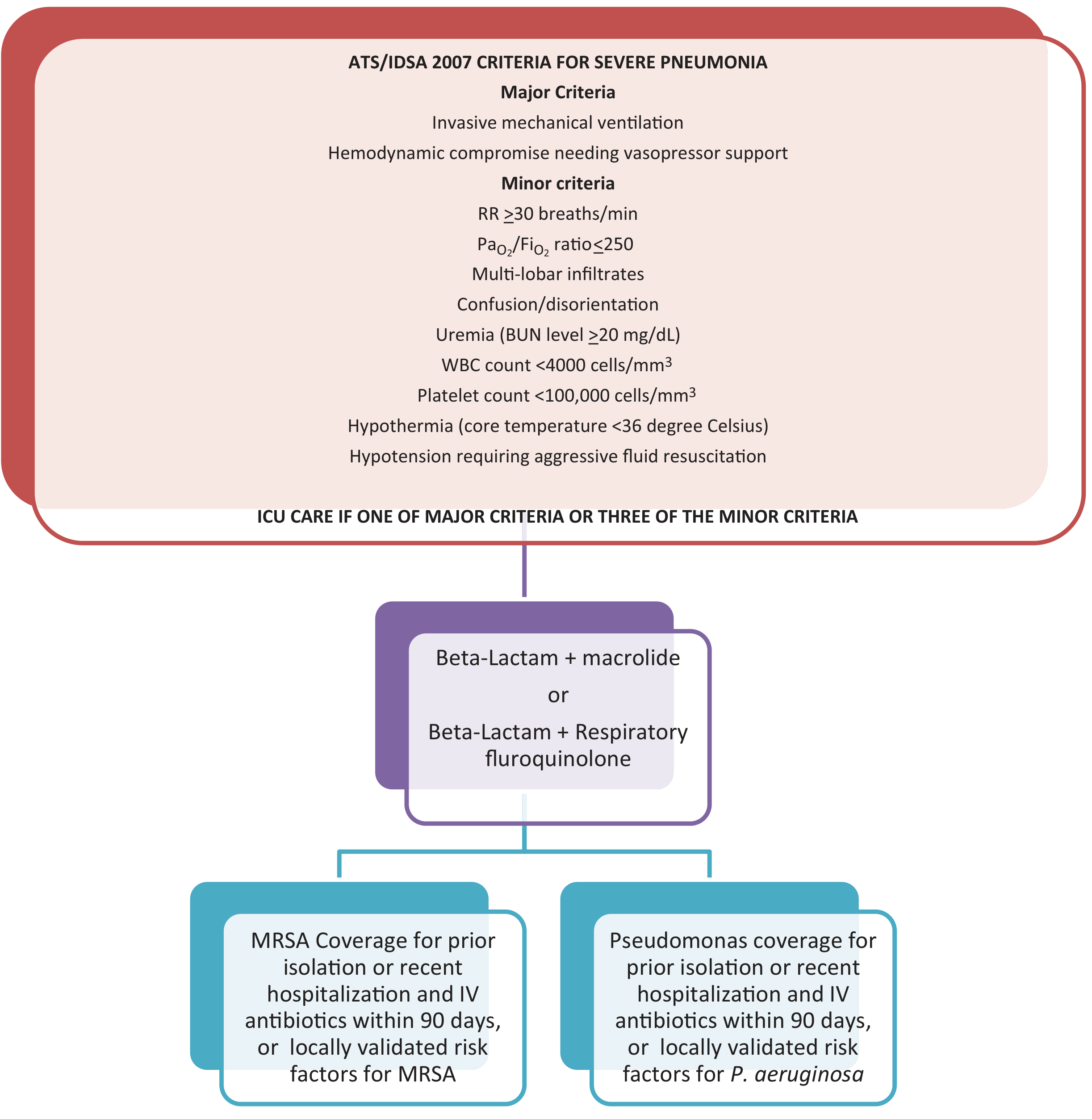
Major criteria:
- Invasive mechanical ventilation required
- Septic shock requiring vasopressors
Minor criteria:
- RR ≥30/min
- PaO₂/FiO₂ ≤250
- Multi-lobar infiltrates
- Confusion/disorientation
- BUN ≥20 mg/dL
- WBC <4000/mm³
- Platelet count <100,000/mm³
- Core temperature <36°C
- Hypotension requiring aggressive fluid resuscitation
8. Treatment
8A. Antibiotic Treatment by Site of Care
Outpatient (No Comorbidities, Low Severity)
Monotherapy for 5 days:
- Amoxicillin (first-line, β-lactam monotherapy now acceptable in guidelines for low-risk)
- Doxycycline
- Azithromycin (where macrolide resistance is low)
Outpatient with Comorbidities (COPD, diabetes, heart/liver/renal disease, or recent antibiotics)
- Respiratory fluoroquinolone (levofloxacin or moxifloxacin) monotherapy, OR
- Amoxicillin/clavulanate or cefpodoxime or cefuroxime + azithromycin/clarithromycin/doxycycline
Inpatient, Non-Severe CAP (No ICU, No MRSA/Pseudomonas Risk)
- Respiratory fluoroquinolone monotherapy (levofloxacin, moxifloxacin), OR
- β-lactam (ampicillin-sulbactam, ceftriaxone, cefotaxime) + macrolide or doxycycline
- Newer options: omadacycline or lefamulin monotherapy (noninferior to fluoroquinolone)
- Note: Fluoroquinolone caution due to risk of C. difficile, tendinopathy, neuropathy, QT prolongation
Severe CAP - ICU
- β-lactam + macrolide (preferred), OR
- β-lactam + respiratory fluoroquinolone
- Add MRSA coverage (vancomycin or linezolid) if: prior MRSA isolation, hospitalization + IV antibiotics in last 90 days, or locally validated risk factors
- Add Pseudomonas coverage (piperacillin-tazobactam, cefepime, or carbapenem) if prior isolation or recent hospitalization with IV antibiotics
Aspiration Pneumonia
- CAP or HAP-directed therapy (anaerobic coverage not routinely needed per current IDSA/ATS 2019 guidelines; gram-negatives predominate)
- Clindamycin or metronidazole if lung abscess or empyema present
MRSA Pneumonia (CA-MRSA, post-influenza, necrotizing)
- Linezolid (preferred - also inhibits toxin production), OR
- Vancomycin ± clindamycin (to inhibit exotoxin production)
Atypical Pneumonia (Mycoplasma, Chlamydia)
- Macrolide (azithromycin) or doxycycline or respiratory fluoroquinolone
Pneumocystis jirovecii Pneumonia (PCP)
-
Trimethoprim-sulfamethoxazole (first-line)
-
Adjunct corticosteroids if PaO₂ <70 mmHg
-
Goldman-Cecil Medicine, p. 993; Fishman's Pulmonary Diseases and Disorders, p. 2202-2204
8B. Duration of Antibiotic Therapy
- Outpatient CAP: 5 days (per current IDSA/ATS 2019 and updated guidelines)
- Inpatient non-severe: 5-7 days
- Severe CAP: 7-10 days (longer for Legionella, bacteremic pneumococcal disease, necrotizing pneumonia, or lung abscess)
- Switch IV to oral when clinically stable: temperature ≤37.8°C, HR <100, RR <24, SpO₂ ≥90%, BP stable, normal mental status
8C. Supportive Treatment
- Oxygen therapy to maintain SpO₂ ≥94% (88-92% in COPD patients)
- IV fluids for dehydration, sepsis
- Analgesia for pleuritic pain (NSAIDs, paracetamol)
- Antipyretics
- Chest physiotherapy in patients with excessive secretions
- NIV/mechanical ventilation for respiratory failure (PaO₂/FiO₂ <250 or severe hypoxemia)
- DVT prophylaxis
- Corticosteroids - adjunctive use in select cases of severe CAP (evidence remains limited but may reduce treatment failure in severe disease)
8D. Complications to Watch For
| Complication | Features |
|---|---|
| Parapneumonic effusion / Empyema | Most common; develops in ~3-5% of CAP; requires thoracentesis ± drainage |
| Lung abscess | Cavitation, prolonged fever, anaerobic organisms; treat with prolonged antibiotics |
| Sepsis / Septic shock | Multi-organ dysfunction; requires ICU, vasopressors |
| Acute respiratory failure / ARDS | PaO₂/FiO₂ <200; mechanical ventilation needed |
| Cardiac events | Atrial fibrillation, heart failure exacerbation, MI - occur in ~20-25% of hospitalized patients |
| Necrotizing pneumonia | CA-MRSA, K. pneumoniae; multiple cavities |
| Meningitis/bacteremia | S. pneumoniae bacteremia in 10-15% |
9. Prevention
- Pneumococcal vaccines: PCV13 (Prevnar 13) - 85% protection against serotype-specific pneumococcal pneumonia; PPSV23 for adults ≥65 or high-risk groups
- Influenza vaccine: annual; reduces secondary bacterial pneumonia after influenza
- COVID-19 vaccine
- Smoking cessation
- Good oral hygiene (reduces aspiration risk)
- Follow-up CXR: at 6 weeks post-discharge in those meeting lung cancer screening criteria to confirm resolution (per NICE NG250, 2025 and ATS guidelines)
10. Follow-Up
Clinical improvement (fever, WBC, respiratory rate) is expected within 48-72 hours of appropriate antibiotics. Radiographic clearance lags behind clinical improvement - may take 4-6 weeks to fully resolve on CXR, especially in elderly patients. Persistent consolidation at 6 weeks warrants CT scan and bronchoscopy to exclude an underlying lesion such as pneumonic-type lung adenocarcinoma. The NICE NG250 guideline (September 2025) specifically recommends this follow-up pathway.
Sources:
- Goldman-Cecil Medicine International Edition (2-Volume Set), Chapter 85
- Harrison's Principles of Internal Medicine, 22nd Edition (2025), Chapter 131
- Fishman's Pulmonary Diseases and Disorders, Chapter 125
- Grainger & Allison's Diagnostic Radiology, Chapter 5
- Murray & Nadel's Textbook of Respiratory Medicine
- ROSEN's Emergency Medicine: Concepts and Clinical Practice
- Symptom to Diagnosis: An Evidence-Based Guide, 4th Ed.
- NICE NG250 Pneumonia: Diagnosis and Management (September 2025)
This is a shared conversation. Sign in to Orris to start your own chat.