
Iron deficiency Anemia explai
iron deficiency anemia blood smear peripheral smear microcytic hypochromic
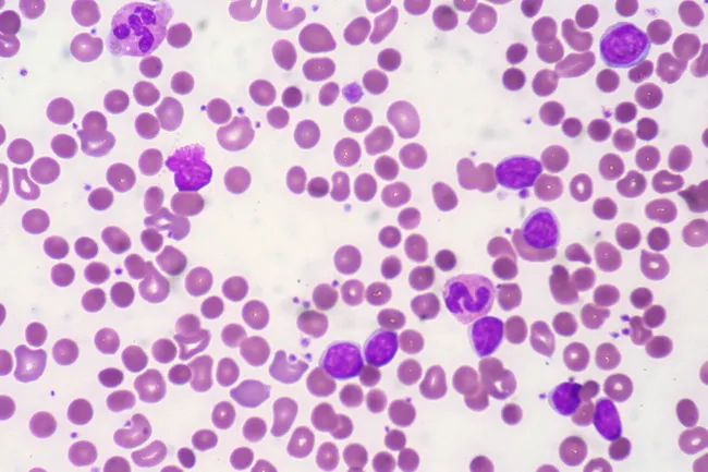
Peripheral blood smear from a patient with known chronic lymphocytic leukemia (CLL) showing autoimmune hemolytic anemia (AIHA). Brightfield microscopy of Wright-Giemsa stained smear at 1000x (oil immersion) reveals two dominant cell populations: numerous small mature lymphocytes typical of CLL and red blood cells showing spherocytic deformation. A smudge cell is evident just left of center, a classical clue for CLL. Red cells display reduced central pallor consistent with spherocytes, indicating intravascular or extravascular hemolysis driven by autoantibodies. The background shows mild polychromasia and rare nucleated RBCs, consistent with a robust reticulocyte response; the corrected reticulocyte count in this case is markedly elevated (>7%). Platelets are not significantly abnormal. These features collectively support autoimmune destruction of erythrocytes in the setting of CLL. Diagnostic significance lies in recognizing AIHA as a common autoimmune complication in CLL, with a clinical picture of sudden Hb decline and compensatory erythropoiesis. Differential considerations include drug-induced hemolysis, other causes of hemolytic anemia, or marrow infiltration by CLL. Clinical correlation includes therapy implications (steroids, rituximab) and close monitoring of hemolysis markers. This image is a valuable teaching resource for hematology morphology, AIHA in CLL, and differential diagnosis in anemia.
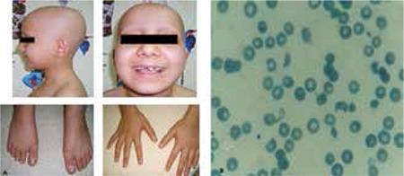
This composite image consists of clinical photographs and a diagnostic peripheral blood smear. Figure 1a (left) shows a pediatric patient with clinical signs including total alopecia (complete absence of scalp hair) and nail pitting affecting both hands and feet, characterized by small punctate depressions on the nail plates. Figure 1b (right) is a microscopic view of a peripheral blood smear demonstrating microangiopathic hemolytic anemia. Visible are numerous schistocytes—fragmented, irregular red blood cells including helmet cells and triangular forms—alongside some normal erythrocytes showing central pallor. The educational focus is on the multi-systemic manifestations of a medical condition, likely a microangiopathic process such as Hemolytic Uremic Syndrome (HUS) or similar systemic pathology, illustrating the correlation between dermatological signs (hair and nail changes) and hematological abnormalities (hemolysis and schistocytosis).
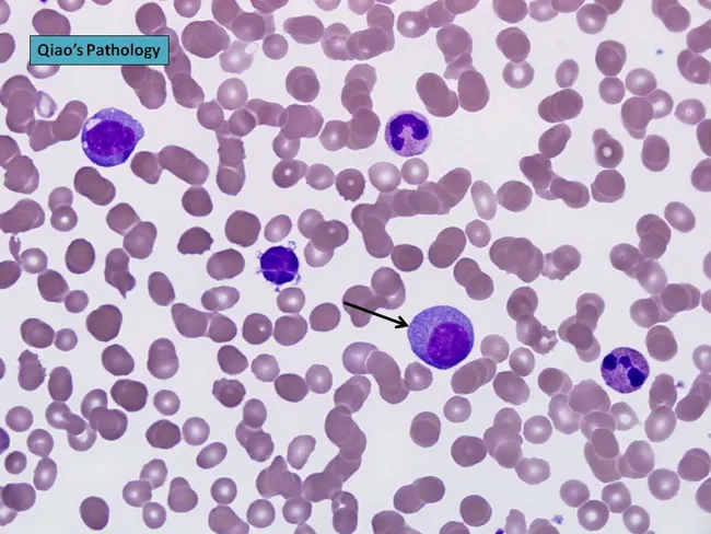
Peripheral blood smear prepared with Wright-Giemsa stain and examined under brightfield illumination using a 100× oil immersion objective reveals a single large plasma cell among a field of mature erythrocytes. The plasma cell displays features typical of clonal plasma cells: basophilic cytoplasm, an eccentrically placed round nucleus, and a conspicuous perinuclear hof; background shows rouleaux formation of red cells. This image exemplifies hematologic involvement by a plasma cell dyscrasia. In plasma cell leukemia, circulating plasma cells constitute more than 20% of leukocytes, a hallmark distinguishing it from conventional multiple myeloma, although both conditions share monoclonal plasma cell proliferation. The presence of circulating plasma cells is associated with younger patient age, higher incidence of anemia, thrombocytopenia, organomegaly, and possible lymphadenopathy; however lytic bone lesions may be less common. Immunophenotypic patterns often include lack of CD56 expression relative to myeloma. Clinically, this morphological finding warrants confirmation with complete blood count, peripheral smear review, quantitative plasma cell percentage, serum protein electrophoresis, and immunofixation to identify monoclonal immunoglobulin isotype (IgD/IgE or light-chain predominance). Early detection of circulating plasma cells supports prognosis assessment and guides therapy decisions in plasma cell dyscrasia management and monitoring, including risk stratification and treatment response evaluation for practice in clinics.
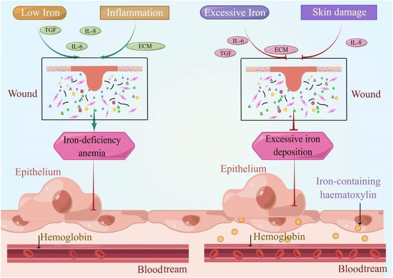
A comparative medical illustration detailing the effects of iron dysregulation on wound healing and systemic states. The left panel depicts a 'Low Iron' and 'Inflammation' scenario where a cutaneous wound is associated with 'Iron-deficiency anemia'. Key signaling molecules shown include TGF, IL-6, IL-8, and ECM components within a wound bed containing fibroblasts and matrix elements. An arrow indicates the downstream impact on the underlying epithelium and hemoglobin within the bloodstream. The right panel depicts an 'Excessive Iron' and 'Skin damage' scenario characterized by 'Excessive iron deposition'. This state is visually distinguished by an inhibitory symbol (red T-bar) directed at the epithelium, suggesting impaired regenerative capacity. Notably, the epithelium in the high-iron state contains yellow granules labeled as 'Iron-containing haematoxylin' (hemosiderin), which serve as a histological marker of iron overload. Both panels show the anatomical relationship between the cutaneous wound, underlying epithelial cells, and the intravascular space (bloodstream) containing red blood cells and hemoglobin, illustrating how iron homeostasis is critical for normal dermatological repair and systemic oxygen transport.
"iron deficiency anemia" diagnosis treatment
iron deficiency anemia microcytic hypochromic red blood cells peripheral smear
Peripheral blood smear from a patient with known chronic lymphocytic leukemia (CLL) showing autoimmune hemolytic anemia (AIHA). Brightfield microscopy of Wright-Giemsa stained smear at 1000x (oil immersion) reveals two dominant cell populations: numerous small mature lymphocytes typical of CLL and red blood cells showing spherocytic deformation. A smudge cell is evident just left of center, a classical clue for CLL. Red cells display reduced central pallor consistent with spherocytes, indicating intravascular or extravascular hemolysis driven by autoantibodies. The background shows mild polychromasia and rare nucleated RBCs, consistent with a robust reticulocyte response; the corrected reticulocyte count in this case is markedly elevated (>7%). Platelets are not significantly abnormal. These features collectively support autoimmune destruction of erythrocytes in the setting of CLL. Diagnostic significance lies in recognizing AIHA as a common autoimmune complication in CLL, with a clinical picture of sudden Hb decline and compensatory erythropoiesis. Differential considerations include drug-induced hemolysis, other causes of hemolytic anemia, or marrow infiltration by CLL. Clinical correlation includes therapy implications (steroids, rituximab) and close monitoring of hemolysis markers. This image is a valuable teaching resource for hematology morphology, AIHA in CLL, and differential diagnosis in anemia.
Peripheral blood smear prepared with Wright-Giemsa stain and examined under brightfield illumination using a 100× oil immersion objective reveals a single large plasma cell among a field of mature erythrocytes. The plasma cell displays features typical of clonal plasma cells: basophilic cytoplasm, an eccentrically placed round nucleus, and a conspicuous perinuclear hof; background shows rouleaux formation of red cells. This image exemplifies hematologic involvement by a plasma cell dyscrasia. In plasma cell leukemia, circulating plasma cells constitute more than 20% of leukocytes, a hallmark distinguishing it from conventional multiple myeloma, although both conditions share monoclonal plasma cell proliferation. The presence of circulating plasma cells is associated with younger patient age, higher incidence of anemia, thrombocytopenia, organomegaly, and possible lymphadenopathy; however lytic bone lesions may be less common. Immunophenotypic patterns often include lack of CD56 expression relative to myeloma. Clinically, this morphological finding warrants confirmation with complete blood count, peripheral smear review, quantitative plasma cell percentage, serum protein electrophoresis, and immunofixation to identify monoclonal immunoglobulin isotype (IgD/IgE or light-chain predominance). Early detection of circulating plasma cells supports prognosis assessment and guides therapy decisions in plasma cell dyscrasia management and monitoring, including risk stratification and treatment response evaluation for practice in clinics.
This composite image consists of clinical photographs and a diagnostic peripheral blood smear. Figure 1a (left) shows a pediatric patient with clinical signs including total alopecia (complete absence of scalp hair) and nail pitting affecting both hands and feet, characterized by small punctate depressions on the nail plates. Figure 1b (right) is a microscopic view of a peripheral blood smear demonstrating microangiopathic hemolytic anemia. Visible are numerous schistocytes—fragmented, irregular red blood cells including helmet cells and triangular forms—alongside some normal erythrocytes showing central pallor. The educational focus is on the multi-systemic manifestations of a medical condition, likely a microangiopathic process such as Hemolytic Uremic Syndrome (HUS) or similar systemic pathology, illustrating the correlation between dermatological signs (hair and nail changes) and hematological abnormalities (hemolysis and schistocytosis).
koilonychia spoon-shaped nails iron deficiency
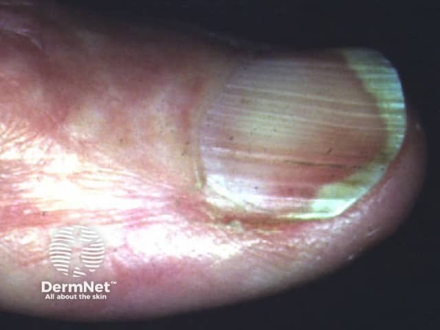
This is a high-resolution clinical photograph of a human fingernail demonstrating koilonychia, also called spoon nails. Imaging modality is clinical photography with macro/close-up technique to enhance surface and curvature details. The primary subject is the fingernail plate and surrounding nail folds, captured from a dorsal perspective for clear visualization of the nail curvature. The nail plate appears thin and concave, with a pronounced central depression producing a spoon-shaped contour. Lateral edges may be slightly tapered, and the distal third of the plate shows mild translucency. The surface is relatively smooth with minimal ridging, and the lunula is less conspicuous in this view. Surrounding cuticle and perionychial skin show mild erythema, possibly secondary to manipulation or irritation; no frank edema or pitting is evident. Clinically, koilonychia is a classic morphological clue associated with iron deficiency anemia and other nutritional or systemic disorders; in many cases nails become spoon-shaped due to chronic iron depletion. The diagnostic significance lies in recognizing a potentially reversible sign when iron stores are restored. This image is useful for dermatology, medical education, nursing training, and clinical scenario discussions focused on anemia screening, differential diagnosis of nail dystrophies, and patient education about nail changes as a diagnostic cue. Consider correlating with CBC and ferritin, and monitoring response to iron therapy.
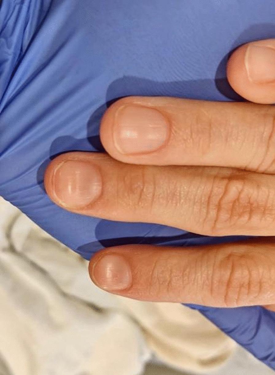
This clinical photograph displays the dorsal surface of three fingers from a patient's hand, demonstrating koilonychia. The nails exhibit a classic 'spoon-shaped' deformity characterized by the loss of normal convexity, resulting in a flattened or centrally concave nail plate with raised lateral and distal edges. The nail plates appear thin and slightly pale, consistent with hypochromic states. The surrounding periungual skin and cuticles appear intact without significant inflammation, paronychia, or splinter hemorrhages. This finding is a pathognomonic physical sign often associated with chronic iron deficiency anemia (Plummer-Vinson syndrome) or other systemic conditions affecting keratinization. The image serves as an educational reference for identifying dermatological manifestations of hematologic disorders during a physical examination.
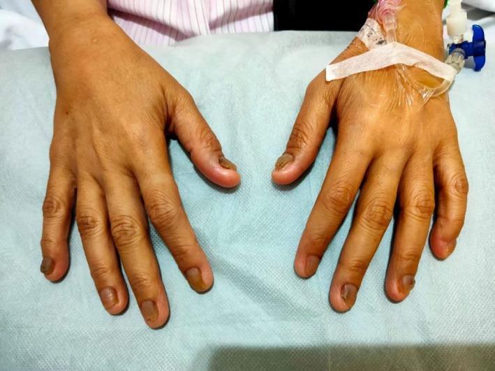
This clinical photograph displays the dorsal surface of both hands of a patient against a medical drape. The primary focus is the prominent nail pathology consistent with koilonychia (spoon nails). The fingernails exhibit a characteristic concave, upward-curving shape, appearing flattened or scooped out. Additionally, there is significant chromonychia characterized by brownish discoloration, with some nails showing hyperpigmented, dark areas near the distal and lateral margins. The nails appear thin and brittle in texture. An intravenous (IV) catheter is secured with white adhesive tape on the dorsum of the right hand, indicating an acute clinical setting. These physical findings are classic cutaneous markers of chronic iron deficiency anemia and are relevant to systemic conditions such as Plummer-Vinson syndrome. The image serves as a diagnostic educational resource for identifying dermatological manifestations of systemic hematologic disorders.
Iron Deficiency Anemia (IDA)
1. Normal Iron Metabolism (Background)
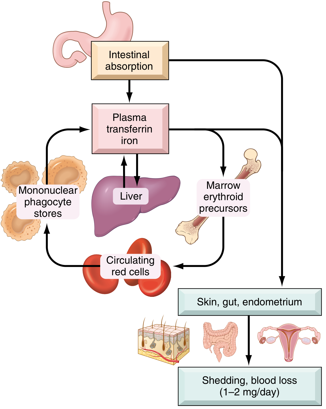
- Total body iron: ~3,450 mg (males) / ~2,450 mg (females)
- ~70% is in hemoglobin (functional pool); ~25% is stored as ferritin and hemosiderin
- Daily diet in the US contains 10-20 mg of iron; only ~1-2 mg is absorbed (10-20% of heme iron vs. 1-2% of non-heme iron)
- Daily losses are fixed at ~1-2 mg (shed epithelial cells, menstrual blood)
- Iron absorbed in the duodenum is transported in plasma bound to transferrin (normally ~1/3 saturated)
- Hepcidin (liver-produced) is the master regulator: it inhibits ferroportin, reducing iron export from enterocytes and macrophages. Iron deficiency suppresses hepcidin, increasing absorption. - Robbins, Cotran & Kumar
2. Causes / Etiology
| Mechanism | Examples |
|---|---|
| Dietary lack | Infants (low iron in milk), vegans, elderly, poverty |
| Impaired absorption | Celiac disease, gastric atrophy, H. pylori, gastrectomy, Roux-en-Y bypass, proton pump inhibitor use |
| Increased requirement | Pregnancy, infancy, adolescence, growth spurts |
| Chronic blood loss | GI bleeding (peptic ulcer, colorectal cancer, hookworm), menorrhagia, urinary/pulmonary losses |
In adult males and postmenopausal females, GI blood loss must be excluded before attributing IDA to any other cause - an occult GI cancer is a key differential. - Sleisenger & Fordtran's GI and Liver Disease
3. Stages of Iron Deficiency
- Negative iron balance - Iron stores (ferritin) deplete; serum ferritin falls. No anemia yet.
- Iron-deficient erythropoiesis - Stores exhausted; transferrin saturation falls <15%; protoporphyrin accumulates in RBCs. MCV begins to fall.
- Iron deficiency anemia - Frank anemia appears; hemoglobin drops, red cells become microcytic and hypochromic.
4. Morphology (What You See on Blood Smear)

- Microcytosis (small RBCs, low MCV)
- Hypochromia (pale cells - zone of central pallor occupies >1/3 of cell diameter, hemoglobin seen only as a thin peripheral rim)
- Poikilocytosis - especially "pencil cells" (small elongated RBCs)
- Bone marrow: increased erythroid precursors; absent stainable iron on Prussian blue stain (diagnostic)
5. Clinical Features
- Fatigue, weakness, exertional dyspnea
- Pallor (conjunctiva, palmar creases)
- Tachycardia, palpitations
-
Koilonychia - spoon-shaped concave nails
-
Pica - craving for non-food substances (clay, ice/pagophagia, flour) - from CNS iron depletion
-
Alopecia (hair loss)
-
Atrophic glossitis (smooth, sore tongue)
-
Angular cheilitis (cracks at mouth corners)
-
Esophageal webs (in severe/chronic cases)
-
Plummer-Vinson Syndrome (triad): microcytic hypochromic anemia + atrophic glossitis + esophageal webs - Robbins, Cotran & Kumar, p.615
6. Laboratory Diagnosis
| Test | Finding in IDA |
|---|---|
| Hemoglobin / Hematocrit | Low (moderate-to-severe reduction) |
| MCV | Low (<80 fL) - microcytic |
| MCH / MCHC | Low - hypochromic |
| Serum iron | Low |
| TIBC (transferrin) | High (liver produces more transferrin) |
| Transferrin saturation | <15% (sensitive indicator) |
| Serum ferritin | Low (<12-15 µg/L) - best single screening test; <15 ng/mL has 99% specificity |
| Serum hepcidin | Low (stores depleted suppress hepcidin) |
| Reticulocyte count | Low or normal (hypoproliferative) |
| Bone marrow Prussian blue | Absent stainable iron (gold standard) |
7. Treatment
- Ferrous sulfate 325 mg (65 mg elemental iron) 2-3x/day on empty stomach
- Response: reticulocyte count rises in 5-7 days, followed by gradual rise in Hb
- Continue for 3-6 months after Hb normalizes to replenish stores
- Side effects: GI upset, constipation (take with food if needed, but reduces absorption)
- Indicated in malabsorption (celiac, post-gastric bypass), inflammatory bowel disease, severe anemia, chronic kidney disease, or post-bariatric surgery
- Options: ferric carboxymaltose, iron sucrose, ferric gluconate
8. High-Risk Groups
- Infants and toddlers (human milk is iron-poor; rapid growth demands)
- Adolescent girls (menstruation + growth)
- Pregnant women (expanding blood volume + fetal demands)
- Premenopausal women with heavy menstrual bleeding
- Males and postmenopausal women - always investigate for GI pathology (cancer until proven otherwise)
- Vegans (only non-heme iron, 1-2% bioavailability)
- Post-bariatric surgery patients (bypass of duodenal absorption site)
Recent Evidence (2024-2026)
Megaloblastic Anemia detail mbbs
megaloblastic anemia hypersegmented neutrophil macrocytic peripheral blood smear
Peripheral blood smear from a patient with known chronic lymphocytic leukemia (CLL) showing autoimmune hemolytic anemia (AIHA). Brightfield microscopy of Wright-Giemsa stained smear at 1000x (oil immersion) reveals two dominant cell populations: numerous small mature lymphocytes typical of CLL and red blood cells showing spherocytic deformation. A smudge cell is evident just left of center, a classical clue for CLL. Red cells display reduced central pallor consistent with spherocytes, indicating intravascular or extravascular hemolysis driven by autoantibodies. The background shows mild polychromasia and rare nucleated RBCs, consistent with a robust reticulocyte response; the corrected reticulocyte count in this case is markedly elevated (>7%). Platelets are not significantly abnormal. These features collectively support autoimmune destruction of erythrocytes in the setting of CLL. Diagnostic significance lies in recognizing AIHA as a common autoimmune complication in CLL, with a clinical picture of sudden Hb decline and compensatory erythropoiesis. Differential considerations include drug-induced hemolysis, other causes of hemolytic anemia, or marrow infiltration by CLL. Clinical correlation includes therapy implications (steroids, rituximab) and close monitoring of hemolysis markers. This image is a valuable teaching resource for hematology morphology, AIHA in CLL, and differential diagnosis in anemia.
This composite image consists of clinical photographs and a diagnostic peripheral blood smear. Figure 1a (left) shows a pediatric patient with clinical signs including total alopecia (complete absence of scalp hair) and nail pitting affecting both hands and feet, characterized by small punctate depressions on the nail plates. Figure 1b (right) is a microscopic view of a peripheral blood smear demonstrating microangiopathic hemolytic anemia. Visible are numerous schistocytes—fragmented, irregular red blood cells including helmet cells and triangular forms—alongside some normal erythrocytes showing central pallor. The educational focus is on the multi-systemic manifestations of a medical condition, likely a microangiopathic process such as Hemolytic Uremic Syndrome (HUS) or similar systemic pathology, illustrating the correlation between dermatological signs (hair and nail changes) and hematological abnormalities (hemolysis and schistocytosis).
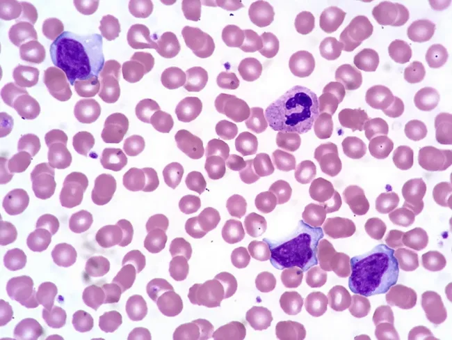
Comprehensive description: This is a brightfield, Wright-Giemsa stained peripheral blood smear examined by light microscopy at high magnification (oil immersion). The specimen shows several activated (atypical) lymphocytes in the peripheral blood, a hallmark of infectious mononucleosis. The lymphocytes have abundant basophilic cytoplasm and irregular, often indented nuclei with clumped chromatin and inconspicuous nucleoli. A normal segmented neutrophil is present for contrast. Erythrocytes exhibit occasional cytoplasmic membrane scalloping around red cells, a feature sometimes observed in EBV-related lymphocytosis. The background cytology is otherwise typical, with uniform erythrocytes and occasional platelets. The activated lymphocytes vary in size and cytoplasmic texture, some appearing plasmacytoid with vesicular basophilia, while others are more indented with coarse chromatin. These morphologic changes reflect an immune response to Epstein-Barr virus infection and are described as reactive lymphocytosis. Clinically, the combination of acute lymphadenitis, fever, a positive Monospot test, and EBV serology supports infectious mononucleosis. The image helps differentiate viral-induced lymphocytosis from bacterial lymphadenitis and from hematologic malignancies when integrated with clinical data. In summary, this smear illustrates the classical hematologic manifestation of EBV-associated mononucleosis. These atypical lymphocytes, sometimes named Downey cells, accompany fever and pharyngitis in EBV infection and aid differentiation from leukemia when combined with serology and clinical data.
Peripheral blood smear prepared with Wright-Giemsa stain and examined under brightfield illumination using a 100× oil immersion objective reveals a single large plasma cell among a field of mature erythrocytes. The plasma cell displays features typical of clonal plasma cells: basophilic cytoplasm, an eccentrically placed round nucleus, and a conspicuous perinuclear hof; background shows rouleaux formation of red cells. This image exemplifies hematologic involvement by a plasma cell dyscrasia. In plasma cell leukemia, circulating plasma cells constitute more than 20% of leukocytes, a hallmark distinguishing it from conventional multiple myeloma, although both conditions share monoclonal plasma cell proliferation. The presence of circulating plasma cells is associated with younger patient age, higher incidence of anemia, thrombocytopenia, organomegaly, and possible lymphadenopathy; however lytic bone lesions may be less common. Immunophenotypic patterns often include lack of CD56 expression relative to myeloma. Clinically, this morphological finding warrants confirmation with complete blood count, peripheral smear review, quantitative plasma cell percentage, serum protein electrophoresis, and immunofixation to identify monoclonal immunoglobulin isotype (IgD/IgE or light-chain predominance). Early detection of circulating plasma cells supports prognosis assessment and guides therapy decisions in plasma cell dyscrasia management and monitoring, including risk stratification and treatment response evaluation for practice in clinics.
"megaloblastic anemia" vitamin B12 folate deficiency
megaloblastic anemia bone marrow megaloblast hypersegmented neutrophil vitamin B12
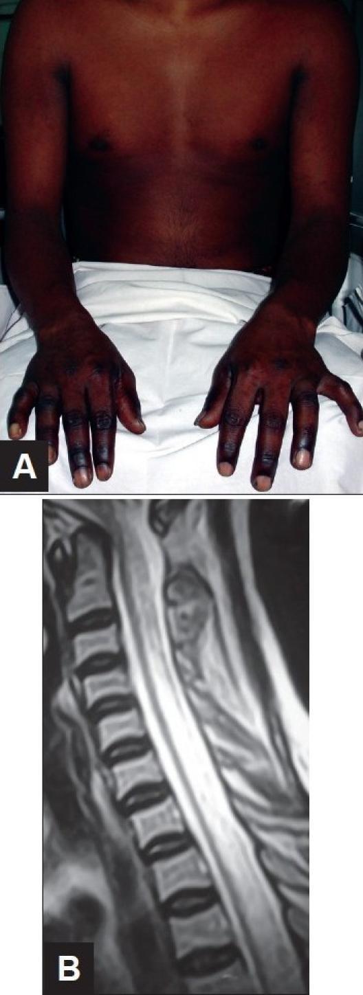
This composite figure demonstrates clinical and diagnostic findings associated with Vitamin B12 deficiency (Subacute Combined Degeneration of the spinal cord). Image A is a clinical photograph of an 18-year-old male showing significant cutaneous hyperpigmentation of the distal upper extremities. The darkening is most pronounced on the dorsal surfaces of the hands and fingers, contrasting with the lighter skin tone of the proximal arms and chest. This is a common dermatologic manifestation of megaloblastic anemia. Image B is a sagittal T2-weighted MRI of the cervical and upper thoracic spine. It reveals a long-segment, linear intramedullary hyperintense signal within the posterior aspect of the spinal cord. This finding represents edema and demyelination characteristic of Subacute Combined Degeneration, which typically affects the posterior and lateral columns while sparing the anterior column. The combination of these visual findings—distal hyperpigmentation and longitudinal posterior cord hyperintensity—is highly suggestive of severe Vitamin B12 deficiency in the context of progressive paraplegia.
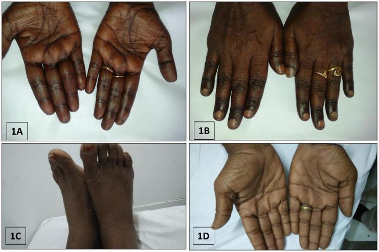
This composite of clinical photographs illustrates various patterns of cutaneous hyperpigmentation associated with megaloblastic anemia due to Vitamin B12 deficiency. Image 1A and 1D display the palmar surfaces of the hands, showing diffuse, brownish-black hyperpigmentation with prominent accentuation along the palmar creases. Image 1B focuses on the dorsal aspect of the hands, highlighting localized brownish-black pigmentation specifically over the knuckle pads and phalangeal joints. Image 1C depicts the dorsal aspect of the feet, exhibiting a similar diffuse, dusky, brownish-black discoloration across the skin surface. These dermatological findings are classic external markers of severe B12 deficiency and often present alongside systemic symptoms like pancytopenia and macrocytosis. The collection serves as an educational tool for recognizing the cutaneous manifestations of nutritional deficiencies and metabolic disorders in hematology and dermatology.
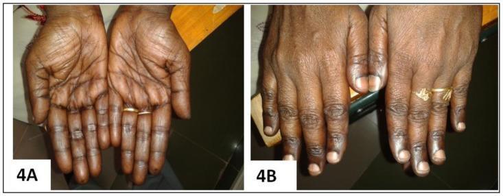
Clinical photograph comparison (Figure 4A and 4B) demonstrating the cutaneous manifestations of Vitamin B12 deficiency and its resolution following treatment. Image 4A (Pre-treatment): Palmar view showing diffuse, brownish-black hyperpigmentation with prominent accentuation along the palmar creases and longitudinal hyperpigmentation of the fingers. The knuckles exhibit characteristic darkening, a classic sign of megaloblastic anemia-associated pigmentary changes. Image 4B (Post-treatment): Dorsal view of the same patient's hands 12 weeks after initiating parenteral cyanocobalamin (Vitamin B12) therapy. There is a marked reversal of the hyperpigmentation, with the skin returning to a more uniform, lighter tone. The previously dusky knuckles and digits show significant clearing, and the nail beds appear healthy. This comparison serves as an educational tool for identifying dermatological markers of nutritional deficiencies and monitoring therapeutic response to Vitamin B12 supplementation.
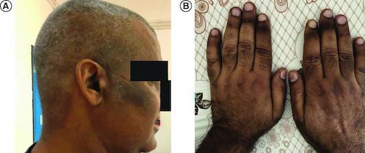
Clinical photograph in two panels (A and B) demonstrating cutaneous hyperpigmentation associated with vitamin B12 deficiency. Panel A shows a lateral view of the head and neck, highlighting patchy, irregular melanoderma on the temporal region and cheek, with a mottled distribution across the scalp. The pigmentation ranges from light tan to dark brown and appears non-elevated and non-scaly. Panel B displays the dorsal surfaces of both hands, revealing generalized darkening with accentuated intensity over the knuckles and phalangeal creases. A proximal-to-distal gradient is visible on the fingers, with the most intense hyperpigmentation occurring proximally. These findings are characteristic of the mucocutaneous manifestations of megaloblastic anemia and vitamin B12 deficiency, serving as a critical clinical sign for differential diagnosis from Addison’s disease.
Megaloblastic Anemia - Detailed MBBS Notes
Definition
Pathophysiology - The Core Mechanism
The Folate-B12 Biochemical Axis
- Dietary folate → absorbed as monoglutamates in the jejunum
- Converted inside cells to tetrahydrofolate (FH4) via dihydrofolate reductase (DHFR)
- FH4 (as N5,10-methylene-FH4) donates a methyl group for:
- dUMP → dTMP (thymidylate synthesis - essential for DNA)
- Purine synthesis
- Homocysteine → methionine conversion
- Vitamin B12 (as methylcobalamin) is required by methionine synthase to convert homocysteine → methionine
- In this reaction, N5-methyl-FH4 → FH4 is regenerated
- Without B12, folate gets "trapped" as N5-methyl-FH4 (the "methylfolate trap" hypothesis)
- Result: FH4 deficiency → impaired dTMP synthesis → defective DNA → megaloblastosis
- Adenosylcobalamin is required by methylmalonyl-CoA mutase for the conversion of methylmalonyl-CoA → succinyl-CoA
- B12 deficiency causes elevated methylmalonic acid (MMA) in blood/urine - a diagnostic marker unique to B12 deficiency (not seen in folate deficiency)
Causes - Comprehensive Classification
Vitamin B12 Deficiency
| Mechanism | Specific Cause |
|---|---|
| Decreased intake | Strict vegetarian/vegan diet, malnutrition |
| Intrinsic factor deficiency | Pernicious anemia (autoimmune), total gastrectomy |
| Malabsorption (ileum) | Ileal resection, Crohn's disease (terminal ileum), lymphoma, systemic sclerosis |
| Competitive uptake | Fish tapeworm (Diphyllobothrium latum), bacterial overgrowth (blind loop syndrome) |
| Drugs | Metformin (reduces IF-B12 complex uptake), PPI (reduces gastric acid needed for B12 release) |
Folate Deficiency
| Mechanism | Specific Cause |
|---|---|
| Decreased intake | Alcoholism (most common cause), poor diet, elderly, infants |
| Impaired absorption | Celiac disease, tropical sprue, Crohn's disease (jejunum) |
| Increased requirement | Pregnancy, rapid cell turnover (hemolysis, cancer, psoriasis) |
| Increased loss | Hemodialysis |
| Impaired utilization | Methotrexate (inhibits DHFR), trimethoprim, pyrimethamine, phenytoin |
Not Responsive to B12 or Folate
- Hydroxyurea, 5-fluorouracil, cytarabine (direct inhibitors of DNA synthesis)
Morphology
Peripheral Blood Smear - Classic Findings
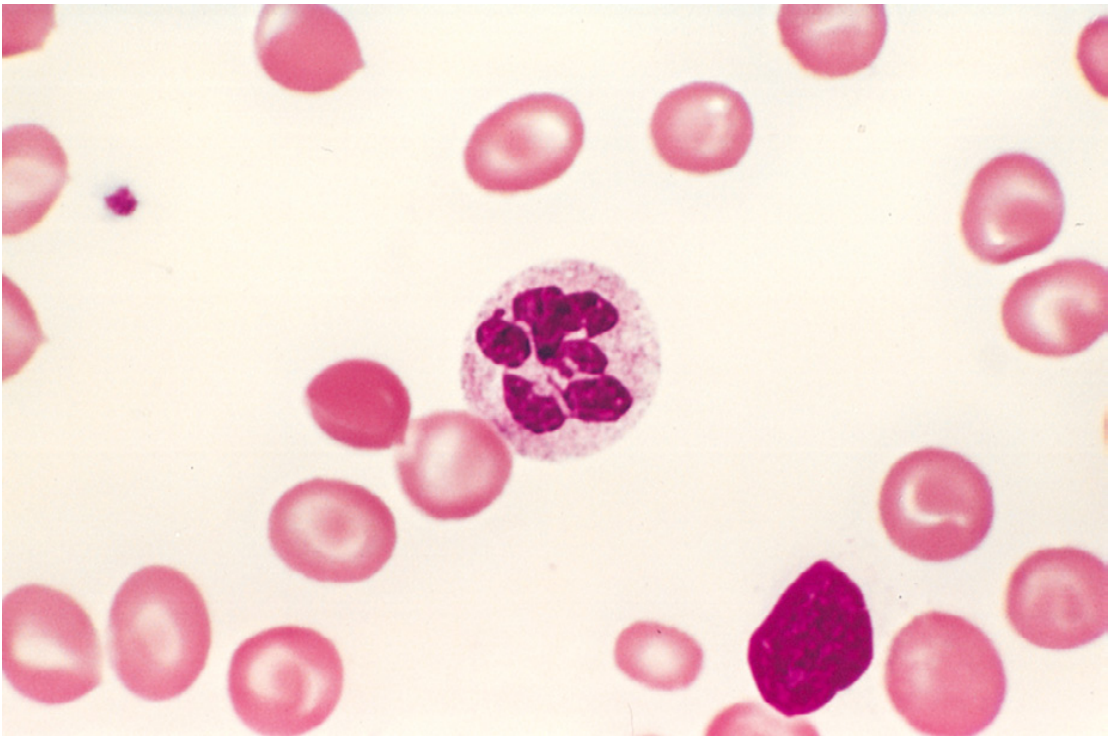
- Macro-ovalocytes (large, oval red cells without central pallor) - pathognomonic
- Hypersegmented neutrophils - ≥5 lobes in one neutrophil, or ≥4% of neutrophils with ≥5 lobes - earliest and most reliable sign
- Marked anisocytosis (variation in size) and poikilocytosis (variation in shape)
- Pancytopenia - low RBC, WBC, and platelets (all rapidly dividing cells affected)
- Low reticulocyte count (ineffective erythropoiesis)
Bone Marrow
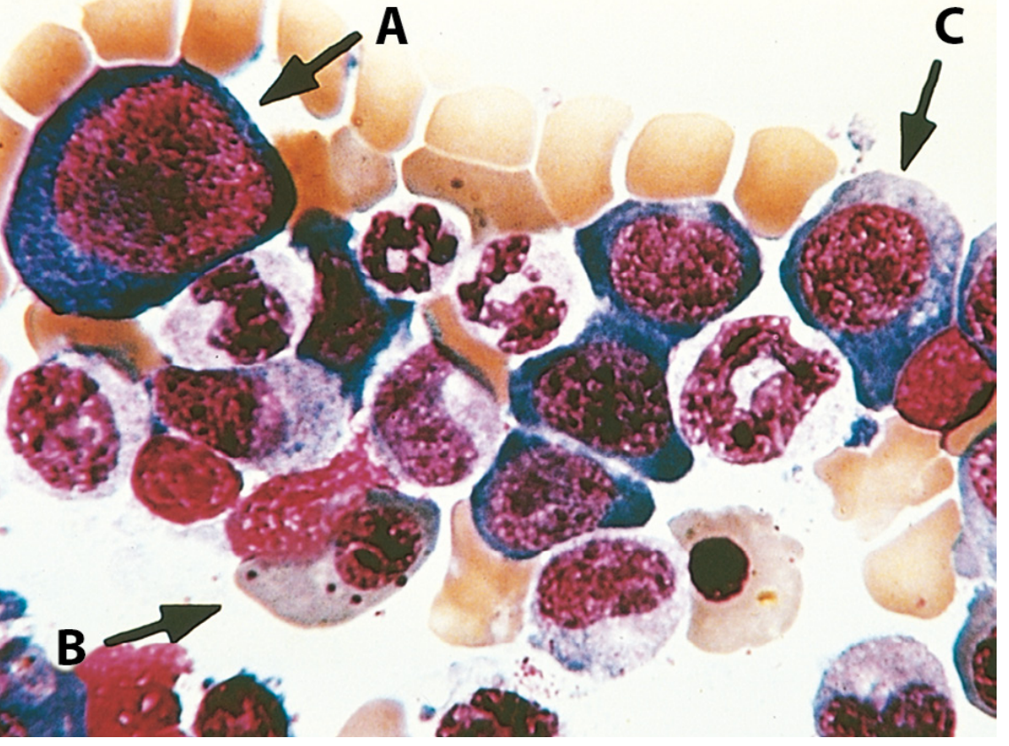
- Markedly hypercellular (erythroid hyperplasia - attempted response)
- Megaloblasts at all stages: large cells with fine, open ("lacy") chromatin despite hemoglobin accumulation in cytoplasm
- Normal normoblasts: nucleus condenses (pyknotic) as cytoplasm fills with hemoglobin
- Megaloblasts: nucleus remains open and immature - nuclear-cytoplasmic asynchrony
- Giant metamyelocytes and band forms (granulocyte precursors also affected)
- Giant, multilobate megakaryocytes
- Most precursors undergo apoptosis in the marrow → ineffective hematopoiesis (marrow looks full but output is low)
Pernicious Anemia (Most Important Subtype)
Vitamin B12 Absorption Pathway (Normal)
- Dietary B12 (animal products) released by pepsin in stomach
- B12 binds haptocorrin (salivary protein) initially
- In duodenum, pancreatic proteases release B12 → binds intrinsic factor (from gastric parietal cells)
- B12-IF complex travels to terminal ileum → binds cubilin receptor → endocytosed
- Inside ileal cells, B12 binds transcobalamin II → transported to liver and bone marrow
Autoimmune Mechanism in Pernicious Anemia
- Type I (blocking antibodies) - ~75% of patients - block B12 binding site on IF
- Type II (binding antibodies) - ~50% - bind IF-B12 complex and prevent ileal absorption
- Anti-parietal cell antibodies - ~90% - against H+/K+ ATPase of parietal cells (less specific)
- Atrophy of fundic glands (chief cells + parietal cells lost)
- Intestinalization (goblet cell metaplasia of gastric mucosa)
- Achlorhydria
Clinical Features of Pernicious Anemia
- Insidious onset, often severe by presentation
- Fatigue, pallor, dyspnea
- Mild jaundice (bilirubin from ineffective erythropoiesis and hemolysis)
- Smooth, beefy-red, painful tongue (atrophic glossitis/Hunter's glossitis)
- Demyelination of posterior columns → loss of vibration and position sense, sensory ataxia (positive Romberg)
- Demyelination of lateral (corticospinal) columns → upper motor neuron signs, spastic paraparesis
- Peripheral neuropathy → symmetrical paresthesias (glove-and-stocking)
- Psychiatric: depression, irritability, dementia ("megaloblastic madness")
- Key point: Neurological damage is NOT seen in folate deficiency. Giving folate to a B12-deficient patient may correct anemia but will worsen neurological disease.
- Reversible skin hyperpigmentation (especially over knuckles and palmar creases) in some patients
Folate Deficiency Anemia
Folate Metabolism (Normal)
- Dietary folate (polyglutamates) absorbed in proximal jejunum (unlike B12, which is absorbed in ileum)
- Converted to FH4 by DHFR
- Body stores last only 3-4 months (vs. B12 stores lasting 3-5 years)
Differences from B12 Deficiency
| Feature | B12 Deficiency | Folate Deficiency |
|---|---|---|
| Store duration | 3-5 years | 3-4 months |
| Site of absorption | Terminal ileum | Proximal jejunum |
| Serum MMA | Elevated | Normal |
| Serum homocysteine | Elevated | Elevated |
| Neurological findings | Subacute combined degeneration | Absent |
| Common causes | Pernicious anemia, vegan diet | Alcoholism, pregnancy, malabsorption |
| Serum folate | Normal or high | Low |
| RBC folate | Normal | Low (better marker - not affected by recent intake) |
Laboratory Diagnosis
| Test | Finding |
|---|---|
| Hemoglobin | Low (often severe) |
| MCV | >100 fL (macrocytic) - often 110-140 fL |
| MCH | High (due to large cells) |
| MCHC | Normal (not truly hyperchromic) |
| WBC | Low (neutropenia) |
| Platelets | Low (thrombocytopenia) |
| Reticulocytes | Low (ineffective erythropoiesis) |
| Serum B12 | Low in B12 deficiency |
| Serum folate | Low in folate deficiency; may be falsely normal in B12 deficiency |
| RBC folate | More reliable marker of folate stores |
| Serum homocysteine | Elevated in BOTH B12 and folate deficiency |
| Serum MMA | Elevated only in B12 deficiency - key differentiator |
| Anti-IF antibodies | Highly specific for pernicious anemia |
| Anti-parietal cell antibodies | Sensitive (~90%) but not specific for PA |
| Bone marrow | Hypercellular with megaloblasts (rarely needed for diagnosis) |
| LDH | Very high (marker of ineffective erythropoiesis) |
| Indirect bilirubin | Mildly elevated |
Treatment
Vitamin B12 Deficiency
- Loading: IM/SC cyanocobalamin or hydroxycobalamin 1 mg/day for 7 days, then weekly x 8 weeks
- Maintenance: 1 mg IM monthly (lifelong in pernicious anemia)
- Hydroxycobalamin retained better; can be given every 2-4 months after loading
- 1000-2000 µg/day oral B12 works via passive diffusion (~0.5-2% absorbed without IF)
- Equally effective as IM in RCTs for hematologic correction
- Preferred by patients who dislike injections - Goldman-Cecil Medicine
- Reticulocyte count rises within 5-7 days
- Hemoglobin normalizes in 1-2 months
- Pancytopenia corrects in first 2 weeks
- Neurological recovery: partial (paresthesias improve; established spinal cord lesions may not fully reverse - duration before treatment determines outcome)
- Gastric atrophy in PA does NOT reverse with B12 therapy
Folate Deficiency
- Folic acid 1-5 mg/day orally for 3-4 months (until stores replenished)
- Pregnancy prophylaxis: 400-800 µg/day folate before conception and throughout the first trimester (prevents neural tube defects)
- Treat underlying cause (alcoholism, celiac disease, etc.)
⚠️ Critical caution: Always rule out B12 deficiency before giving folate alone. Folate can correct the anemia but mask B12 deficiency while the neurological damage of SACD progresses. - Robbins, Cotran & Kumar Pathologic Basis of Disease
B12 vs. Folate Deficiency - Quick Summary Table
| B12 | Folate | |
|---|---|---|
| Sources | Meat, eggs, dairy | Green vegetables, cereals, liver |
| Daily requirement | 2-3 µg | 50-100 µg |
| Body stores | 3-5 years | 3-4 months |
| Absorption site | Terminal ileum (with IF) | Proximal jejunum |
| Top cause | Pernicious anemia | Alcoholism |
| Neuro features | SACD (posterior + lateral columns) | None |
| Serum MMA | Elevated | Normal |
| Treatment | B12 IM or high-dose oral | Folic acid oral |
High-Yield MBBS Points
- Most common cause worldwide: Folate deficiency (alcoholism, malnutrition); pernicious anemia is the classic B12 cause
- Fastest developing B12 deficiency: Strict vegans (low intake), but takes years due to large liver stores
- Fastest developing folate deficiency: Alcoholics, pregnant women (stores last only months)
- Earliest peripheral smear finding: Hypersegmented neutrophil (appears before macro-ovalocytes)
- SACD: Never seen in folate deficiency - exclusive to B12 deficiency
- MMA is the key differentiator: Only elevated in B12 deficiency
- Homocysteine: Elevated in both - not a differentiator
- Methotrexate causes megaloblastosis by inhibiting DHFR (blocks FH4 regeneration) - reversed by leucovorin (folinic acid), not folic acid
- Pernicious anemia association: Other autoimmune diseases (thyroid, vitiligo, Addison's), increased gastric cancer risk
- Schilling test (radioactive B12 absorption test): historically used to diagnose PA - now largely replaced by anti-IF antibodies