
Perianal abcess and fistula indetail
Reading File
Now let me search the medical library for perianal abscess and fistula content in parallel.
Finding Sources
Finding Sources
Searching PubMed
"perianal abscess" AND "fistula in ano"
Good - I have several relevant sources. Let me now read the detailed content from the key blocks in parallel.
Reading File
Reading File
Reading File
Good. Let me read more content from Goldman-Cecil and Mulholland's Surgery, plus get the fistula anatomy diagram and Park's classification content.
Reading File
Reading File
Finding Sources
Loading Image
Excellent. Now let me get the Parks classification diagram and Sabiston/Bailey & Love content, plus the Mulholland content on fistula management.
Loading Image
Reading File
Reading File
I now have rich content from multiple authoritative sources. Let me also get the abscess types/locations image and the Sabiston fistula classification section.
Reading File
Reading File
Now let me fetch the Parks classification diagram and Goodsall's rule diagram, and read the Sabiston treatment section.
Loading Image
Loading Image
Loading Image
Reading File
I now have comprehensive content from multiple authoritative surgical textbooks along with excellent images. Let me compile the detailed response.
Perianal Abscess and Fistula-in-Ano - A Detailed Review
PART 1: PERIANAL ABSCESS
Definition and Pathophysiology
A perianal abscess is a collection of pus in the perianal tissues and is one of the most painful anal conditions encountered in clinical practice. The pain is severe, disabling, and progressive. The most widely accepted theory is the cryptoglandular hypothesis: infection originates at the dentate line in the anal crypts, spreads through the intersphincteric plane (where the anal glands lie), and then tracks through the path of least resistance into the perianal spaces, creating a closed environment ideal for mixed bacterial proliferation.
- Pfenninger & Fowler's Procedures for Primary Care, p. 781
- Mulholland & Greenfield's Surgery, p. 3557
Etiology
| Category | Examples |
|---|---|
| Cryptoglandular (most common) | Infected anal glands at dentate line |
| Inflammatory | Crohn disease, tuberculosis, actinomycosis, lymphogranuloma venereum |
| Traumatic | Impalement, foreign body, anal fissure |
| Iatrogenic | Episiotomy, hemorrhoidectomy, prostatectomy, radiation |
| Malignancy | Rectal/anal carcinoma, leukemia, lymphoma |
- Sabiston Textbook of Surgery, Box 97.1
Locations and Incidence
The four anatomical spaces where abscesses can form are well illustrated below:
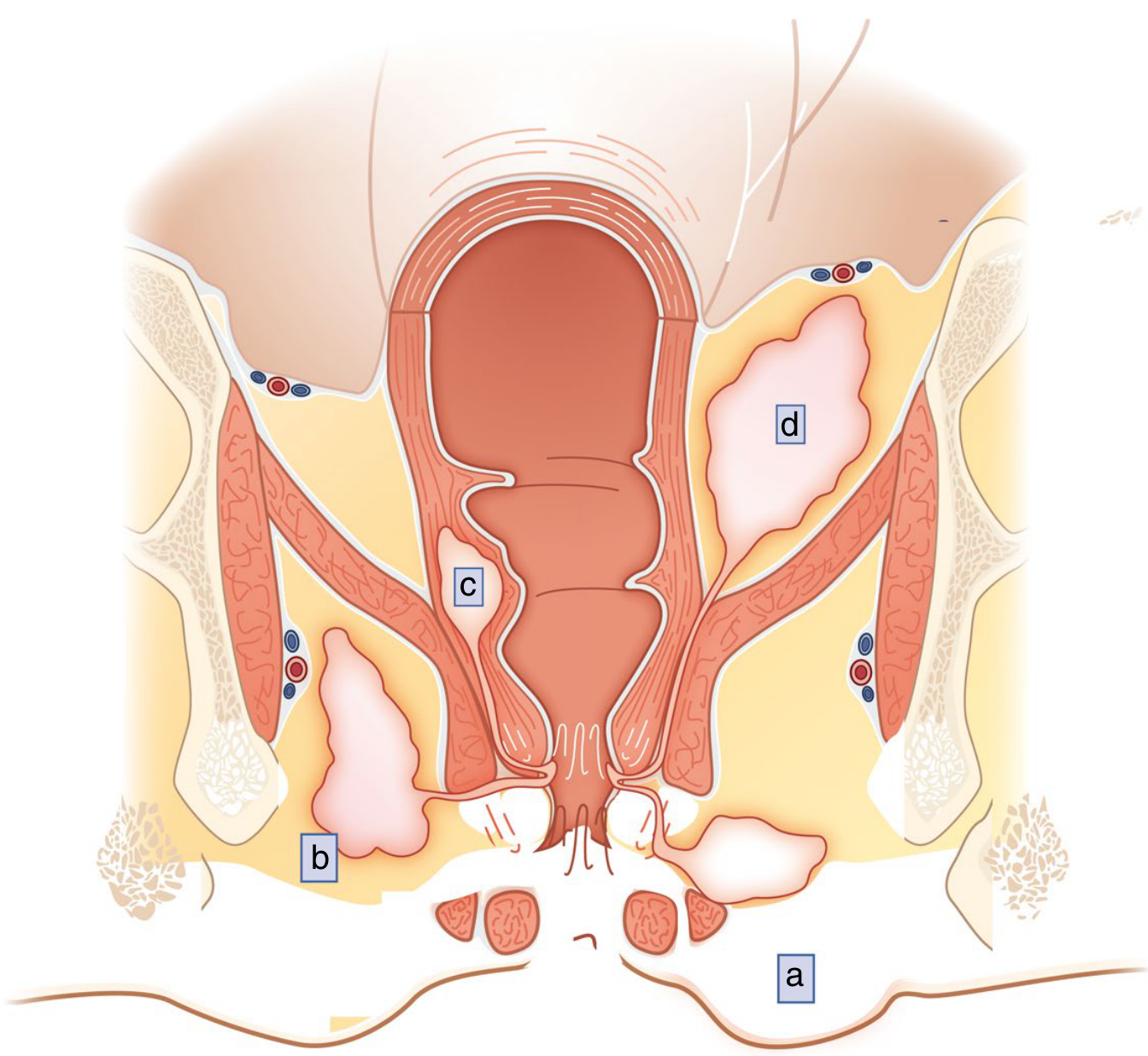
Common extensions of anorectal abscesses - Sabiston Textbook of Surgery, Fig. 97.13
| Location | Incidence | Notes |
|---|---|---|
| Perianal (superficial) | ~60% | Adjacent to anal verge; most common |
| Ischiorectal | ~25% | 2-3 cm from anal verge, outside sphincters |
| Intersphincteric | ~5-10% | Between internal and external sphincters; may not be visible externally; diagnosed on digital exam |
| Supralevator | Least common | More correctly "perirectal"; always look for intra-abdominal/pelvic source (appendicitis, diverticulitis, PID) |
- Pfenninger & Fowler's Procedures for Primary Care, p. 781
Clinical Features
- Pain: Severe, throbbing perianal pain - the hallmark symptom. Aggravated by sitting, coughing, sneezing, and straining.
- Fever and marked leukocytosis depending on severity
- External signs: Swelling, erythema, induration, fluctuance in superficial abscesses
- Deeper abscesses may have no external signs - tenderness and fullness on digital rectal exam only
- Associated symptoms: tenesmus, difficulty voiding
Important clinical pitfall: A common error is diagnosing "cellulitis" when there is no fluctuance. Many of these patients simply have a deeper abscess - antibiotics alone will be insufficient.
Diagnosis
-
Primarily clinical - history and examination
-
Digital rectal exam reveals fluctuant mass (even for intersphincteric abscesses not visible externally)
-
Thorough DRE and anoscopy are often deferred in the acute setting due to pain
-
CT pelvis or pelvic MRI when physical findings are limited or diagnosis is uncertain
-
Mulholland & Greenfield's Surgery, p. 3557
-
Sabiston Textbook of Surgery, p. 2161
Treatment
The treatment of choice is prompt incision and drainage (I&D).
- Antibiotics do not adequately penetrate abscess cavities
- Antibiotic therapy alone is inadequate and must never substitute for drainage
- Extension of a local infection can lead to sepsis and complex long-term problems
Procedure:
- Drainage starting at the most fluctuant area, staying as close to the anus as possible (to shorten any subsequent fistula tract)
- Incision size tailored to abscess size - large enough for adequate drainage
- A cruciate incision with removal of corners ensures ongoing drainage and prevents premature skin closure
- Loculations within the cavity are broken up carefully (avoid aggressive disruption that can injure the sphincter or pudendal nerve)
- If abscess is >5 cm, consider counter-incisions bridged with Penrose drains or vessel loops
- Deeper abscesses: Pezzar or Malecot drainage catheter left in place for days to weeks
Special cases:
- Large/deep abscesses (postanal, horseshoe): best managed in the operating room with sedation
- Intersphincteric abscesses without external fluctuation: drain through intersphincteric groove or into anal canal via internal sphincter incision (NOT externally, to avoid iatrogenic fistula)
- Supralevator abscesses from intersphincteric extension: drain internally through rectal wall
- Horseshoe abscess: modified Hanley technique - posterior midline drainage of the deep postanal space plus drainage of both ischiorectal spaces
Post-drainage antibiotics (e.g., metronidazole 500 mg q8h + ciprofloxacin 500 mg q12h for 7 days) may reduce subsequent fistula formation. Antibiotics are especially recommended in:
-
Immunocompromised patients (poorly controlled HIV, diabetes, transplant recipients, chemotherapy patients)
-
Extensive cellulitis or severe systemic symptoms
-
High-risk for endovascular infection (cardiac shunts, prosthetic valves)
-
Goldman-Cecil Medicine, p. 1535
-
Sabiston Textbook of Surgery, p. 2162
Complications of Untreated/Inadequately Treated Abscess
- Necrotizing fasciitis - life-threatening
- Perineal sepsis / Fournier's gangrene - medical emergency with classic triad of pain, fever, and inability to void
- Fistula formation - the chronic sequela
- Recurrence - most common complication after I&D; most often due to an unrecognized associated fistula
PART 2: FISTULA-IN-ANO
Definition and Epidemiology
A fistula (from Latin for "pipe") is an abnormal communication between two anatomical structures. An anal fistula represents the chronic form of a perianal abscess in which the tract from the infected anal gland to the abscess cavity does not adequately heal, leaving a persistent channel from the anal canal to the perianal skin.
-
Occurs in 30-50% of patients following a perianal abscess (Goldman-Cecil) - though the figure of <20% after a single episode is cited by Pfenninger; recurrence risk rises to >50% with recurrent abscess at the same location
-
Incidence: approximately 8.6 per 100,000
-
2-3 times more common in men than women
-
After appropriate surgery, ~85% are healed at 6 years
-
Goldman-Cecil Medicine, p. 1535
Pathogenesis
After abscess drainage, the internal opening at the dentate line (where the infected gland originated) may remain patent, leaving:
- A source of recurrent infection
- A tract from the internal gland to the abscess cavity/drainage site
This tract, lined with granulation tissue, becomes the fistula. Multiple or atypical fistulae should always raise suspicion for Crohn disease.
Parks Classification (Relationship to Sphincter Complex)
The Parks classification is the most widely used system worldwide:
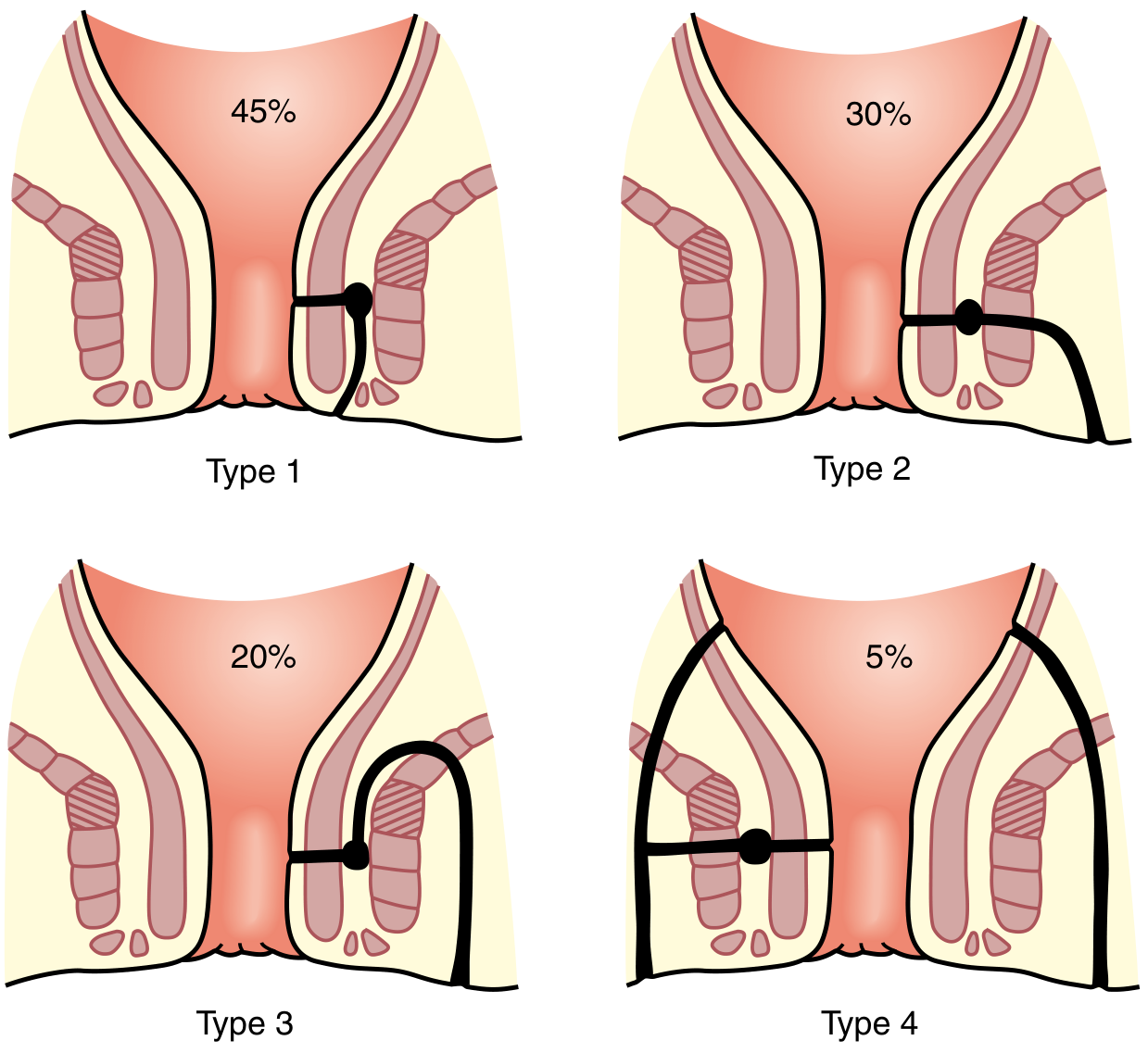
Parks Classification of fistula-in-ano - Sabiston Textbook of Surgery, Fig. 97.16
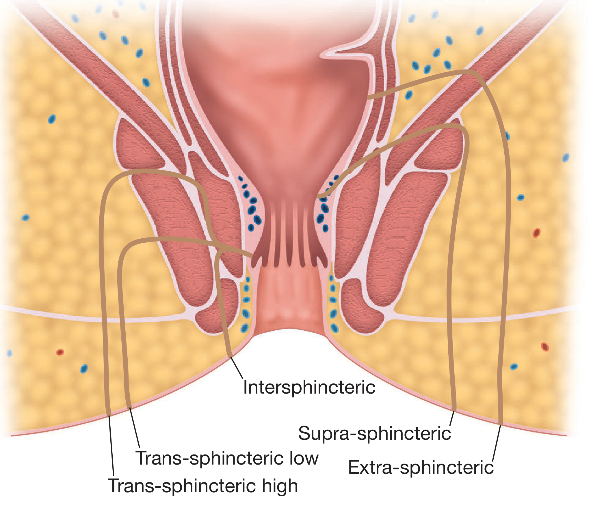
Coronal section view of Parks' classification - Bailey & Love's Surgery, Fig. 80.29
| Type | Description | Incidence |
|---|---|---|
| Type 1 - Intersphincteric | Tract lies between internal and external sphincters; most common and simplest | ~45-70% |
| Type 2 - Transsphincteric | Traverses both internal and external sphincters; subdivided into low (distal EAS) and high (proximal EAS) | ~20-30% |
| Type 3 - Suprasphincteric | Originates at dentate line, loops over the entire sphincter complex | ~20% |
| Type 4 - Extrasphincteric | Internal opening remote from dentate line; typically from pelvic abscess (ruptured appendix, diverticulitis, Crohn disease) | ~5% |
Horseshoe fistula: External openings with tracts on both sides of the midsagittal plane, most commonly with a single internal opening in the posterior midline.
- Goldman-Cecil Medicine, p. 1535
- Sabiston Textbook of Surgery, p. 2163
Goodsall's Rule
A key intraoperative guideline for predicting internal opening location:
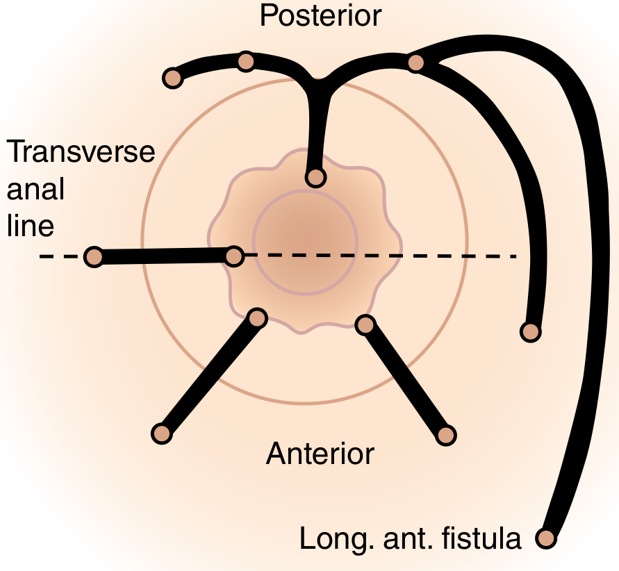
Goodsall's Rule - Sabiston Textbook of Surgery, Fig. 97.17
-
Anterior external openings (anterior to the transverse anal line): track radially (straight) into the nearest crypt
-
Posterior external openings (posterior to transverse anal line): curve in a horseshoe fashion to a posterior midline internal opening
-
Exception: anterior openings >3 cm from the anal verge may also track to the posterior midline
-
Sabiston Textbook of Surgery, p. 2163
Clinical Presentation
-
Recurrent abscesses in the same perianal location
-
Persistent purulent drainage from an abscess site that has not fully healed
-
Intermittent cycle: area seems to heal for weeks, then a "boil" reforms at the same site, ruptures, and relieves symptoms
-
External opening visible on perianal skin
-
Fibrous tract may be palpable along the course toward the anal canal
-
Internal opening occasionally visible on anoscopy (not essential for diagnosis)
-
Goldman-Cecil Medicine, p. 1535
Investigation
-
Clinical examination is the primary tool
-
MRI pelvis (T2-weighted) is the gold standard imaging - accurately defines fistula anatomy, identifies secondary tracts, and assesses sphincter involvement
-
Sabiston Textbook of Surgery, Fig. 97.15 - MRI showing intersphincteric and transsphincteric fistulae
Intraoperative:
- Fistula probe passed gently through the external opening (avoid creating false passages)
- Injection of dilute hydrogen peroxide, methylene blue, or milk into the external opening to identify the internal opening
- Anoscopy to inspect the dentate line and crypts
Treatment
The goals of fistula treatment are:
- Treat any undrained infection
- Define fistula anatomy
- Remove/ablate the epithelialized tract
- Avoid or minimize fecal incontinence
- Prevent recurrence
Treatment is always surgical. The approach depends on the type and amount of sphincter muscle involved.
1. Fistulotomy (Lay-Open)
- Indication: Simple, low-lying fistulae (intersphincteric or low transsphincteric) involving <1/3 of the external anal sphincter
- The entire tract is laid open and allowed to heal by secondary intention
- Recurrence rate: 2-8%
- Incontinence risk: <5% in patients with normal preoperative sphincter function
- Caution: Anterior fistulae in women require particular care (shorter anterior sphincter, previous obstetric injury)
2. Seton Placement
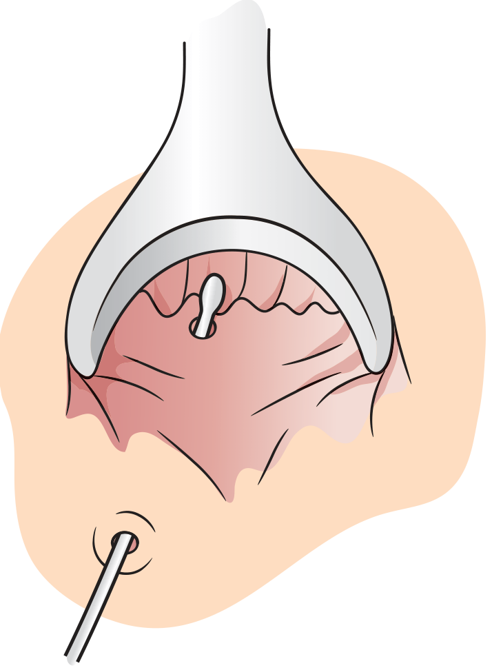
Fistula probe in a simple tract - Pfenninger & Fowler's Procedures
- Indication: High or complex fistulae where fistulotomy would risk significant incontinence
- A suture, silastic vessel loop, or rubber band is passed through the fistula tract, out the anus, and secured to itself
- Draining seton: Keeps external opening patent, prevents recurrent abscess, allows the tract to fibrose over weeks
- Cutting seton: Progressively tightened at 2-4 week intervals to slowly erode through sphincter muscle with concurrent fibrosis - success up to 94%, but incontinence risk 23% or more
3. Endoanal/Endorectal Advancement Flap
- A flap of mucosa + underlying internal sphincter is raised; the internal fistula opening is sutured closed; the flap covers the internal opening
- Success rate: 66-87%
- Preferred for complex/high fistulae in sphincter-preserving surgery
- Failed flaps can be repeated after interval healing
4. LIFT Procedure (Ligation of Intersphincteric Fistula Tract)
- After seton-induced fibrosis, an incision is made in the intersphincteric groove, the fistula tract is isolated, both ends ligated, and the tract divided
- Success rate: 40-95% (variable)
- Sphincter-sparing technique
5. Fibrin Glue / Fistula Plug
- Bioresorbable plug or fibrin glue obliterates the tract; sphincter-sparing
- Generally lower long-term success rates but may be appropriate in selected complex cases
6. Primary Fistulotomy at Time of Abscess Drainage
-
Controversial - inflammation and edema can make accurate assessment of sphincter involvement difficult, potentially underestimating muscle involvement
-
May be considered by experienced surgeons in selected cases
-
Sabiston Textbook of Surgery, pp. 2163-2165
-
Mulholland & Greenfield's Surgery, pp. 3561-3563
-
Goldman-Cecil Medicine, p. 1535
Special Situations
Crohn Disease-Associated Fistulae
-
Anal fistulae occur in 13-54% of Crohn disease patients
-
Asymptomatic fistulae do not need treatment
-
Often multiple, complex, recurrent
-
First step: unroof branching tracts to create a single track
-
Anti-TNF therapy (e.g., infliximab, adalimumab) for medical management
-
Fistulotomy is generally avoided except for most superficial fistulae
-
Long-term draining setons + medical therapy is the preferred approach
-
Temporary diversion may facilitate healing
-
Proctectomy may be necessary for severe, refractory perianal disease
-
Mulholland & Greenfield's Surgery, p. 3563
Horseshoe Abscesses/Fistulae
- Modified Hanley technique: posterior midline drainage of deep postanal space + drainage of both ischiorectal spaces
Preoperative Planning Considerations
- Preexisting continence status
- Stool consistency
- History of sphincter injury or prior surgery
- Amount of sphincter likely to be divided
- Anterior location in females
- Underlying Crohn disease or other predisposing factors
Summary Table
| Feature | Perianal Abscess | Fistula-in-Ano |
|---|---|---|
| Nature | Acute infection | Chronic sequela of abscess |
| Presentation | Severe pain, swelling, fever | Recurrent abscess, persistent drainage |
| Most common type | Perianal (60%) | Intersphincteric (45-70%) |
| Diagnosis | Clinical ± CT/MRI | Clinical ± MRI (gold standard) |
| Treatment | I&D (urgent) | Surgical (fistulotomy/seton/flap/LIFT) |
| Key complication | Necrotizing fasciitis, fistula | Incontinence, recurrence |
| Antibiotics | Adjunct only (post-I&D in high risk) | Not primary treatment |
Sources:
- Goldman-Cecil Medicine, International Edition, pp. 1535-1536
- Sabiston Textbook of Surgery, 21st ed., pp. 2161-2165
- Mulholland & Greenfield's Surgery, 7th ed., pp. 3557-3563
- Pfenninger & Fowler's Procedures for Primary Care, 3rd ed., pp. 781-783
- Bailey & Love's Short Practice of Surgery, 28th ed., Fig. 80.29
This is a shared conversation. Sign in to Orris to start your own chat.