
Write a lecture on these *TOPIC: DETECTION METHODS OF MICROBES* *Topic Content:* Principles of microbial detection (e.g., culture media, microscopy, biochemical tests) *Learning Objectives:* - List of common methods of microbial detection - Defines culture, staining, and sensitivity testing - Discuss various principles of microbial detection - Demonstrated staining, sensitivity and culture
"Bacteriological Techniques"[MeSH Terms] AND "Microbial Sensitivity Tests"[MeSH Terms]
Gram stain gram-positive cocci and gram-negative bacilli microscopy
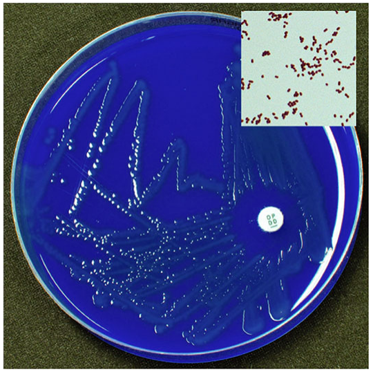
disk diffusion antibiotic susceptibility zone of inhibition Mueller-Hinton agar
Lecture: Detection Methods of Microbes
Learning objectives
- List the common methods used to detect microbes.
- Define culture, staining and sensitivity testing.
- Explain the principle behind each detection method.
- Carry out (demonstrate) a Gram stain, a culture streak and a disk diffusion sensitivity test.
1. Why detect microbes?
- Is a microbe present?
- Which microbe is it?
- How much of it is there?
- Which drugs will kill it?
2. Common methods of microbial detection
| Group | Methods | Main purpose |
|---|---|---|
| Microscopy | Gram stain, acid-fast (Ziehl-Neelsen) stain, negative stain (India ink/nigrosin), wet mount, fluorescence (auramine-rhodamine), dark-field | Rapid, direct look at morphology |
| Culture | Blood agar, MacConkey, selective, enrichment and differential media; broth culture; blood culture bottles | Isolate and grow the organism |
| Biochemical tests | Catalase, coagulase, oxidase, urease, indole, citrate, sugar fermentation, triple sugar iron (TSI) | Identify species |
| Sensitivity testing | Disk diffusion, broth/agar dilution (MIC), gradient strip (E-test) | Guide drug choice |
| Immunological | Antigen detection, ELISA, latex agglutination, serology | Detect antigen or antibody |
| Molecular | PCR, real-time PCR, sequencing, MALDI-TOF mass spectrometry | Rapid, sensitive identification, resistance genes |
3. Key definitions
- Culture: Growing microbes in the laboratory on or in nutrient media under suitable conditions (temperature, atmosphere, time). The aim is to multiply the organism so it can be isolated, identified and tested.
- Staining: Applying dyes to a smear to make microbes visible and to differentiate them by structure. Examples are the Gram stain and acid-fast stain.
- Sensitivity (susceptibility) testing: Exposing an isolated organism to antimicrobial drugs in vitro to see whether the drugs inhibit its growth. Results are reported as susceptible, intermediate or resistant.
- MIC (minimum inhibitory concentration): The lowest concentration of an antibiotic that inhibits visible growth of an organism in vitro (Henry's Clinical Diagnosis and Management by Laboratory Methods).
4. Principles of detection
4.1 Microscopy and staining
- Crystal violet stains all cells.
- Gram's iodine forms a crystal violet-iodine complex.
- Alcohol or acetone decolorizes Gram-negative cells but not Gram-positive cells.
- Safranin counterstain colours the decolorized Gram-negative cells red.
- Result: Gram-positive cells are purple-blue and Gram-negative cells are red/pink.
- The difference depends on cell wall structure. In Gram-positive bacteria the thick peptidoglycan layer traps the complex. In Gram-negative bacteria the decolorizer disrupts the outer membrane and the dye washes out (Jawetz, 28E, p. 43-44; Medical Microbiology 9e).
- Gram-positive cocci in clusters suggest staphylococci. Gram-positive cocci in chains suggest streptococci.
- Gram stain does not identify species.
- Some nonviable Gram-positive bacteria can stain Gram-negative.
- Note pus cells. Many squamous epithelial cells suggest a poor-quality sputum or wound sample (Jawetz, 28E, Ch. 47).
- Mycobacteria have mycolic acid-rich walls. They retain hot carbolfuchsin even after acid-alcohol decolorization.
- Acid-fast bacilli appear red against a blue (methylene blue) background. Other organisms take the counterstain colour (Jawetz, 28E, p. 44).
- Negative stain (nigrosin or India ink) is used for capsules, e.g., Cryptococcus.
- Fluorescent stains (auramine-rhodamine) are used for mycobacteria.
- Wet mount and dark-field microscopy show motility and spirochaetes.
4.2 Culture media
| Type | Function | Example |
|---|---|---|
| Basal / non-selective | Grows most organisms | Nutrient agar; blood agar (5% sheep blood is the standard clinical medium) |
| Enriched | Supports fastidious organisms | Chocolate agar (Haemophilus, Neisseria) |
| Selective | Inhibits unwanted flora, allows target growth | TCBS for Vibrio cholerae; bismuth sulfite for Salmonella |
| Differential | Colony colour or appearance separates organisms | MacConkey, EMB (lactose fermenters versus non-fermenters) |
| Enrichment (liquid) | Favours a small number of target organisms | Alkaline peptone water for Vibrio |
| Transport | Preserves organisms in transit | Stuart's, Amies |
- Colony size, shape, colour and surface.
- Haemolysis on blood agar (alpha, beta or gamma).
- Lactose fermentation on MacConkey (pink = lactose fermenter, pale = non-fermenter).
- Growth conditions: aerobic, anaerobic or CO₂ incubation.
4.3 Biochemical tests (identification)
| Test | Principle | Typical use |
|---|---|---|
| Catalase | Catalase splits H₂O₂ into O₂ (bubbles) | Staphylococci (positive) versus streptococci (negative) |
| Coagulase | Clots plasma | S. aureus (positive) |
| Oxidase | Cytochrome oxidase oxidizes a dye (purple) | Pseudomonas, Neisseria (positive) |
| Urease | Urea → ammonia, raises pH | Proteus, H. pylori |
| Indole | Tryptophanase makes indole | E. coli (positive) |
| Citrate | Uses citrate as sole carbon source | Klebsiella (positive), E. coli (negative) |
| Sugar fermentation / TSI | Acid production changes indicator colour | Enterobacterales |
4.4 Sensitivity (susceptibility) testing
- Disk diffusion (Kirby-Bauer). Antibiotic diffuses from a paper disk into inoculated agar and forms a concentration gradient. A clear zone of inhibition appears where growth is prevented. The zone diameter is compared with CLSI breakpoints to give S, I or R. It is qualitative and does not give an MIC directly.
- Broth or agar dilution. The organism is tested against doubling dilutions of the drug. The lowest concentration with no visible growth is the MIC.
- Gradient diffusion (E-test). A strip with a continuous drug gradient is placed on the agar. The MIC is read where the ellipse of inhibition meets the strip.
- Automated systems and molecular tests. These are fast, and PCR can detect resistance genes (e.g., mecA, rpoB). They do not give an MIC.
4.5 Immunological and molecular methods (overview)
- Antigen detection and serology. Antibodies bind microbial antigens (or the reverse). Examples are latex agglutination, ELISA and rapid antigen tests. They are useful when organisms are hard to culture.
- PCR. Amplifies microbial DNA or RNA. It is very sensitive, fast, and does not need live organisms.
- MALDI-TOF. Matches a protein mass spectrum to a database. It identifies cultured colonies in minutes.
5. Demonstrations
Demonstration A: Gram stain
- Make a thin smear on a clean slide. Air dry, then heat fix (or methanol fix).
- Flood with crystal violet for 10-30 seconds. Rinse with water.
- Apply Gram's iodine for 10-30 seconds. Rinse.
- Decolorize with 30% acetone-alcohol (10-30 seconds), until the stain stops running off. Rinse. This is the most critical step. Over-decolorizing makes Gram-positive cells look Gram-negative.
- Counterstain with safranin for 10-30 seconds. Rinse and dry.
- Examine under oil immersion (100×).
Demonstration B: Ziehl-Neelsen acid-fast stain (brief)
- Heat-fix the smear. Cover with carbolfuchsin and heat gently for 5 minutes. Do not boil or let it dry.
- Rinse. Decolorize with 3% acid-alcohol until only a faint pink remains.
- Rinse. Counterstain with methylene blue for 1 minute.
- Examine under oil immersion. Acid-fast bacilli are red on a blue background.
Demonstration C: Culture (streak plate for isolation)
- Label the plate (name, date, specimen) on the base.
- Flame the loop until red hot, then cool it.
- Pick up the inoculum and spread it over the first area (quadrant 1) of a blood agar plate.
- Flame and cool the loop. Drag it from quadrant 1 into quadrant 2. Repeat for quadrants 3 and 4.
- Incubate inverted at 35-37 °C for 18-24 hours (CO₂ if needed).
- Read the plate. Describe colonies, haemolysis and, on MacConkey, lactose fermentation.
- Pick a single isolated colony for Gram stain, biochemical tests and sensitivity testing.
Demonstration D: Disk diffusion sensitivity test
- Pick 3-5 similar colonies and suspend them in saline. Adjust to 0.5 McFarland turbidity (standard inoculum).
- Dip a sterile swab, press out excess fluid, and swab the entire surface of a Mueller-Hinton agar plate in three directions. Allow it to dry for 3-5 minutes.
- Apply antibiotic disks with sterile forceps, spaced evenly, and press gently so they contact the agar.
- Incubate inverted at 35 °C for 16-18 hours.
- Measure each zone of inhibition in mm, including the disk diameter.
- Compare with CLSI tables and report S, I or R.
- An inoculum that is too heavy gives smaller zones.
- Too light an inoculum gives larger zones.
- Delay between applying disks and incubation changes results.
- Wrong agar depth or pH changes results.
6. Strengths and limits at a glance
| Method | Strength | Limitation |
|---|---|---|
| Gram stain | Fast, cheap, guides early therapy | Needs about 10⁵ organisms/mL; cannot identify species |
| Culture | Gold standard, gives an isolate for AST | Slow (1-3 days or more); fastidious organisms may fail; prior antibiotics reduce yield |
| Biochemical tests | Cheap, reliable identification | Takes extra time after culture |
| Disk diffusion | Simple, low cost | Qualitative; strict standardization needed |
| MIC methods | Quantitative | More labour or cost |
| Molecular | Very fast, sensitive | Costly; detects DNA from dead organisms; does not always show phenotypic resistance |
7. Summary points
- Microbial detection moves from direct microscopy to culture, then identification (biochemical tests), then sensitivity testing.
- Staining makes microbes visible and differentiates them. The Gram stain divides most bacteria by cell wall structure, and the acid-fast stain identifies mycobacteria.
- Culture isolates and grows organisms. Media can be non-selective, enriched, selective or differential.
- Sensitivity testing guides therapy. Disk diffusion is qualitative, and dilution or gradient methods give an MIC.
- Standardization at every step (inoculum, media, timing) determines whether results can be trusted.
8. Review questions
- Define culture, staining and sensitivity testing.
- Why does a Gram-negative bacterium lose crystal violet while a Gram-positive one keeps it?
- What is the difference between a selective and a differential medium? Give one example of a medium that is both.
- Why is acid-alcohol used in the Ziehl-Neelsen stain?
- List three variables that must be standardized in disk diffusion testing.
- A smear shows Gram-positive cocci in clusters. Which two bench tests would you do next, and what results would you expect for S. aureus?
Make a PPT presentation of this lecture
~/
mkdir -p /tmp/workspace/microbe-detection-ppt/assets /tmp/workspace/microbe-detection-ppt/output && cd /tmp/workspace/microbe-detection-ppt && python /tmp/skills/shared/scripts/fetch_images.py "https://cdn.orris.care/cdss_images/HARRISON_1763035966873_8460ba69-de47-4ffd-8e86-a36f06bb7d86.png" > assets/img.json; python - <<'E' import json,base64 d=json.load(open('assets/img.json')) for x in d: print(x['url'][-20:],x['error']) if x['base64']: h,b=x['base64'].split(',',1) if ',' in x['base64'] else x['base64'].split(';base64,') open('assets/fig.png','wb').write(base64.b64decode(b)) from PIL import Image print(Image.open('assets/fig.png').size) E
~/microbe-detection-ppt/build.py
from pptx import Presentation
from pptx.util import Inches, Pt
from pptx.dml.color import RGBColor
from pptx.enum.text import PP_ALIGN, MSO_ANCHOR
from pptx.enum.shapes import MSO_SHAPE

NAVY = RGBColor(0x0B, 0x25, 0x45)
TEAL = RGBColor(0x13, 0x8A, 0x8C)
LIGHT = RGBColor(0xF2, 0xF7, 0xF9)
CORAL = RGBColor(0xE0, 0x5A, 0x3A)
GREY = RGBColor(0x44, 0x4F, 0x5A)
WHITE = RGBColor(0xFF, 0xFF, 0xFF)
PALE_TEAL = RGBColor(0xDC, 0xEF, 0xEF)
FONT = "Calibri"

prs = Presentation()
prs.slide_width = Inches(13.333)
prs.slide_height = Inches(7.5)
BLANK = prs.slide_layouts[6]
page = [0]


def rect(slide, x, y, w, h, fill, shape=MSO_SHAPE.RECTANGLE, line=None):
    s = slide.shapes.add_shape(shape, Inches(x), Inches(y), Inches(w), Inches(h))
    s.fill.solid()
    s.fill.fore_color.rgb = fill
    if line is None:
        s.line.fill.background()
    else:
        s.line.color.rgb = line
        s.line.width = Pt(1)
    s.shadow.inherit = False
    return s


def text(slide, x, y, w, h, content, size=18, bold=False, color=GREY,
         align=PP_ALIGN.LEFT, anchor=MSO_ANCHOR.TOP, spacing=None):
    """content: str or list of str / (str, dict) paragraphs."""
    tb = slide.shapes.add_textbox(Inches(x), Inches(y), Inches(w), Inches(h))
    tf = tb.text_frame
    tf.word_wrap = True
    tf.vertical_anchor = anchor
    tf.margin_left = tf.margin_right = Inches(0.05)
    tf.margin_top = tf.margin_bottom = Inches(0.03)
    items = content if isinstance(content, list) else [content]
    for i, it in enumerate(items):
        opts = {}
        if isinstance(it, tuple):
            it, opts = it
        p = tf.paragraphs[0] if i == 0 else tf.add_paragraph()
        p.alignment = opts.get("align", align)
        if spacing:
            p.space_after = Pt(spacing)
        r = p.add_run()
        r.text = it
        r.font.name = FONT
        r.font.size = Pt(opts.get("size", size))
        r.font.bold = opts.get("bold", bold)
        r.font.color.rgb = opts.get("color", color)
    return tb


def bullets(slide, x, y, w, h, items, size=18, color=GREY, gap=8):
    tb = slide.shapes.add_textbox(Inches(x), Inches(y), Inches(w), Inches(h))
    tf = tb.text_frame
    tf.word_wrap = True
    tf.margin_left = tf.margin_right = Inches(0.05)
    for i, it in enumerate(items):
        p = tf.paragraphs[0] if i == 0 else tf.add_paragraph()
        p.space_after = Pt(gap)
        if isinstance(it, tuple):
            head, rest = it
            r1 = p.add_run()
            r1.text = "\u2022  " + head
            r1.font.bold = True
            r1.font.color.rgb = NAVY
            r1.font.size = Pt(size)
            r1.font.name = FONT
            r2 = p.add_run()
            r2.text = " " + rest
            r2.font.size = Pt(size)
            r2.font.color.rgb = color
            r2.font.name = FONT
        else:
            r = p.add_run()
            r.text = "\u2022  " + it
            r.font.size = Pt(size)
            r.font.color.rgb = color
            r.font.name = FONT
    return tb


def new_slide(title, source=None, notes=None):
    s = prs.slides.add_slide(BLANK)
    page[0] += 1
    rect(s, 0, 0, 13.333, 7.5, WHITE)
    rect(s, 0, 0, 0.25, 7.5, TEAL)
    text(s, 0.6, 0.35, 12.2, 0.8, title, size=32, bold=True, color=NAVY,
         anchor=MSO_ANCHOR.MIDDLE)
    rect(s, 0.65, 1.2, 1.4, 0.06, CORAL)
    if source:
        text(s, 0.6, 7.0, 11.2, 0.35, source, size=11, color=RGBColor(0x7A, 0x86, 0x90))
    text(s, 12.2, 7.0, 0.8, 0.35, str(page[0] + 1), size=11,
         color=RGBColor(0x7A, 0x86, 0x90), align=PP_ALIGN.RIGHT)
    if notes:
        s.notes_slide.notes_text_frame.text = notes
    return s


def table(slide, x, y, w, col_w, rows, size=14, row_h=0.55, header_size=None):
    nrows, ncols = len(rows), len(rows[0])
    gs = slide.shapes.add_table(nrows, ncols, Inches(x), Inches(y), Inches(w),
                                Inches(row_h * nrows))
    t = gs.table
    tblPr = gs._element.graphic.graphicData.tbl.tblPr
    # remove built-in style banding look by explicit fills
    for ci, cw in enumerate(col_w):
        t.columns[ci].width = Inches(cw)
    for ri, row in enumerate(rows):
        t.rows[ri].height = Inches(row_h)
        for ci, val in enumerate(row):
            c = t.cell(ri, ci)
            c.fill.solid()
            if ri == 0:
                c.fill.fore_color.rgb = NAVY
            else:
                c.fill.fore_color.rgb = LIGHT if ri % 2 else WHITE
            c.margin_left = c.margin_right = Inches(0.1)
            c.margin_top = c.margin_bottom = Inches(0.04)
            c.vertical_anchor = MSO_ANCHOR.MIDDLE
            tf = c.text_frame
            tf.word_wrap = True
            p = tf.paragraphs[0]
            r = p.add_run()
            r.text = val
            r.font.name = FONT
            r.font.size = Pt(header_size or size) if ri == 0 else Pt(size)
            r.font.bold = (ri == 0) or (ci == 0)
            r.font.color.rgb = WHITE if ri == 0 else (NAVY if ci == 0 else GREY)
    return t


def card(slide, x, y, w, h, head, body, head_fill=TEAL, body_size=16):
    rect(slide, x, y, w, h, LIGHT)
    rect(slide, x, y, w, 0.6, head_fill)
    text(slide, x + 0.1, y, w - 0.2, 0.6, head, size=19, bold=True, color=WHITE,
         anchor=MSO_ANCHOR.MIDDLE)
    text(slide, x + 0.15, y + 0.7, w - 0.3, h - 0.8, body, size=body_size, color=GREY)


def steps(slide, x, y, w, items, size=15, h=1.0, fill=PALE_TEAL):
    n = len(items)
    gap = 0.15
    bw = (w - gap * (n - 1)) / n
    for i, (head, body) in enumerate(items):
        bx = x + i * (bw + gap)
        rect(slide, bx, y, bw, h, fill)
        circ = rect(slide, bx + 0.08, y + 0.08, 0.38, 0.38, TEAL, MSO_SHAPE.OVAL)
        tf = circ.text_frame
        tf.margin_left = tf.margin_right = tf.margin_top = tf.margin_bottom = 0
        p = tf.paragraphs[0]
        p.alignment = PP_ALIGN.CENTER
        r = p.add_run()
        r.text = str(i + 1)
        r.font.size = Pt(14)
        r.font.bold = True
        r.font.color.rgb = WHITE
        r.font.name = FONT
        text(slide, bx + 0.5, y + 0.05, bw - 0.55, 0.45, head, size=size, bold=True,
             color=NAVY, anchor=MSO_ANCHOR.MIDDLE)
        text(slide, bx + 0.1, y + 0.52, bw - 0.2, h - 0.55, body, size=size - 2)


# ---------------- 1. Title ----------------
s = prs.slides.add_slide(BLANK)
rect(s, 0, 0, 13.333, 7.5, NAVY)
rect(s, 0, 0, 0.35, 7.5, TEAL)
rect(s, 1.0, 3.85, 1.6, 0.07, CORAL)
text(s, 1.0, 1.6, 11, 0.5, "MICROBIOLOGY LECTURE", size=18, bold=True, color=RGBColor(0x7F, 0xD1, 0xD2))
text(s, 1.0, 2.1, 11.3, 1.7, "Detection Methods of Microbes", size=52, bold=True, color=WHITE,
     anchor=MSO_ANCHOR.MIDDLE)
text(s, 1.0, 4.1, 11, 1.0,
     "Principles of microbial detection: culture media, microscopy and staining, biochemical tests, sensitivity testing",
     size=22, color=RGBColor(0xD5, 0xE3, 0xEA))
text(s, 1.0, 6.4, 11, 0.4, "Includes practical demonstrations: Gram stain, streak culture, disk diffusion",
     size=16, color=RGBColor(0x9F, 0xB6, 0xC4))
s.notes_slide.notes_text_frame.text = ("Welcome. This lecture follows the laboratory pathway from specimen to "
                                       "report and ends with three bench demonstrations.")

# ---------------- 2. Objectives ----------------
s = new_slide("Learning objectives",
              notes="Read the four objectives and tell students the practicals are at the end.")
objs = [
    ("List", "the common methods of microbial detection"),
    ("Define", "culture, staining and sensitivity testing"),
    ("Discuss", "the principles behind each detection method"),
    ("Demonstrate", "staining, sensitivity testing and culture"),
]
for i, (h, b) in enumerate(objs):
    y = 1.65 + i * 1.3
    rect(s, 0.9, y, 11.6, 1.1, LIGHT)
    rect(s, 0.9, y, 0.12, 1.1, TEAL)
    text(s, 1.3, y, 2.6, 1.1, h, size=26, bold=True, color=NAVY, anchor=MSO_ANCHOR.MIDDLE)
    text(s, 3.9, y, 8.4, 1.1, b, size=22, anchor=MSO_ANCHOR.MIDDLE)

# ---------------- 3. Why detect ----------------
s = new_slide("Why detect microbes?",
              notes="Four clinical questions drive every microbiology request. Then show the workflow.")
qs = [("Is a microbe present?", "Presence"), ("Which microbe is it?", "Identity"),
      ("How much is there?", "Quantity"), ("Which drugs kill it?", "Susceptibility")]
for i, (q, tag) in enumerate(qs):
    x = 0.9 + i * 3.0
    rect(s, x, 1.6, 2.8, 1.6, PALE_TEAL)
    text(s, x + 0.1, 1.65, 2.6, 0.4, tag.upper(), size=13, bold=True, color=TEAL)
    text(s, x + 0.1, 2.05, 2.6, 1.1, q, size=20, bold=True, color=NAVY)
text(s, 0.9, 3.6, 11, 0.5, "Routine laboratory pathway", size=22, bold=True, color=NAVY)
flow = ["Specimen", "Direct microscopy\n(stain)", "Culture", "Identification\n(biochemical)",
        "Sensitivity\ntesting", "Report"]
bw, gap = 1.7, 0.25
for i, f in enumerate(flow):
    x = 0.9 + i * (bw + gap)
    b = rect(s, x, 4.3, bw, 1.3, NAVY if i not in (0, 5) else TEAL, MSO_SHAPE.ROUNDED_RECTANGLE)
    tf = b.text_frame
    tf.word_wrap = True
    tf.vertical_anchor = MSO_ANCHOR.MIDDLE
    for j, line in enumerate(f.split("\n")):
        p = tf.paragraphs[0] if j == 0 else tf.add_paragraph()
        p.alignment = PP_ALIGN.CENTER
        r = p.add_run()
        r.text = line
        r.font.size = Pt(16)
        r.font.bold = True
        r.font.color.rgb = WHITE
        r.font.name = FONT
    if i < len(flow) - 1:
        text(s, x + bw - 0.02, 4.55, gap + 0.04, 0.6, "\u203A", size=30, bold=True, color=CORAL,
             align=PP_ALIGN.CENTER, anchor=MSO_ANCHOR.MIDDLE)
text(s, 0.9, 5.95, 11.5, 0.8,
     "Each step narrows the answer: stain gives a quick clue, culture gives the isolate, "
     "biochemical tests name it, sensitivity testing guides treatment.", size=18)

# ---------------- 4. Common methods ----------------
s = new_slide("Common methods of microbial detection",
              source="Sources: Jawetz, Melnick & Adelberg's Medical Microbiology 28E; Sherris & Ryan's Medical Microbiology 8E",
              notes="This answers objective 1. Walk across the six groups.")
rows = [
    ["Group", "Methods", "Main purpose"],
    ["Microscopy", "Gram, acid-fast, negative stain, wet mount, fluorescence, dark-field", "Rapid look at morphology"],
    ["Culture", "Blood agar, MacConkey, selective and enrichment media, broth, blood culture", "Isolate and grow organism"],
    ["Biochemical tests", "Catalase, coagulase, oxidase, urease, indole, citrate, TSI", "Identify species"],
    ["Sensitivity testing", "Disk diffusion, broth or agar dilution (MIC), E-test", "Guide drug choice"],
    ["Immunological", "Antigen detection, ELISA, latex agglutination, serology", "Detect antigen or antibody"],
    ["Molecular", "PCR, real-time PCR, sequencing, MALDI-TOF", "Fast, sensitive identification"],
]
table(s, 0.9, 1.6, 11.6, [2.5, 6.0, 3.1], rows, size=15, row_h=0.72)

# ---------------- 5. Definitions ----------------
s = new_slide("Key definitions",
              source="MIC definition: Henry's Clinical Diagnosis and Management by Laboratory Methods",
              notes="This answers objective 2. Ask students to restate each definition in one sentence.")
card(s, 0.9, 1.6, 3.7, 3.5, "Culture",
     "Growing microbes on or in nutrient media under suitable temperature, atmosphere and time, "
     "so they can be isolated, identified and tested.", body_size=17)
card(s, 4.8, 1.6, 3.7, 3.5, "Staining",
     "Applying dyes to a smear to make microbes visible and to separate them by structure. "
     "Examples: Gram stain, acid-fast stain.", head_fill=NAVY, body_size=17)
card(s, 8.7, 1.6, 3.7, 3.5, "Sensitivity testing",
     "Exposing an isolate to antimicrobial drugs in vitro to see if growth is inhibited. "
     "Reported as susceptible, intermediate or resistant.", head_fill=CORAL, body_size=17)
rect(s, 0.9, 5.4, 11.5, 1.3, PALE_TEAL)
text(s, 1.1, 5.45, 11.1, 1.2,
     [("MIC (minimum inhibitory concentration)", {"bold": True, "color": NAVY, "size": 20}),
      ("The lowest concentration of an antibiotic that inhibits visible growth of an organism in vitro.",
       {"size": 18})], anchor=MSO_ANCHOR.MIDDLE)

# ---------------- 6. Microscopy principles ----------------
s = new_slide("Principles: microscopy and dyes",
              source="Jawetz, Melnick & Adelberg's Medical Microbiology 28E, p. 43 and Ch. 47",
              notes="Emphasise the sensitivity threshold: about 10^5 organisms per mL are needed to see bacteria on a smear.")
text(s, 0.9, 1.6, 6.2, 0.5, "How dyes work", size=24, bold=True, color=NAVY)
bullets(s, 0.9, 2.2, 6.3, 4.6, [
    ("Basic stains", "have a coloured cation (e.g., methylene blue) that binds the negatively charged phosphate groups of nucleic acids."),
    ("Acidic stains", "do not stain the cells, so they colour the background (negative staining, e.g., nigrosin)."),
    ("Special stains", "can show flagella, capsules, spores, granules and cell walls."),
], size=18, gap=12)
rect(s, 7.7, 1.6, 4.8, 4.9, NAVY)
text(s, 7.95, 1.8, 4.3, 0.5, "SENSITIVITY OF A SMEAR", size=14, bold=True, color=RGBColor(0x7F, 0xD1, 0xD2))
text(s, 7.95, 2.3, 4.3, 1.2, "10\u2075", size=66, bold=True, color=WHITE)
text(s, 7.95, 3.6, 4.3, 0.9, "organisms per mL are needed before bacteria are likely to be seen on a smear",
     size=17, color=WHITE)
text(s, 7.95, 4.9, 4.3, 1.4,
     "Solid media can grow from 10\u00B2 to 10\u00B3 organisms/mL, so culture is far more sensitive than microscopy.",
     size=15, color=RGBColor(0xD5, 0xE3, 0xEA))

# ---------------- 7. Gram stain ----------------
s = new_slide("Gram stain: a differential stain",
              source="Jawetz, Melnick & Adelberg's Medical Microbiology 28E, p. 43-44; Medical Microbiology 9E",
              notes="Stress that the difference is cell wall structure. Over-decolorizing makes Gram-positive cells look Gram-negative.")
steps(s, 0.9, 1.6, 11.6, [
    ("Crystal violet", "Stains all cells purple"),
    ("Gram's iodine", "Forms crystal violet-iodine complex"),
    ("Decolorize", "Alcohol or acetone washes out Gram-negative cells"),
    ("Safranin", "Counterstain colours Gram-negative cells red"),
], size=16, h=1.9)
rect(s, 0.9, 3.9, 5.6, 2.8, LIGHT)
rect(s, 0.9, 3.9, 0.1, 2.8, RGBColor(0x6A, 0x3D, 0x9A))
text(s, 1.2, 3.95, 5.2, 0.5, "Gram-positive: purple-blue", size=21, bold=True, color=RGBColor(0x6A, 0x3D, 0x9A))
text(s, 1.2, 4.5, 5.2, 2.1,
     "Thick peptidoglycan layer traps the crystal violet-iodine complex, so the cell resists decolorizing.",
     size=17)
rect(s, 6.9, 3.9, 5.6, 2.8, LIGHT)
rect(s, 6.9, 3.9, 0.1, 2.8, CORAL)
text(s, 7.2, 3.95, 5.2, 0.5, "Gram-negative: red-pink", size=21, bold=True, color=CORAL)
text(s, 7.2, 4.5, 5.2, 2.1,
     "Decolorizer disrupts the outer membrane and the thin peptidoglycan layer lets the dye wash out.",
     size=17)

# ---------------- 8. Interpreting + acid fast ----------------
s = new_slide("Reading smears and the acid-fast stain",
              source="Jawetz, Melnick & Adelberg's Medical Microbiology 28E, Ch. 2 and Ch. 47",
              notes="Gram stain suggests but does not prove identity. Mycobacteria need a special stain because of mycolic acid.")
card(s, 0.9, 1.6, 5.7, 5.1, "Interpreting a Gram smear", "", head_fill=NAVY)
bullets(s, 1.05, 2.4, 5.4, 4.2, [
    "Gram-positive cocci in clusters suggest staphylococci",
    "Gram-positive cocci in chains suggest streptococci",
    "Species cannot be identified from a smear",
    "Some nonviable Gram-positive cells stain Gram-negative",
    "Note pus cells; many squamous cells suggest a poor sputum or wound sample",
], size=16, gap=8)
card(s, 6.9, 1.6, 5.6, 5.1, "Acid-fast (Ziehl-Neelsen) stain", "")
bullets(s, 7.05, 2.4, 5.3, 4.2, [
    "Mycobacteria have mycolic acid-rich walls",
    "They keep hot carbolfuchsin despite acid-alcohol decolorizing",
    "Acid-fast bacilli appear red on a blue background",
    "Fluorescent auramine-rhodamine is an alternative",
    "Negative stain (nigrosin, India ink) shows capsules, e.g., Cryptococcus",
], size=16, gap=8)

# ---------------- 9. Culture media ----------------
s = new_slide("Principles: culture media",
              source="Jawetz, Melnick & Adelberg's Medical Microbiology 28E, Ch. 1, 16 and 47",
              notes="Principle: given nutrients, pH, temperature and atmosphere, one cell grows into one visible colony.")
text(s, 0.9, 1.45, 11.6, 0.6,
     "Principle: with the right nutrients, pH, temperature and gas atmosphere, a single cell grows into a visible colony.",
     size=17, color=NAVY, bold=True)
rows = [
    ["Type", "Function", "Example"],
    ["Basal / non-selective", "Grows most organisms", "Nutrient agar; blood agar (5% sheep blood)"],
    ["Enriched", "Supports fastidious organisms", "Chocolate agar (Haemophilus, Neisseria)"],
    ["Selective", "Inhibits unwanted flora", "TCBS (V. cholerae); bismuth sulfite (Salmonella)"],
    ["Differential", "Colony colour separates organisms", "MacConkey, EMB (lactose fermenters)"],
    ["Enrichment (liquid)", "Favours few target organisms", "Alkaline peptone water (Vibrio)"],
    ["Transport", "Preserves organisms in transit", "Stuart's, Amies"],
]
table(s, 0.9, 2.15, 11.6, [2.9, 3.7, 5.0], rows, size=15, row_h=0.62)
text(s, 0.9, 6.55, 11.6, 0.4,
     "Read: colony shape and colour, haemolysis (alpha, beta, gamma), lactose fermentation (pink vs pale).",
     size=15)

# ---------------- 10. Biochemical ----------------
s = new_slide("Principles: biochemical tests",
              notes="Each species has a characteristic enzyme set. We add a substrate and watch for colour, gas or precipitate.")
text(s, 0.9, 1.45, 11.6, 0.6,
     "Principle: each species has a characteristic set of enzymes. A substrate is added and a colour change, gas or precipitate is read.",
     size=17, color=NAVY, bold=True)
rows = [
    ["Test", "Principle", "Typical use"],
    ["Catalase", "Catalase splits H\u2082O\u2082 into O\u2082 (bubbles)", "Staphylococci +, streptococci \u2212"],
    ["Coagulase", "Clots plasma", "S. aureus +"],
    ["Oxidase", "Cytochrome oxidase oxidizes dye (purple)", "Pseudomonas, Neisseria +"],
    ["Urease", "Urea to ammonia, raises pH", "Proteus, H. pylori"],
    ["Indole", "Tryptophanase makes indole", "E. coli +"],
    ["Citrate", "Citrate used as sole carbon source", "Klebsiella +, E. coli \u2212"],
    ["Sugar fermentation / TSI", "Acid production changes indicator colour", "Enterobacterales"],
]
table(s, 0.9, 2.2, 11.6, [2.9, 5.0, 3.7], rows, size=15, row_h=0.58)

# ---------------- 11. Sensitivity methods ----------------
s = new_slide("Principles: sensitivity testing methods",
              source="Henry's Clinical Diagnosis and Management by Laboratory Methods (susceptibility testing chapter)",
              notes="The isolate is exposed to a standard drug concentration and growth inhibition is measured.")
card(s, 0.9, 1.6, 2.75, 4.2, "Disk diffusion",
     "Kirby-Bauer. Drug diffuses from a disk into inoculated agar. Zone diameter is compared with CLSI breakpoints. Qualitative.", body_size=15)
card(s, 3.8, 1.6, 2.75, 4.2, "Dilution",
     "Broth or agar dilution with doubling drug concentrations. Lowest concentration with no visible growth is the MIC.", head_fill=NAVY, body_size=15)
card(s, 6.7, 1.6, 2.75, 4.2, "Gradient strip",
     "E-test. Strip with a continuous drug gradient. MIC is read where the inhibition ellipse meets the strip.", head_fill=CORAL, body_size=15)
card(s, 9.6, 1.6, 2.9, 4.2, "Automated / molecular",
     "Fast, high volume. PCR detects resistance genes (e.g., mecA, rpoB) but gives no MIC.", head_fill=GREY, body_size=15)
rect(s, 0.9, 6.0, 11.6, 0.8, PALE_TEAL)
text(s, 1.1, 6.0, 11.2, 0.8,
     "Results are reported as Susceptible (S), Intermediate (I) or Resistant (R).",
     size=18, bold=True, color=NAVY, anchor=MSO_ANCHOR.MIDDLE)

# ---------------- 12. Standardization ----------------
s = new_slide("Standardization and when to test",
              source="Henry's Clinical Diagnosis and Management by Laboratory Methods, Key Points",
              notes="CLSI standards are what make results reproducible between laboratories.")
text(s, 0.9, 1.55, 5.6, 0.5, "Variables that must be standardized", size=22, bold=True, color=NAVY)
bullets(s, 0.9, 2.2, 5.6, 4.5, [
    "Media (e.g., Mueller-Hinton agar)",
    "Inoculum density (0.5 McFarland)",
    "Incubation time and temperature",
    "Atmosphere",
    "Supplements for fastidious organisms, e.g., defibrinated sheep blood",
], size=18, gap=10)
text(s, 7.0, 1.55, 5.5, 0.5, "Why test?", size=22, bold=True, color=NAVY)
bullets(s, 7.0, 2.2, 5.5, 4.5, [
    "Test when susceptibility cannot be predicted reliably from the organism's identity",
    "Cumulative antibiograms guide empirical therapy before results are ready",
    "They help pharmacists and stewardship teams track resistance trends",
], size=18, gap=10)

# ---------------- 13. Immuno / molecular ----------------
s = new_slide("Immunological and molecular methods",
              notes="Overview only. These complement culture, especially for organisms that are slow or hard to grow.")
card(s, 0.9, 1.6, 3.7, 4.6, "Antigen / serology",
     "Antibodies bind microbial antigens, or the reverse. Examples: latex agglutination, ELISA, rapid antigen tests. Useful when organisms are hard to culture.", body_size=17)
card(s, 4.8, 1.6, 3.7, 4.6, "PCR",
     "Amplifies microbial DNA or RNA. Very sensitive and fast, and does not need live organisms.", head_fill=NAVY, body_size=17)
card(s, 8.7, 1.6, 3.7, 4.6, "MALDI-TOF",
     "Matches a protein mass spectrum to a database. Identifies cultured colonies in minutes.", head_fill=CORAL, body_size=17)

# ---------------- 14. Demo A: Gram ----------------
s = new_slide("Demonstration A: Gram stain",
              source="Procedure per Jawetz, Melnick & Adelberg's Medical Microbiology 28E, Table 47-1",
              notes="Wear gloves and coat; discard slides in disinfectant. Decolorizing is the most critical step.")
steps(s, 0.9, 1.6, 11.6, [
    ("Smear", "Thin smear, air dry, heat fix"),
    ("Crystal violet", "10-30 s, rinse"),
    ("Iodine", "10-30 s, rinse"),
    ("Decolorize", "30% acetone-alcohol, 10-30 s, rinse"),
    ("Safranin", "10-30 s, rinse, dry"),
], size=15, h=1.7)
rect(s, 0.9, 3.65, 5.6, 3.1, LIGHT)
text(s, 1.1, 3.7, 5.2, 0.5, "Key points", size=21, bold=True, color=NAVY)
bullets(s, 1.1, 4.25, 5.3, 2.5, [
    "Do not blot between rinses",
    "Over-decolorizing makes Gram-positive cells look Gram-negative",
    "Examine under oil immersion (100x)",
], size=16, gap=6)
rect(s, 6.9, 3.65, 5.6, 3.1, PALE_TEAL)
text(s, 7.1, 3.7, 5.2, 0.5, "Expected result", size=21, bold=True, color=NAVY)
bullets(s, 7.1, 4.25, 5.3, 2.5, [
    "Gram-positive cocci: purple",
    "Gram-negative bacilli: pink-red",
    "Report shape, arrangement and pus cells",
], size=16, gap=6)

# ---------------- 15. Demo B/C ----------------
s = new_slide("Demonstration B and C: Ziehl-Neelsen and streak culture",
              source="Ziehl-Neelsen steps per Jawetz 28E, Table 47-1",
              notes="Streaking dilutes the inoculum so single cells give isolated colonies and a pure culture.")
card(s, 0.9, 1.6, 5.7, 5.1, "B. Ziehl-Neelsen stain", "", head_fill=NAVY)
bullets(s, 1.05, 2.4, 5.4, 4.2, [
    "Heat-fix the smear",
    "Carbolfuchsin with gentle heat, 5 min (do not boil or dry)",
    "Rinse; decolorize with 3% acid-alcohol until faint pink",
    "Counterstain with methylene blue, 1 min",
    "Result: red acid-fast bacilli on blue background",
], size=16, gap=8)
card(s, 6.9, 1.6, 5.6, 5.1, "C. Streak plate (blood agar)", "")
bullets(s, 7.05, 2.4, 5.3, 4.2, [
    "Label the base of the plate",
    "Flame and cool the loop; inoculate quadrant 1",
    "Reflame; drag from quadrant 1 into 2, then 3 and 4",
    "Incubate inverted, 35-37 \u00B0C, 18-24 h",
    "Read colonies and haemolysis; pick one colony for Gram stain, biochemical tests and AST",
], size=16, gap=8)

# ---------------- 16. Demo D with figure ----------------
s = new_slide("Demonstration D: disk diffusion",
              source="Figure: Harrison's Principles of Internal Medicine. Method per Henry's and CLSI standards",
              notes="Blood agar with a penicillin disk and a clear zone; inset shows Gram-positive cocci in clusters.")
steps_items = [
    "Suspend 3-5 colonies in saline; adjust to 0.5 McFarland",
    "Swab entire Mueller-Hinton plate in 3 directions; dry 3-5 min",
    "Apply disks with sterile forceps; press gently",
    "Incubate inverted at 35 \u00B0C, 16-18 h",
    "Measure zones (mm); compare with CLSI tables; report S, I or R",
]
for i, t in enumerate(steps_items):
    y = 1.6 + i * 0.82
    c = rect(s, 0.9, y + 0.08, 0.5, 0.5, TEAL, MSO_SHAPE.OVAL)
    p = c.text_frame.paragraphs[0]
    c.text_frame.margin_left = c.text_frame.margin_right = 0
    p.alignment = PP_ALIGN.CENTER
    r = p.add_run()
    r.text = str(i + 1)
    r.font.size = Pt(16)
    r.font.bold = True
    r.font.color.rgb = WHITE
    r.font.name = FONT
    text(s, 1.55, y, 5.6, 0.75, t, size=16, anchor=MSO_ANCHOR.MIDDLE)
rect(s, 0.9, 5.8, 6.3, 1.0, RGBColor(0xFD, 0xEE, 0xE9))
text(s, 1.05, 5.8, 6.0, 1.0,
     "Errors: heavy inoculum gives small zones; light inoculum gives large zones; wrong agar depth or pH alters results.",
     size=14, color=CORAL, anchor=MSO_ANCHOR.MIDDLE, bold=True)
s.shapes.add_picture("/tmp/workspace/microbe-detection-ppt/assets/fig.png", Inches(7.9), Inches(1.5),
                     height=Inches(4.7))
text(s, 7.9, 6.25, 4.7, 0.6,
     "Blood agar with penicillin disk and zone of inhibition; inset: Gram-positive cocci in clusters",
     size=12, color=RGBColor(0x7A, 0x86, 0x90))

# ---------------- 17. Strengths and limits ----------------
s = new_slide("Strengths and limits at a glance",
              notes="Use this table to link the methods together as a pathway.")
rows = [
    ["Method", "Strength", "Limitation"],
    ["Gram stain", "Fast, cheap, guides early therapy", "Needs about 10\u2075 organisms/mL; no species ID"],
    ["Culture", "Gold standard; gives isolate for AST", "Slow; fastidious organisms may fail; prior antibiotics reduce yield"],
    ["Biochemical tests", "Cheap, reliable identification", "Extra time after culture"],
    ["Disk diffusion", "Simple, low cost", "Qualitative; strict standardization"],
    ["MIC methods", "Quantitative", "More labour or cost"],
    ["Molecular", "Very fast, sensitive", "Costly; detects DNA of dead organisms; may not show phenotypic resistance"],
]
table(s, 0.9, 1.6, 11.6, [2.6, 4.2, 4.8], rows, size=15, row_h=0.72)

# ---------------- 18. Summary ----------------
s = new_slide("Summary",
              notes="Recap the five take-home points.")
pts = [
    ("Pathway:", "microscopy, then culture, then identification, then sensitivity testing."),
    ("Staining:", "Gram stain splits bacteria by cell wall; acid-fast stain finds mycobacteria."),
    ("Culture:", "isolates and grows organisms on non-selective, enriched, selective or differential media."),
    ("Sensitivity:", "disk diffusion is qualitative; dilution and gradient methods give the MIC."),
    ("Standardization:", "inoculum, media and timing decide whether results can be trusted."),
]
for i, (h, b) in enumerate(pts):
    y = 1.55 + i * 1.03
    rect(s, 0.9, y, 11.6, 0.9, LIGHT)
    rect(s, 0.9, y, 0.1, 0.9, TEAL)
    text(s, 1.2, y, 2.8, 0.9, h, size=20, bold=True, color=NAVY, anchor=MSO_ANCHOR.MIDDLE)
    text(s, 4.0, y, 8.4, 0.9, b, size=17, anchor=MSO_ANCHOR.MIDDLE)

# ---------------- 19. Review questions ----------------
s = new_slide("Review questions",
              notes="Answers: 6) catalase (negative would suggest streptococci; positive for staphylococci) and coagulase (positive for S. aureus).")
qs = [
    "Define culture, staining and sensitivity testing.",
    "Why does a Gram-negative bacterium lose crystal violet while a Gram-positive one keeps it?",
    "How does a selective medium differ from a differential medium? Name an example of each.",
    "Why is acid-alcohol used in the Ziehl-Neelsen stain?",
    "List three variables that must be standardized in disk diffusion testing.",
    "A smear shows Gram-positive cocci in clusters. Which two bench tests come next, and what results fit S. aureus?",
]
for i, q in enumerate(qs):
    y = 1.5 + i * 0.9
    c = rect(s, 0.9, y + 0.1, 0.55, 0.55, NAVY, MSO_SHAPE.OVAL)
    c.text_frame.margin_left = c.text_frame.margin_right = 0
    p = c.text_frame.paragraphs[0]
    p.alignment = PP_ALIGN.CENTER
    r = p.add_run()
    r.text = str(i + 1)
    r.font.size = Pt(18)
    r.font.bold = True
    r.font.color.rgb = WHITE
    r.font.name = FONT
    text(s, 1.65, y, 10.8, 0.8, q, size=19, anchor=MSO_ANCHOR.MIDDLE)

prs.save("/tmp/workspace/microbe-detection-ppt/output/Detection_Methods_of_Microbes.pptx")
print("saved", len(prs.slides))