
Ears summary easy points from snells clinical anatomy
Reading File
Now let me search the library for Snell's Clinical Anatomy ear content in parallel.
Finding Sources
Finding Sources
Snell's is not in the library, but Gray's Anatomy for Students and Scott-Brown's Otorhinolaryngology are excellent anatomy sources. Let me pull from Gray's (closest anatomy equivalent) and Scott-Brown's, plus check all books for ear anatomy content.
Finding Sources
Reading File
Gray's Anatomy for Students has a detailed ear section. Let me read more of it.
Reading File
Reading File
Reading File
Reading File
Reading File
I now have all the essential content needed for a complete, well-organized ear summary. Let me fetch the key diagram images too.
Loading Image
Loading Image
I have enough content for a thorough, well-organized summary. Here it is:
👂 Ear - Summary (Clinical Anatomy Easy Points)
Source: Gray's Anatomy for Students (the closest equivalent to Snell's in the library - same clinical anatomy style and detail)
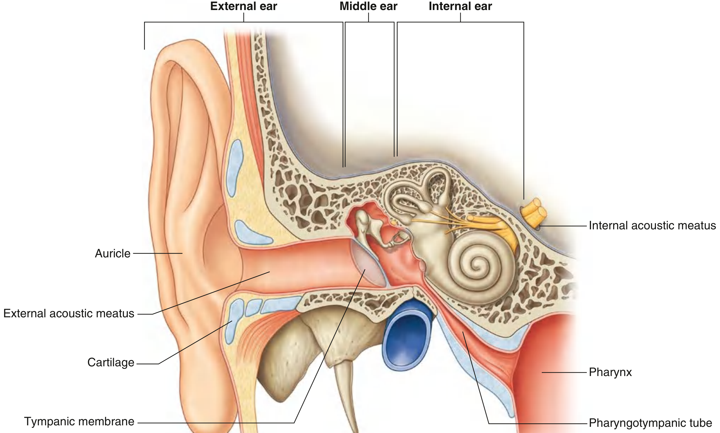
1. Overview - Three Parts
| Part | Location | Function |
|---|---|---|
| External ear | Outside + canal | Captures and conducts sound |
| Middle ear | Petrous temporal bone cavity | Transmits vibrations via ossicles |
| Internal ear | Petrous temporal bone (medial) | Converts sound to electrical signals + balance |
2. External Ear
Auricle (Pinna)
- Cartilage covered with skin (except the lobule - only part with NO cartilage)
- Key landmarks: helix (outer rim), antihelix (inner parallel rim), tragus (anterior to meatus opening), antitragus (opposite tragus), concha (hollow center)
- Muscles: intrinsic (change shape) + extrinsic (anterior, superior, posterior auricular) - all innervated by facial nerve [VII]
Sensory Innervation of the Auricle
- Auriculotemporal nerve (V3) - anterosuperior skin
- Greater auricular nerve (C2, C3) - lower auricle and lobule
- Lesser occipital nerve (C2) - superior auricle
- Auricular branch of vagus [X] - some of the posterior surface
- Facial nerve [VII] - small contribution
External Acoustic Meatus (EAM)
- Length: ~2.5 cm
- Outer 1/3 - cartilaginous, inner 2/3 - bony (tympanic part of temporal bone)
- Skin of cartilaginous part has ceruminous glands (wax) and hair follicles
- The meatus has a slight S-shaped curve - to examine, pull pinna upward and backward in adults
- Sensory supply: auriculotemporal nerve [V3] (anterior + superior walls), auricular branch of vagus [X] (posterior + inferior walls)
- Clinical note: foreign body in EAM can stimulate vagal branch and cause cough/vomiting (Arnold's reflex)
3. Tympanic Membrane
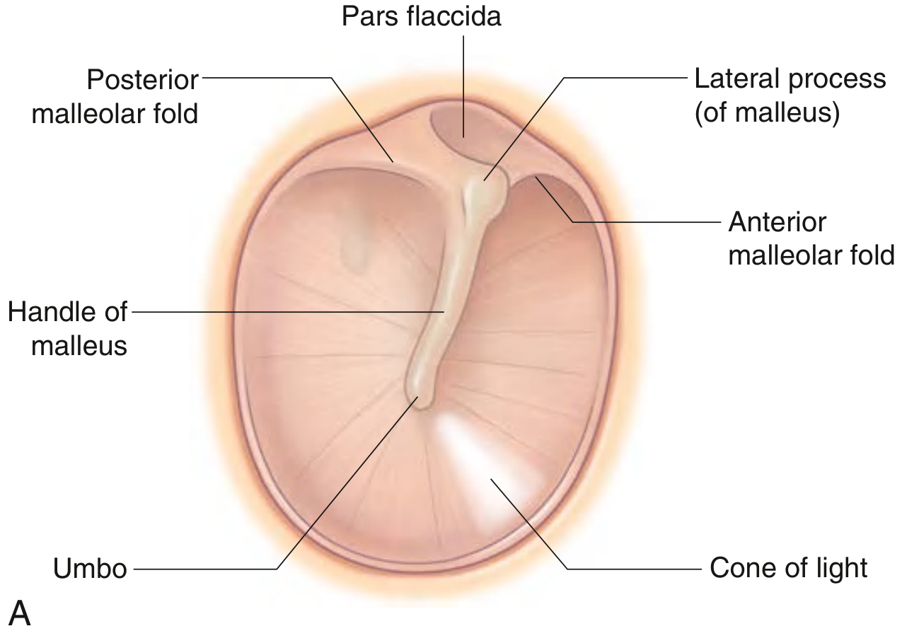
- Separates external meatus from middle ear
- Oriented obliquely - slopes medially from top to bottom, and posteriorly to anteriorly
- Structure: fibrocartilaginous ring peripherally + connective tissue core + skin outside + mucous membrane inside
- Umbo = center of concavity, where the handle of malleus attaches
- Cone of light = bright otoscopic reflection, anteroinferior to umbo
- Pars flaccida (Shrapnell's membrane) = thin, slack superior portion above the malleolar folds
- Pars tensa = thick, taut remainder of the membrane
- Handle of malleus runs superior to umbo; the lateral process of malleus creates a small bulge at the top
4. Middle Ear (Tympanic Cavity)
Extent
- Extends from tympanic membrane laterally to the medial (labyrinthine) wall
- The epitympanic recess is the space above the tympanic membrane
Boundaries (6 walls - easy mnemonic: "The Mighty Jaw Joins Lateral Master")
| Wall | Name | Important Structure |
|---|---|---|
| Roof | Tegmental wall | Tegmen tympani (thin bone separating middle ear from middle cranial fossa) |
| Floor | Jugular wall | Separates from internal jugular vein; tympanic branch of CN IX enters here |
| Lateral | Membranous wall | Mostly the tympanic membrane |
| Medial | Labyrinthine wall | Oval window (stapes), round window, promontory (cochlear 1st turn), facial canal |
| Anterior | Carotid wall | Pharyngotympanic tube opening, tensor tympani muscle |
| Posterior | Mastoid wall | Aditus to mastoid antrum; pyramidal eminence (for stapedius muscle) |
Communications
- Anteriorly via pharyngotympanic tube (Eustachian tube) → nasopharynx
- Posteriorly via aditus → mastoid antrum → mastoid air cells
5. Pharyngotympanic (Eustachian) Tube
- Length ~3.5 cm (adults); shorter and more horizontal in children = more prone to middle ear infections
- Medial 2/3 is cartilaginous; lateral 1/3 is bony
- Opens during swallowing/yawning to equalize middle ear pressure
- Lined with mucous membrane continuous with the nasopharynx (infection can spread easily)
- Innervation: tympanic plexus (via CN IX)
- Arterial supply: ascending pharyngeal artery + branches of maxillary artery (middle meningeal + artery of pterygoid canal)
6. Auditory Ossicles
Three bones forming a chain: Tympanic membrane → Malleus → Incus → Stapes → Oval window (internal ear)
| Ossicle | Key Features |
|---|---|
| Malleus | Largest; head (in epitympanic recess), neck, handle (attached to tympanic membrane), lateral process, anterior process |
| Incus | Middle bone; body + short crus + long crus + lenticular process (connects to stapes) |
| Stapes | Smallest bone in body; head, two crura, footplate (in oval window) |
Muscles of the Ossicles
| Muscle | Bone moved | Nerve | Action |
|---|---|---|---|
| Tensor tympani | Malleus (pulls handle medially) | Medial pterygoid nerve (CN V3) | Stiffens chain, damps vibration |
| Stapedius | Stapes | Facial nerve [VII] | Stiffens chain, protects from loud sounds (stapedius reflex) |
- Paralysis of stapedius (facial nerve palsy) → hyperacusis (sounds painfully loud)
7. Innervation of Middle Ear
- Tympanic nerve (branch of CN IX, glossopharyngeal) → enters middle ear → forms tympanic plexus
- Tympanic plexus supplies mucosa of middle ear, pharyngotympanic tube, and mastoid area
- Lesser petrosal nerve branches from tympanic plexus → exits via foramen ovale → otic ganglion → postganglionic parasympathetics to parotid gland
- Facial nerve [VII] runs in the facial canal on the medial wall of the middle ear - at risk in middle ear surgery
- Chorda tympani (branch of VII) crosses the middle ear between malleus and incus
8. Internal Ear
Located in the petrous part of the temporal bone, between:
- Middle ear (lateral)
- Internal acoustic meatus (medial)
Two Labyrinths
| Bony Labyrinth | Membranous Labyrinth | |
|---|---|---|
| Fluid | Perilymph (like CSF - high Na⁺) | Endolymph (like intracellular fluid - high K⁺) |
| Parts | Vestibule, 3 semicircular canals, cochlea | Utricle, saccule, 3 semicircular ducts, cochlear duct |
Bony Labyrinth Parts
- Vestibule - central cavity, contains utricle and saccule
- 3 Semicircular canals - anterior, posterior, lateral (each at right angles to each other)
- Cochlea - snail-shaped, 2.5 turns; modiolus = central bony axis
Membranous Labyrinth - Balance Organs
| Structure | Responds to |
|---|---|
| Utricle (macula) | Linear acceleration in horizontal plane + sideways head tilts |
| Saccule (macula) | Linear acceleration in vertical plane (up-down, forward-backward) |
| Semicircular ducts (cristae in ampullae) | Rotational (angular) movement in any direction |
Hearing Organ - Cochlear Duct
- The cochlear duct divides the cochlea into: scala vestibuli (above) and scala tympani (below)
- Cochlear duct filled with endolymph; scalae filled with perilymph
- Scala vestibuli starts at oval window (stapes footplate); scala tympani ends at round window
- Organ of Corti sits on the basilar membrane in the floor of the cochlear duct - the actual hearing receptor
- The vestibular membrane (Reissner's) forms the roof of the cochlear duct
Endolymphatic Sac
- Endolymphatic duct passes through the vestibular aqueduct → expands into the endolymphatic sac on the posterior surface of petrous temporal bone
- Functions in resorption of endolymph
9. Vestibulocochlear Nerve [CN VIII]
- Runs in the internal acoustic meatus alongside facial nerve [VII]
- Divides into:
- Cochlear nerve - hearing
- Vestibular nerve (superior + inferior divisions) - balance
- Cell bodies for vestibular part: vestibular ganglion (Scarpa's ganglion)
- Cell bodies for cochlear part: spiral ganglion (in modiolus)
10. Quick Clinical Pearls
| Fact | Clinical Relevance |
|---|---|
| Pars flaccida is the weak part of tympanic membrane | Cholesteatoma typically starts here |
| Chorda tympani crosses middle ear | Damaged in ear surgery → loss of taste on anterior 2/3 tongue |
| Children's Eustachian tube is short + horizontal | Prone to acute otitis media |
| Facial nerve in bony canal on medial wall | At risk in mastoiditis or middle ear surgery |
| Stapedius reflex | Tests CN VII integrity; loss → hyperacusis |
| Arnold's reflex (vagal branch in EAM) | Ear instrumentation can cause cough or even cardiac vagal reflex |
| Round window covered by secondary tympanic membrane | If ruptured → perilymph fistula, sensorineural hearing loss |
| Endolymph resorbed by endolymphatic sac | Disruption → endolymphatic hydrops (Meniere's disease) |
Source: Gray's Anatomy for Students, Head & Neck chapter (Ear section, pp. 1093-1106). Note: Snell's Clinical Anatomy was not available in the library, but Gray's for Students covers the same anatomy at an equivalent level.
This is a shared conversation. Sign in to Orris to start your own chat.