
Synapse
Reading File
A synapse is a junction between two neurons (or a neuron and an effector cell) where signals are transmitted from one cell to the next. Here is a comprehensive overview:
Searching sources...
Finding Sources
Searching the Web
synapse structure function neurotransmission overview
Reading File
Reading File
Reading File
Loading Image
Loading Image
Finding Sources
The Synapse
A synapse is a specialized junction that allows a neuron to transmit signals to another neuron or to a target effector cell (muscle, gland). The human brain contains an estimated 100 trillion synapses - making them the fundamental computational units of the nervous system.
Types of Synapses
1. Chemical Synapse (the most common)
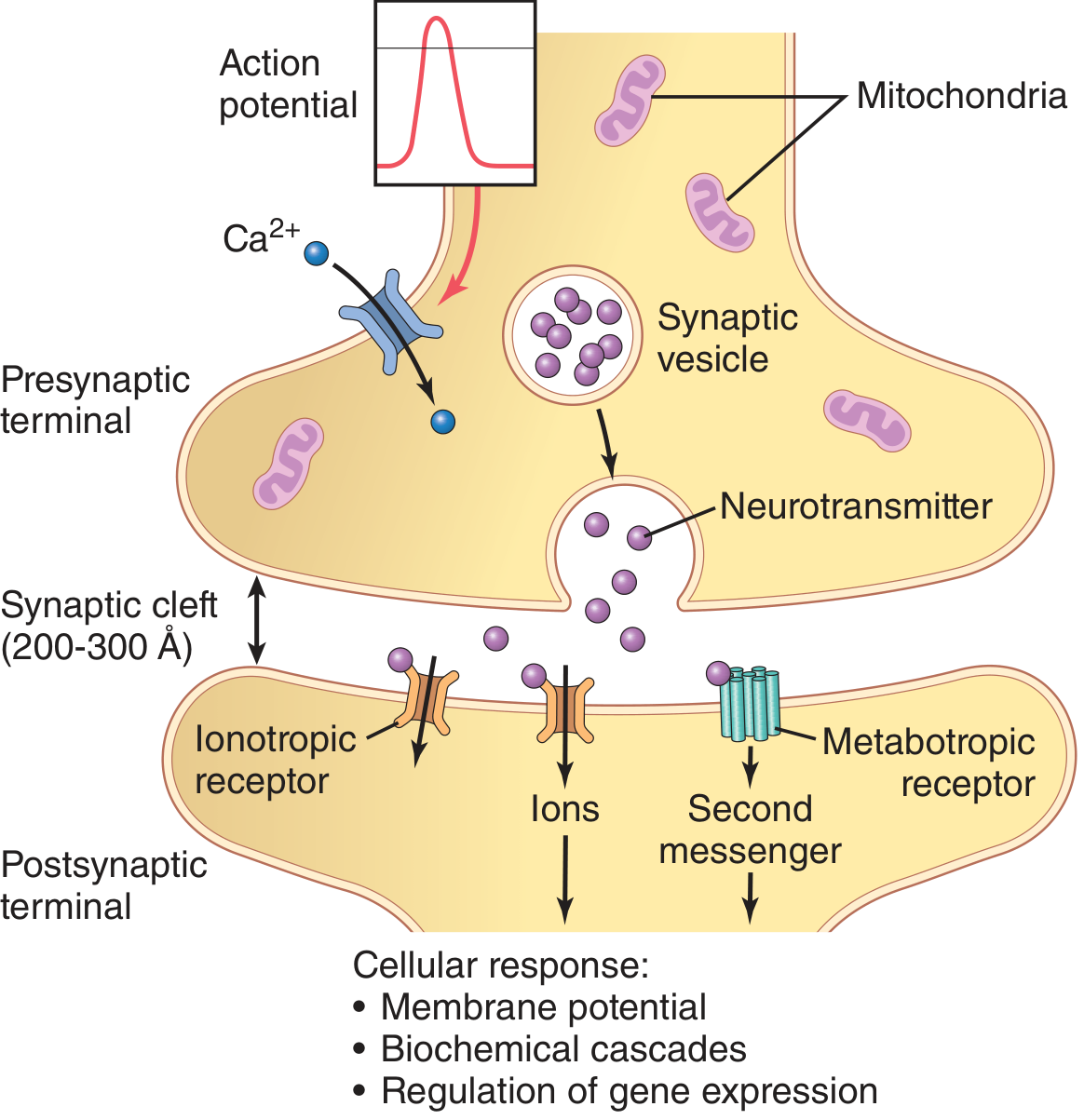
The chemical synapse converts an electrical signal into a chemical one and back again. Its key structural components are:
| Component | Description |
|---|---|
| Presynaptic terminal | Axon bouton filled with synaptic vesicles and mitochondria |
| Synaptic cleft | Gap of 200-300 Å between membranes |
| Synaptic vesicles | Membrane-bound packets storing neurotransmitter |
| Postsynaptic membrane | Contains receptor proteins (ionotropic or metabotropic) |
Steps of chemical synaptic transmission:
- An action potential propagates down the presynaptic axon and arrives at the terminal
- Voltage-gated Ca²+ channels open in the presynaptic membrane
- Ca²+ influx causes synaptic vesicles to migrate, dock, and fuse with the membrane (exocytosis)
- Neurotransmitter is released into the synaptic cleft
- It diffuses across and binds to receptors on the postsynaptic membrane
- Depending on receptor type, this opens ion channels or activates second messengers
- The result is a change in postsynaptic membrane potential
The time delay from arrival of the presynaptic action potential to onset of the postsynaptic response is approximately 0.5 ms, most of which is consumed by the Ca²+-triggered release process.
- Katzung's Basic and Clinical Pharmacology, 16th Ed.
- Guyton and Hall Textbook of Medical Physiology
2. Electrical Synapse
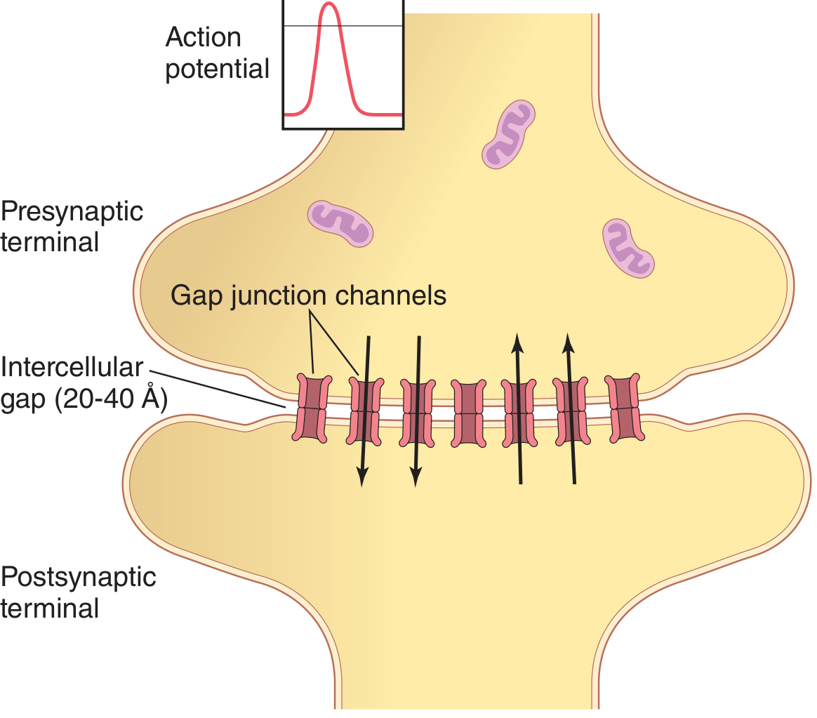
Electrical synapses use gap junction channels to directly pass ionic current between adjacent cells. The intercellular gap is only 20-40 Å (much narrower than a chemical synapse). Key features:
-
Bidirectional - signals can flow in either direction (unlike chemical synapses)
-
No synaptic delay - near-instantaneous transmission
-
Used to synchronize neuronal firing in large groups of interconnected neurons
-
Found in hypothalamic hormone-secreting neurons to coordinate simultaneous hormone release
-
Less amenable to pharmacological modulation than chemical synapses
-
Guyton and Hall Textbook of Medical Physiology
Postsynaptic Receptor Types
| Receptor Type | Mechanism | Speed | Example |
|---|---|---|---|
| Ionotropic | Directly gates an ion channel | Fast (ms) | NMDA, AMPA, GABA-A, nAChR |
| Metabotropic | Activates G-protein -> second messengers | Slow (sec-min) | mGluR, muscarinic, adrenergic |
Postsynaptic Potentials
- EPSP (Excitatory Postsynaptic Potential): A small depolarization generated by excitatory transmitters (e.g., glutamate). Multiple EPSPs can summate spatially or temporally to reach threshold and fire an action potential.
- IPSP (Inhibitory Postsynaptic Potential): A small hyperpolarization generated by inhibitory transmitters (e.g., GABA). An IPSP can prevent an otherwise suprathreshold EPSP from generating a spike.
One-Way Conduction
A defining feature of chemical synapses is that they transmit signals in one direction only - from the presynaptic (transmitting) neuron to the postsynaptic (receiving) cell. This directionality allows the nervous system to route signals to specific targets for precise control of sensation, movement, memory, and gland secretion.
Clinical Relevance
The synapse is the primary site of action of many drugs and toxins:
- Anesthetics, benzodiazepines, barbiturates - potentiate GABA at inhibitory synapses
- SSRIs, SNRIs - block reuptake of monoamine neurotransmitters in the cleft
- Botulinum toxin - blocks vesicle fusion and neurotransmitter release at the neuromuscular junction
- Organophosphates - inhibit acetylcholinesterase, prolonging ACh action in the cleft
- Synaptic dysfunction underlies conditions such as Alzheimer's disease, schizophrenia, depression, myasthenia gravis, and epilepsy
Synaptic Plasticity
The synapse is not static. Repeated activation strengthens synaptic connections through long-term potentiation (LTP), the cellular basis of learning and memory. Conversely, long-term depression (LTD) weakens under-used connections. This plasticity, combined with the electrical-to-chemical signal conversion, is what gives the brain its computational flexibility.
- Neuroscience: Exploring the Brain, 5th Ed.
Muscle contraction
Muscle contraction involves the sliding filament mechanism where actin and myosin filaments slide past each other, powered by ATP hydrolysis. It is initiated by a neural signal, Ca²+ release from the sarcoplasmic reticulum, and troponin-tropomyosin complex displacement.
Searching sources...
Finding Sources
Searching the Web
muscle contraction mechanism sliding filament theory steps
Reading File
Reading File
Reading File
Finding Sources
Reading File
Reading File
Loading Image
Loading Image
Loading Image
Muscle Contraction
1. Structural Basis - The Sarcomere
Skeletal muscle is built from myofibrils, each made up of repeating units called sarcomeres - the functional unit of contraction. Each sarcomere contains:
- ~1,500 thick filaments (myosin) - each with protruding cross-bridge heads
- ~3,000 thin filaments (actin) - two helical strands of F-actin with active binding sites
- Tropomyosin - wraps along actin grooves, covering active sites at rest
- Troponin complex - three subunits (TnI, TnT, TnC) attached to tropomyosin; TnC binds Ca²+
The A band (dark) contains myosin + overlapping actin. The I band (light) contains only actin. Z disks anchor actin filaments and define sarcomere boundaries.
2. The Actin-Tropomyosin-Troponin System
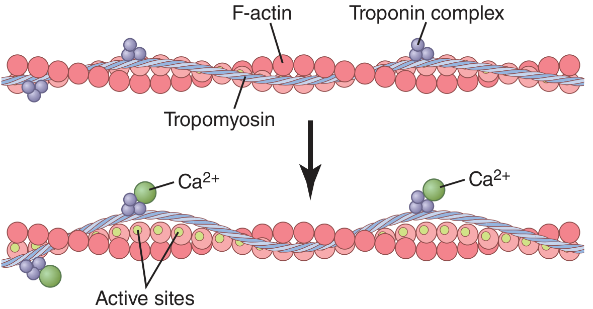
At rest: Tropomyosin lies over the active sites on actin, physically blocking myosin head binding. The troponin-tropomyosin complex acts as an "off switch."
When Ca²+ rises: Ca²+ binds to troponin C (up to 4 ions per molecule). This causes a conformational change that pulls tropomyosin deeper into the actin groove, uncovering the active sites and switching the system "on."
- Guyton and Hall Textbook of Medical Physiology
3. The Cross-Bridge Cycle (Walk-Along / Ratchet Theory)
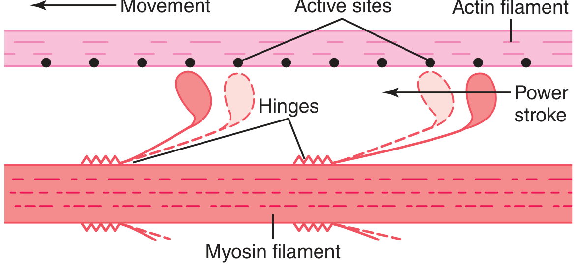
The cycle proceeds as follows:
| Step | Event |
|---|---|
| 1. Cocking | Myosin head cleaves ATP → ADP + Pi remain bound. Head extends perpendicular to actin ("cocked" high-energy state). |
| 2. Attachment | Head binds to exposed actin active site, forming a cross-bridge. |
| 3. Power stroke | Release of Pi triggers head to tilt ~45°, pulling the actin filament toward the sarcomere center. ADP is released. Force is generated. |
| 4. Rigor | Myosin head is firmly bound (low-energy position). This is the rigor state. |
| 5. Detachment | New ATP binds to myosin head, causing it to detach from actin. |
| 6. Re-cocking | ATP hydrolysis re-cocks the head, ready for the next cycle. |
Each cycle moves the filament ~10 nm. Because hundreds of cross-bridges act simultaneously and asynchronously, smooth, sustained force is produced.
Rigor mortis occurs after death when ATP is depleted: myosin heads cannot detach from actin, locking muscles in a rigid state.
4. Excitation-Contraction (E-C) Coupling
This is how a nerve signal is translated into a mechanical contraction:
- Motor nerve fires → releases acetylcholine at the neuromuscular junction
- Action potential generated in the sarcolemma spreads into the interior via T-tubules
- T-tubule depolarization activates dihydropyridine receptors (DHPR), voltage sensors in the T-tubule wall
- DHPR mechanically (or via Ca²+ signal) opens ryanodine receptors (RyR) on the sarcoplasmic reticulum (SR)
- Ca²+ floods from the SR into the cytosol, raising [Ca²+] from ~10⁻⁷ M to ~10⁻⁵ M
- Ca²+ binds troponin C → tropomyosin shifts → actin active sites exposed → cross-bridge cycling begins → contraction
Relaxation occurs when the SR Ca²+-ATPase (SERCA) pumps Ca²+ back into the SR, [Ca²+] falls, troponin reassumes its inhibitory conformation, and cross-bridges stop cycling.
5. Cardiac vs. Skeletal E-C Coupling
Cardiac muscle has a key difference: it uses Ca²+-induced Ca²+ release (CICR). During the plateau of the cardiac action potential, Ca²+ enters through L-type (dihydropyridine) channels and acts as a trigger to release a much larger amount of Ca²+ from the SR via ryanodine receptors. The magnitude of cardiac tension is therefore directly proportional to intracellular Ca²+ concentration - the basis for how drugs like digoxin and catecholamines modulate cardiac contractility.
- Costanzo Physiology, 7th Ed.
6. ATP Sources for Contraction
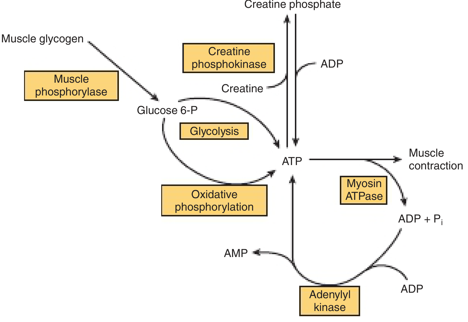
Resting muscle ATP only sustains contraction for a few seconds. Four pathways replenish it:
| Source | Speed | Notes |
|---|---|---|
| Creatine phosphate | Fastest | Immediate buffer; creatine kinase transfers phosphate to ADP |
| Glycolysis | Fast | Anaerobic; uses blood glucose or glycogen; produces lactate |
| Oxidative phosphorylation | Sustained | Primary source during prolonged exercise; requires O₂ |
| Adenylyl kinase | Emergency | 2 ADP → ATP + AMP |
Muscle glycogen is mobilized by glycogen phosphorylase, which is itself activated by Ca²+, epinephrine, and AMP - linking the start of contraction directly to fuel mobilization.
- Harper's Illustrated Biochemistry, 32nd Ed.
Summary Flow
Nerve impulse → ACh release → Sarcolemmal AP → T-tubule depolarization
    → SR Ca²+ release → Ca²+ binds TnC → Tropomyosin shifts
    → Actin sites exposed → Myosin cross-bridges attach
    → Power stroke (ATP → ADP + Pi) → Filaments slide → Sarcomere shortens
    → SERCA pumps Ca²+ back → Relaxation
Spermatogenesis
Spermatogenesis is the process by which diploid spermatogonia develop into haploid spermatozoa in the seminiferous tubules of the testis. It takes approximately 64-74 days in humans and involves mitosis, meiosis, and spermiogenesis.
Searching sources...
Finding Sources
Searching the Web
spermatogenesis stages steps hormonal regulation overview
Reading File
Reading File
Reading File
Loading Image
Loading Image
Reading File
Loading Image
Loading Image
Reading File
Spermatogenesis
Spermatogenesis is the continuous process by which male gametes (spermatozoa) are produced in the seminiferous tubules of the testis. It begins at puberty (~age 13) and continues throughout life, with one complete cycle taking approximately 74 days.
1. The Testicular Architecture
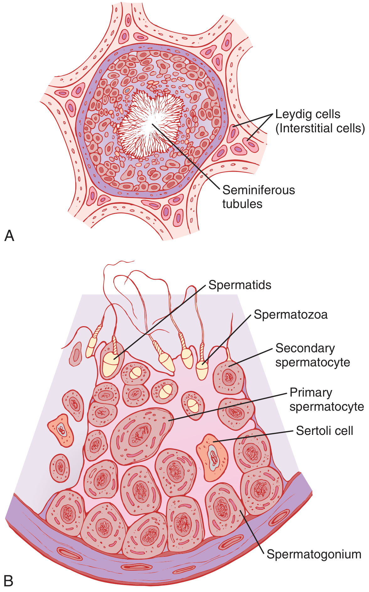
- Seminiferous tubules: site of sperm production
- Sertoli cells: large "nurse cells" lining the tubules; support, nourish, and regulate germ cell development; form the blood-testis barrier
- Leydig (interstitial) cells: located between tubules; produce testosterone under LH stimulation
2. Stages of Spermatogenesis
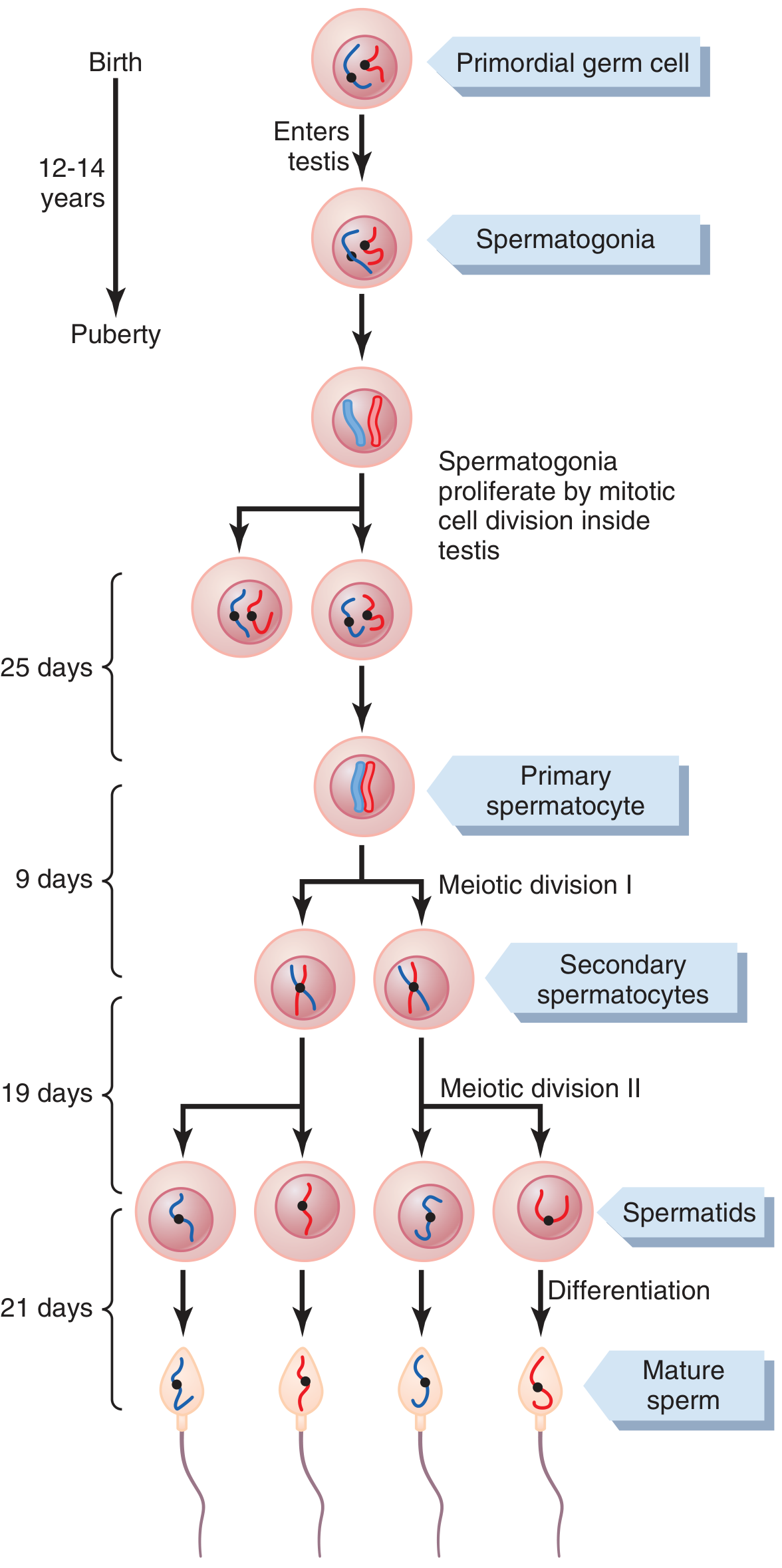
Phase 1 - Proliferation (Mitosis) ~25 days
Primordial germ cells migrate into the testis during embryonic development and become spermatogonia, which lie along the basal lamina. At puberty, spermatogonia begin active mitotic divisions, producing two populations:
- Type A spermatogonia - self-renewing stem cells (maintain the germ cell pool)
- Type B spermatogonia - committed cells that enter meiosis
Type B spermatogonia grow and differentiate into primary spermatocytes - the largest germ cells in the tubule, and still diploid (46 chromosomes).
Note: Descendant cells remain connected by cytoplasmic bridges through the late spermatid stage, ensuring synchronized differentiation of each clone. A single spermatogonium can ultimately yield ~512 spermatids.
Phase 2 - Meiosis ~28 days
Meiosis I (~9 days): Each primary spermatocyte (2n) undergoes the reductive division, producing two secondary spermatocytes (each haploid, n = 23, but still with paired chromatids). This is where crossing-over (genetic recombination) occurs.
Meiosis II (~19 days): Secondary spermatocytes divide again without DNA replication, yielding four spermatids - small, round, haploid (23 chromosomes) cells.
Net result: 1 spermatogonium → 4 haploid spermatids
Phase 3 - Spermiogenesis ~21 days
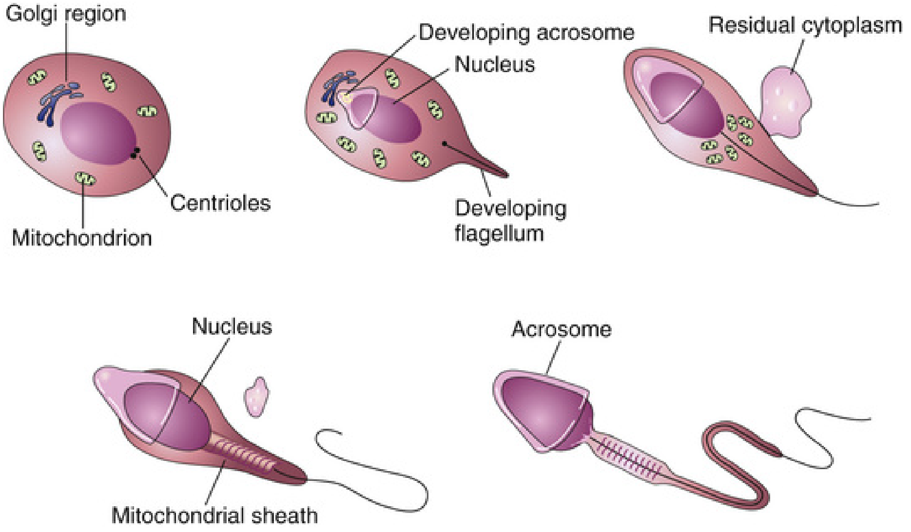
Spermiogenesis is the morphological transformation of round spermatids into mature spermatozoa. No further cell division occurs. Key events:
| Event | Detail |
|---|---|
| Acrosome formation | Golgi apparatus condenses to form the acrosome cap over the anterior nucleus |
| Nuclear condensation | Nucleus elongates and chromatin condenses tightly |
| Flagellum development | Centrioles form the axoneme (9+2 microtubule arrangement) |
| Mitochondrial sheath | Mitochondria aggregate around the proximal tail to form the energy-supplying mid-piece |
| Cytoplasm shedding | Most cytoplasm is discarded as the "residual body," phagocytosed by Sertoli cells |
3. Structure of the Mature Spermatozoon
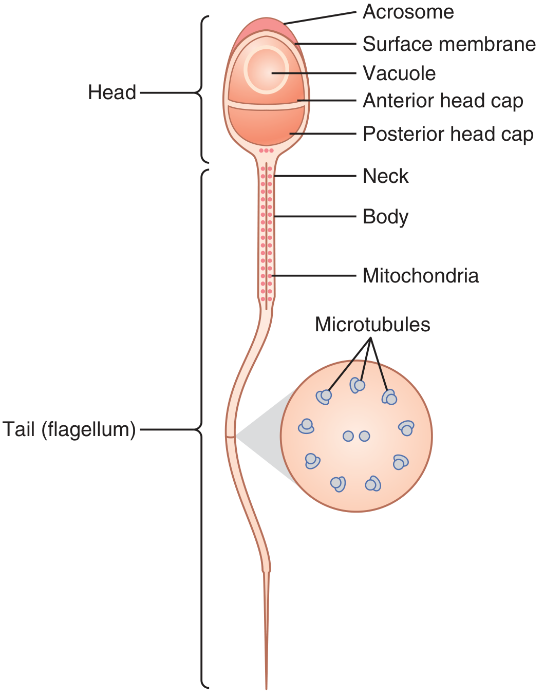
| Region | Contents/Function |
|---|---|
| Head | Condensed nucleus (haploid DNA) |
| Acrosome | Lysosome-like cap; contains hyaluronidase and proteolytic enzymes for zona pellucida penetration |
| Neck | Junction of head and tail; contains centrioles |
| Mid-piece (body) | Mitochondrial sheath; generates ATP for flagellar movement |
| Principal tail | Axoneme of 11 microtubules (9+2 arrangement); powers motility |
Sperm travel at 1-4 mm/min via flagellar movement powered by ATP from mid-piece mitochondria.
4. Hormonal Regulation
The hypothalamic-pituitary-gonadal (HPG) axis drives and maintains spermatogenesis:
Hypothalamus → GnRH (pulsatile)
    ↓
Anterior Pituitary
    ├── LH → Leydig cells → Testosterone
    └── FSH → Sertoli cells → Spermiogenesis, ABP, Inhibin
| Hormone | Source | Role |
|---|---|---|
| GnRH | Hypothalamus | Pulsatile release; drives LH and FSH secretion |
| LH | Anterior pituitary | Stimulates Leydig cells to secrete testosterone |
| Testosterone | Leydig cells | Essential for germ cell growth and early spermatogenesis; acts via androgen receptors on Sertoli cells |
| FSH | Anterior pituitary | Stimulates Sertoli cells; required for spermiogenesis (spermatid → sperm) |
| Estrogens | Sertoli cells (aromatase converts testosterone) | Required for spermiogenesis |
| Inhibin | Sertoli cells | Negative feedback on FSH secretion |
| Growth hormone | Anterior pituitary | Promotes early division of spermatogonia; deficiency causes infertility |
| Androgen-binding protein (ABP) | Sertoli cells | Maintains high androgen concentration in tubular fluid |
Negative feedback: Testosterone inhibits LH (via hypothalamus/pituitary); Inhibin inhibits FSH specifically.
5. Maturation in the Epididymis
Sperm leaving the seminiferous tubules are not yet motile or capable of fertilization. They pass through the ~6-meter epididymis over several days, during which:
- They acquire motility (within 18-24 hours in the epididymis)
- Final functional maturation ("capacitation" occurs later, in the female tract)
- Inhibitory proteins in epididymal fluid suppress premature activation
- Sperm can be stored in the epididymis and vas deferens for up to one month while remaining fertile
- Two testes produce up to 120 million sperm per day
6. Temperature Requirement
Spermatogenesis requires a temperature 2-3°C below body temperature (~34°C), which is why the testes are located in the scrotum outside the abdominal cavity. Elevated testicular temperature (cryptorchidism, varicocele, fever) increases germ cell apoptosis, causes DNA damage, and disrupts the blood-testis barrier, leading to impaired sperm production - a common cause of male infertility.
Summary Flow
Primordial germ cells (embryo) → Spermatogonia (seminiferous tubule, basal lamina)
    → [Mitosis] Primary spermatocytes (2n, diploid)
    → [Meiosis I] Secondary spermatocytes (n, haploid)
    → [Meiosis II] Spermatids (n, haploid) × 4
    → [Spermiogenesis] Mature spermatozoa
    → [Epididymis] Functionally mature, motile sperm
- Guyton and Hall Textbook of Medical Physiology
- The Developing Human: Clinically Oriented Embryology
- Ganong's Review of Medical Physiology, 26th Ed.
Spermatogenesis
I just covered spermatogenesis in detail in the previous response above. Here is a quick recap of the key points:
Spermatogenesis - Summary
Location: Seminiferous tubules of the testis
Duration: ~74 days total
Stages
| Stage | Process | Duration | Ploidy |
|---|---|---|---|
| Proliferation | Spermatogonia divide by mitosis | ~25 days | Diploid (2n) |
| Meiosis I | Primary → 2 Secondary spermatocytes | ~9 days | Haploid (n) |
| Meiosis II | Secondary spermatocytes → 4 Spermatids | ~19 days | Haploid (n) |
| Spermiogenesis | Spermatids → Mature spermatozoa (no division) | ~21 days | Haploid (n) |
Key Cells & Structures
- Sertoli cells - nurse and support developing sperm; secrete ABP, inhibin, estrogens
- Leydig cells - produce testosterone (stimulated by LH)
- Acrosome - cap on sperm head containing hyaluronidase and proteases for egg penetration
Hormonal Control
- GnRH (hypothalamus) → LH + FSH (anterior pituitary)
- LH → Leydig cells → Testosterone (essential for spermatogonial division)
- FSH → Sertoli cells → drives spermiogenesis
- Inhibin → negative feedback on FSH
After the Testis
Sperm pass through the epididymis (~6 m long) over several days, acquiring motility. Final capacitation occurs in the female reproductive tract.
Scroll up for the full detailed explanation with diagrams. Let me know if you want to focus on any particular aspect - meiosis, hormonal regulation, sperm structure, or clinical correlations like male infertility.
..
Looks like that might have been an accidental message. No worries!
Is there a topic you'd like to explore? I'm happy to continue with any subject - medical, scientific, or otherwise. Just let me know.
This is a shared conversation. Sign in to Orris to start your own chat.