
CLABSI Care Bundle Hand hygiene Maximal sterile barrier precautions Chlorhexidine skin preparation Optimal catheter site Daily review and early removal For seminar refer from mbbs standard textbooks and sources for 2nd year
CLABSI care bundle components evidence based practice 2nd year MBBS microbiology
CLABSI central line care bundle prevention diagram
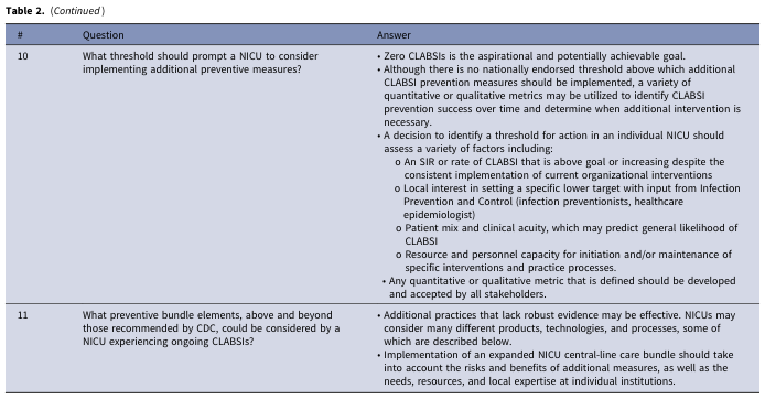
Table 2. (Continued) <table><thead><tr><th>#</th><th>Question</th><th>Answer</th></tr></thead><tbody><tr><td>10</td><td>What threshold should prompt a NICU to consider implementing additional preventive measures?</td><td>• Zero CLABSIs is the aspirational and potentially achievable goal.<br>• Although there is no nationally endorsed threshold above which additional CLABSI prevention measures should be implemented, a variety of quantitative or qualitative metrics may be utilized to identify CLABSI prevention success over time and determine when additional intervention is necessary.<br>• A decision to identify a threshold for action in an individual NICU should assess a variety of factors including:<br>○ An SIR or rate of CLABSI that is above goal or increasing despite the consistent implementation of current organizational interventions<br>○ Local interest in setting a specific lower target with input from Infection Prevention and Control (infection preventionists, healthcare epidemiologist)<br>○ Patient mix and clinical acuity, which may predict general likelihood of CLABSI<br>○ Resource and personnel capacity for initiation and/or maintenance of specific interventions and practice processes.<br>• Any quantitative or qualitative metric that is defined should be developed and accepted by all stakeholders.</td></tr><tr><td>11</td><td>What preventive bundle elements, above and beyond those recommended by CDC, could be considered by a NICU experiencing ongoing CLABSIs?</td><td>• Additional practices that lack robust evidence may be effective. NICUs may consider many different products, technologies, and processes, some of which are described below.<br>• Implementation of an expanded NICU central-line care bundle should take into account the risks and benefits of additional measures, as well as the needs, resources, and local expertise at individual institutions.</td></tr></tbody></table>
This medical illustration is a simplified anatomical schematic showing the connectivity of the parahippocampal cingulum bundle within the human brain. The diagram utilizes a sagittal cross-section to map neuroanatomical relationships. Key regions identified include the medial prefrontal cortex, parietal cortex, retrosplenial cortex, anterior thalamus, hippocampus, and parahippocampal cortex. The parahippocampal cingulum bundle is highlighted as a central hub with blue arrows representing axonal pathways. Solid blue lines denote direct anatomical connections linking the bundle to the retrosplenial cortex, parahippocampal cortex, hippocampus, and anterior thalamus. A dashed blue line signifies an indirect connection to the medial prefrontal cortex via other cingulum subdivisions. This visual aid is designed for neuroscience and neurology education to illustrate white matter tract pathways essential for cognitive functions such as autobiographical memory recall. The schematic facilitates the understanding of how various cortical and subcortical regions communicate through specific white matter bundles.
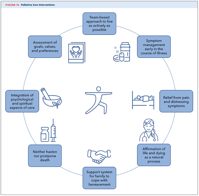
Summary : This figure presents the key interventions and principles of palliative care, organized in a circular diagram to emphasize their interconnectedness and holistic approach. conceptual diagram: # Central Theme : • A stylized human figure in a yoga pose at the center, symbolizing holistic well-being and active living. # Palliative Care Interventions (Nodes) : • Team-based approach to live as actively as possible • Symptom management early in the course of illness • Relief from pain and distressing symptoms • Affirmation of life and dying as a natural process • Support system for family to cope with bereavement • Neither hasten nor postpone death • Integration of psychological and spiritual aspects of care • Assessment of goals, values, and preferences # Layout : • Circular arrangement of eight blue boxes, each with an icon representing the intervention. • Boxes are evenly spaced around the central figure, visually connecting all aspects of care. # Design Encodings : • Blue color scheme for all boxes and icons. • Simple line icons for each intervention (e.g., handshake, medicine, mortar and pestle, bed, heart in hands). • No arrows; the circular layout implies equal importance and interrelation. # Analysis : • The diagram emphasizes that palliative care is multidimensional, involving medical, psychological, spiritual, and family support. • The circular structure suggests that all interventions are equally important and work together to support both patients and families. • The central figure highlights the goal of maintaining quality of life and active living, even in the context of serious illness.
hand hygiene WHO five moments technique
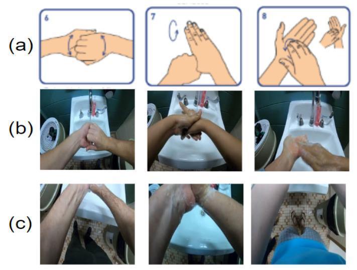
This educational image demonstrates World Health Organization (WHO) hand-hygiene protocols through three distinct formats: (a) illustrative diagrams, (b) clear clinical photographs of hand washing, and (c) egocentric/first-person view frames. Row (a) shows standardized hand rubbing techniques: interlacing fingers to clean the backs of fingers, rotational rubbing of the thumb, and rotational rubbing of the fingertips in the opposing palm. Row (b) presents real-world execution of these steps over a clinical sink, showing ideal camera visibility for monitoring hand and arm movements. Row (c) displays perspective challenges where the hands are partially or fully out of the camera view, emphasizing the impact of body size and movement on automated monitoring systems. The visual content focuses on infection control, aseptic technique, and the prevention of cross-contamination in medical and food-handling environments. Key educational concepts include the 12-step hygiene protocol, critical rubbing motions, and the technical requirements for assessing clinical compliance via imaging.
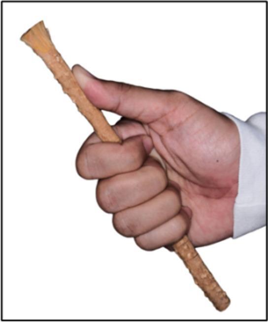
This clinical photograph demonstrates the specific 'five-finger grip technique' used for oral hygiene with a Salvadora persica chewing stick, also known as a miswak. The image displays a human hand holding the cylindrical, light-brown fibrous stick. The thumb is extended along the shaft to provide leverage and stability, while the four remaining fingers are curled around the body of the stick to secure it in a power grip. The distal end of the miswak is prepared for use, showing frayed, brush-like natural fibers intended for mechanical plaque removal and gingival massage. The grip is designed to allow controlled, precise vertical movements against tooth surfaces while minimizing the risk of gingival trauma. This visual serves as an educational guide for traditional oral hygiene practices within dental public health and clinical trials comparing traditional versus modern mechanical plaque control methods.
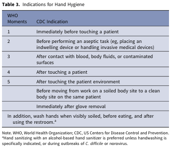
Table 3. Indications for Hand Hygiene <table><thead><tr><th>WHO Moments</th><th>CDC Indication</th></tr></thead><tbody><tr><td>1</td><td>Immediately before touching a patient</td></tr><tr><td>2</td><td>Before performing an aseptic task (eg, placing an indwelling device or handling invasive medical devices)</td></tr><tr><td>3</td><td>After contact with blood, body fluids, or contaminated surfaces</td></tr><tr><td>4</td><td>After touching a patient</td></tr><tr><td>5</td><td>After touching the patient environment</td></tr><tr><td></td><td>Before moving from work on a soiled body site to a clean body site on the same patient</td></tr><tr><td></td><td>Immediately after glove removal</td></tr><tr><td colspan="2">In addition, wash hands when visibly soiled, before eating, and after using the restroom.ᵃ</td></tr></tbody></table> Note. WHO, World Health Organization; CDC, US Centers for Disease Control and Prevention. ᵃHand sanitizing with an alcohol-based hand sanitizer is preferred unless handwashing is specifically indicated, or during outbreaks of C. difficile or norovirus.
CLABSI Care Bundle - Seminar Notes
For 2nd Year MBBS | Microbiology / Community Medicine / Surgery
INTRODUCTION
- Sabiston Textbook of Surgery describes CLABSI as occurring when skin flora travels along the catheter and colonizes it, making its way into the bloodstream
- CLABSI carries a 15% mortality rate
- It is among the most common and most preventable hospital-acquired (nosocomial) infections
- Associated with significant morbidity, prolonged ICU stays, and increased healthcare costs
- CRBSI (Catheter-Related Bloodstream Infection): Clinical definition requiring specific lab tests (catheter tip culture, quantitative blood cultures, differential time to positivity) - used for diagnosing and treating individual patients
- CLABSI: Surveillance definition used by CDC/NHSN to monitor and track infection rates at an institutional/national level - may overestimate true CRBSI incidence
- Current Surgical Therapy 14e
PATHOGENESIS
- Extraluminal (skin) contamination - most common for short-term non-tunneled catheters; skin flora migrates along the external catheter surface
- Intraluminal / hub contamination - predominates in longer-term catheters; microbes introduced during catheter manipulation
- Hematogenous seeding from a distant focus of infection
- Contaminated infusions (least common)
THE CARE BUNDLE CONCEPT
- Hand Hygiene
- Maximal Sterile Barrier Precautions
- Chlorhexidine Skin Preparation
- Optimal Catheter Site Selection
- Daily Review of Necessity and Early Removal
BUNDLE ELEMENT 1: HAND HYGIENE
WHO "My Five Moments for Hand Hygiene"
| WHO Moment | Indication |
|---|---|
| 1 | Before touching a patient |
| 2 | Before an aseptic task (placing a device, handling invasive equipment) |
| 3 | After contact with blood, body fluids, or contaminated surfaces |
| 4 | After touching a patient |
| 5 | After touching the patient environment |
Methods
- Alcohol-based hand rub (ABHR): Preferred for routine hand antisepsis when hands are not visibly soiled. Effective against most bacteria, fungi, and enveloped viruses
- Soap and water: Mandatory when hands are visibly dirty, contaminated with blood/body fluids, or after caring for C. difficile / norovirus patients
For CVC insertion specifically
- Proceduralist AND all assistants must perform hand hygiene before the procedure
- Hand hygiene must be repeated before and after all IV system manipulation (maintenance bundle)
- Park's Textbook of Preventive and Social Medicine identifies hospital-acquired infections as indicators of poor quality health service delivery, and hand hygiene compliance is the cornerstone of prevention
BUNDLE ELEMENT 2: MAXIMAL STERILE BARRIER (MSB) PRECAUTIONS
Components of MSB
| What is worn/used | Details |
|---|---|
| Surgical cap | Covers all hair |
| Surgical mask | Full face mask (covering nose and mouth) |
| Sterile gown | Long-sleeved, worn by the proceduralist |
| Sterile gloves | Worn by the proceduralist |
| Large sterile drape | Covers the entire patient from head to foot, not just the insertion site |
- Mulholland and Greenfield's Surgery 7e - "maximal sterile barrier precautions (handwashing, sterile gloves, large drape, sterile gown, mask, and cap) during insertion"
- Roberts and Hedges' Clinical Procedures in Emergency Medicine - "Sterile barrier precautions with cap, face mask, sterile gown, and gloves should be used at all times during insertion of CVCs"
BUNDLE ELEMENT 3: CHLORHEXIDINE SKIN PREPARATION
Preferred Agent: Chlorhexidine-Alcohol
- Chlorhexidine gluconate 2% in 70% isopropyl alcohol (e.g., ChloraPrep) is the agent of choice
- Randomized trials have confirmed that chlorhexidine-alcohol provides greater protection against catheter-related infections than povidone-iodine with alcohol
- Mechanism: Chlorhexidine disrupts cell membranes of gram-positive and gram-negative bacteria; alcohol provides rapid killing
- Unlike povidone-iodine, chlorhexidine has residual activity - it binds to skin proteins and continues to inhibit bacterial regrowth after application
- Goldman-Cecil Medicine and Barash, Cullen, and Stoelting's Clinical Anesthesia 9e
Application Technique
- Apply with back-and-forth scrubbing motion
- Allow to air dry completely before needle insertion (at least 30 seconds for alcohol, longer for chlorhexidine to be effective)
- Do not blot or fan dry - this reduces efficacy
Alternatives
- Povidone-iodine (10% solution or in alcohol): Used when chlorhexidine is contraindicated (allergy, neonates/premature infants under 2 months)
- Do not use povidone-iodine over dry skin - it requires moisture to activate
Additional Skin Maintenance Measures
- Use chlorhexidine-impregnated dressings over the insertion site
- Daily chlorhexidine bathing of patients with CVCs (Fuster and Hurst's The Heart 15e)
BUNDLE ELEMENT 4: OPTIMAL CATHETER SITE SELECTION
Infection Risk by Site (lowest to highest)
| Site | CLABSI Risk | Notes |
|---|---|---|
| Subclavian vein | Lowest | Preferred for infection control; drier skin, easier to maintain sterile dressing |
| Internal jugular (IJ) | Intermediate | Easier to place under ultrasound; similar risk to femoral depending on body habitus |
| Femoral vein | Highest | Avoid in adult patients whenever possible; groin flora, proximity to perineum |
- Sabiston Textbook of Surgery: "choice of catheter site (in order of infection risk: femoral vein > internal jugular vein > subclavian vein)"
- However, subclavian insertion carries the highest risk of pneumothorax - risk-benefit analysis is required per patient
- Femoral site is also associated with higher rates of DVT
- Ultrasound guidance for all CVC placements is now standard of care (Current Surgical Therapy 14e)
Additional Selection Considerations
- Avoid sites near non-intact skin (burned, infected), open wounds, or tracheostomy sites
- Use catheters with the minimum number of lumens necessary - each extra lumen = extra access point = extra infection risk
- Consider antimicrobial-impregnated catheters (minocycline/rifampin or chlorhexidine/silver sulfadiazine) for expected catheter duration >5-7 days in high-risk patients
- Use PICC lines (peripherally inserted central catheters) for prolonged IV access when feasible, as they have lower CLABSI rates
- Mulholland and Greenfield's Surgery 7e and Current Surgical Therapy 14e
BUNDLE ELEMENT 5: DAILY REVIEW OF NECESSITY AND EARLY REMOVAL
Key Principle
Daily Review Checklist - Questions to Ask Each Day
- Is the central line still clinically indicated?
- Can the indication be served by a peripheral IV or oral route?
- Is there a plan for when the CVC will be removed?
- Is the insertion site clean, with intact dressing?
- Are all lumens actually in use?
Documentation Required (per CVC Maintenance Bundle)
- Location of CVC
- Date of insertion
- Duration of use
- Date of anticipated removal
- Any complications
- Roberts and Hedges' Clinical Procedures: bundling policy includes "daily review of the need for the line, with prompt removal of unnecessary lines"
- Comprehensive Clinical Nephrology 7e: "Daily review of the need for the line, with prompt removal of unnecessary lines"
INSERTION BUNDLE vs MAINTENANCE BUNDLE
Insertion Bundle
| Element | Recommendation |
|---|---|
| Hand hygiene | Before and after each patient contact |
| MSB precautions | Full sterile gown, gloves, cap, mask, large drape |
| Skin antisepsis | Chlorhexidine-alcohol (or povidone-iodine if allergic) |
| Catheter type | Minimum ports; antimicrobial-impregnated if >7 days |
| Site selection | Subclavian preferred; femoral avoided |
| Ultrasound guidance | Standard of care for all central lines |
| Sterile dressing | Applied immediately post-placement |
| CVC insertion kit/cart | All supplies available in one organized kit |
| Checklist use | Ensures all steps are followed; procedure stopped if protocol violated |
Maintenance Bundle
| Element | Recommendation |
|---|---|
| Daily review | Document and remove unnecessary CVCs promptly |
| Hand hygiene | Before all IV system manipulation |
| Hub disinfection | "Scrub the hub" before and after each access (alcohol swab, 15 seconds) |
| Dressings | Change gauze every 48 hours; transparent dressing every 5-7 days; or when soiled/loose |
| Chlorhexidine dressing | Chlorhexidine-impregnated dressing around insertion site |
| Needleless connectors | Minimize open-lumen exposure |
| IV tubing | Change tubing per protocol (every 72-96 hours; within 24 hours after blood/lipids) |
ORGANISMS AND TREATMENT
- Coagulase-negative Staphylococci (e.g., S. epidermidis) - most common
- Staphylococcus aureus (including MRSA)
- Gram-negative bacilli (E. coli, Klebsiella pneumoniae, Acinetobacter baumannii)
- Enterococcus spp.
- Candida spp. (especially in immunocompromised, TPN patients)
- Remove catheter if possible for ALL short-term catheters
- Catheter salvage is only acceptable in patients with severely limited access or long-term tunneled catheters (antimicrobial lock therapy + systemic antibiotics)
- Empiric antibiotics (vancomycin + gram-negative coverage) pending culture results
- Duration depends on the organism isolated
NOSOCOMIAL INFECTIONS - 2nd YEAR MBBS CONTEXT
- Nosocomial (hospital-acquired) infections are infections acquired >48 hours after admission that were not present or incubating at the time of admission
- They are both endemic (baseline occurrence) and epidemic (outbreak-related)
- Surveillance criteria for vascular catheter infection: "inflammation, lymphangitis, or purulent discharge at the insertion site of the catheter"
- High frequency of nosocomial infections = indicator of poor quality health care delivery
- Factors promoting nosocomial infections: immunocompromised hosts, invasive procedures, antibiotic resistance from selective pressure
CHECKLIST FOR CVC INSERTION (Sample - Current Surgical Therapy 14e)
- Identify patient and obtain informed consent
- Verify correct site (right/left; IJ/subclavian/femoral)
- All team members wear hat and mask and perform hand hygiene
- Prep insertion site with ChloraPrep (or Betadine if allergic) and allow to dry
- Proceduralist dons sterile gown and sterile gloves
- Drape entire patient in sterile fashion
- Maintain sterile field and aseptic technique
- Stop procedure if contamination of sterile field, CVC, or instruments occurs
- Attach caps or IV tubing to lumens before removing sterile drape
- Apply sterile dressing and label with date and time
- Wash hands
- Dispose of all sharps in proper receptacles
- Document insertion details in medical record
KEY FACTS TO REMEMBER FOR EXAMS
| Point | Detail |
|---|---|
| CLABSI definition | Bloodstream infection with CVC in place >48 h, no other source |
| Mortality rate | ~15% |
| Landmark study | Pronovost Keystone Study, 103 Michigan ICUs, 66% reduction in CLABSI |
| Best skin prep agent | Chlorhexidine-alcohol (>0.5% chlorhexidine in alcohol) |
| Highest infection risk site | Femoral vein |
| Lowest infection risk site | Subclavian vein |
| Scheduled catheter change | Does NOT reduce CLABSI - not recommended |
| Most common organisms | CoNS (S. epidermidis), S. aureus, Gram-negatives, Candida |
| Maintenance bundle key | Daily review + early removal; scrub the hub |
SOURCES (MBBS Standard Textbooks)
- Sabiston Textbook of Surgery (The Biological Basis of Modern Surgical Practice) - Chapter: Hospital Acquired Infections / CLABSI
- Current Surgical Therapy 14e (Cameron) - Chapter: CRBSI and CLABSI, Catheter Care Bundles (Tables 1 & 2), CVC Insertion Checklist
- Park's Textbook of Preventive and Social Medicine - Chapter: Hospital-Acquired Infections / Nosocomial Infections
- Harrison's Principles of Internal Medicine 22e (2025, McGraw Hill) - Catheter insertion bundle elements
- Mulholland and Greenfield's Surgery: Scientific Principles and Practice 7e - Hospital Acquired Infections
- Goldman-Cecil Medicine - Evidence-based recommendations for CLABSI prevention
- Barash, Cullen & Stoelting's Clinical Anesthesia 9e - Catheter-associated bloodstream infections
- Comprehensive Clinical Nephrology 7e - Best practices to minimize catheter-associated bloodstream infection
- CDC / Keystone Study (Dr. Peter Pronovost, Johns Hopkins) - Evidence base for the five-element bundle
CAUTI Care Bundle Indications for catheter Aseptic insertion Hand hygiene Closed drainage system Catheter below bladder level Daily assessment and early removal
CAUTI care bundle components indications catheter aseptic insertion closed drainage daily assessment 2nd year MBBS
urinary catheter Foley catheter anatomy drainage bag closed system
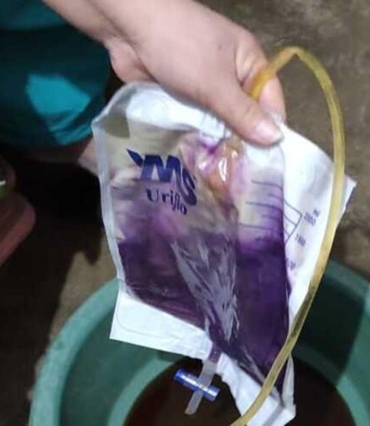
Clinical photograph illustrating Purple Urine Bag Syndrome (PUBS) in a patient with a chronic indwelling Foley catheter. The image shows a standard medical drainage bag containing urine with a distinctive, deep purple discoloration. The intensity of the pigment is most concentrated at the bottom of the bag, showing a layering effect where the color transitions to a lighter lavender and brownish-yellow tint at the top. The drainage system features a translucent, yellowish catheter tube and a blue-and-white T-tap drainage valve at the base of the graduated collection bag. This phenomenon is a rare manifestation typically associated with urinary tract infections caused by tryptophan-metabolizing bacteria (such as E. coli, Klebsiella, or Proteus species) in patients with alkaline urine and chronic constipation. The purple pigment results from the chemical reaction of indigo and indirubin, which are metabolic byproducts of dietary tryptophan, with the plastic materials of the catheter and bag.
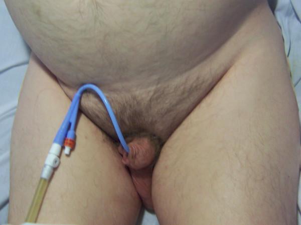
This clinical photograph displays the lower abdomen and genital region of a male patient, demonstrating a correctly placed indwelling Foley catheter. The catheter, which is blue, enters the urethral meatus of the penis and exhibits a short, curved external segment consistent with proper intravesical positioning of the balloon. The external portion of the catheter connects to a white dual-port Y-connector, with one port capped in orange for balloon inflation and the other connected to clear drainage tubing containing yellow-colored urine. The patient's anatomy shows a rounded lower abdomen with fair skin tone and normal pubic hair distribution. This image serves as a clinical reference for the 'short catheter sign,' which confirms that the catheter balloon is correctly situated within the urinary bladder rather than the urethra, as would be indicated by a 'long catheter sign' where a significantly longer segment of tubing is visible outside the body.
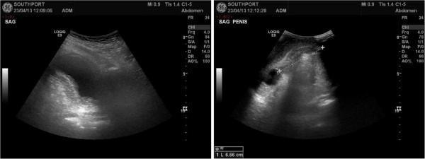
This diagnostic ultrasound image consists of two panels illustrating the clinical complication of an incorrectly positioned Foley catheter. The left panel, a sagittal abdominal scan (labeled 'SAG'), displays a view of the urinary bladder which is notably empty, with no Foley catheter balloon visualized within the lumen. The right panel, labeled 'SAG PENIS', shows a sagittal ultrasound view of the penile anatomy. In this image, a rounded, anechoic (dark) structure representing the inflated balloon of a Foley catheter is clearly visible, malpositioned within the membranous urethra rather than the bladder. Electronic calipers on the right panel measure the distance from the tip of the penis to the center of the balloon, recorded at 6.66 cm. This comparison demonstrates 'non-drainage' of urine caused by urethral catheterization failure, where the balloon is inflated prematurely in the urethra instead of the bladder lumen. The clinical significance involves the risk of urethral trauma and secondary hematuria.
CAUTI Care Bundle - Seminar Notes
For 2nd Year MBBS | Microbiology / Community Medicine / Surgery
INTRODUCTION
Definition
- A UTI developing in a patient with a urinary catheter in place or within 48 hours of catheter removal
- CDC diagnostic threshold: >10² CFU/mL in catheter specimen
- IDSA threshold: ≥10³ CFU/mL of a uropathogen
- Sabiston Textbook of Surgery, Goldman-Cecil Medicine, Washington Manual of Medical Therapeutics
Why CAUTI Matters
- Most common HAI - exceeds CLABSI, VAP, and SSI in sheer numbers
- Responsible for significant morbidity: bacteremia, pyelonephritis, urosepsis
- 10-25% of patients with catheter-associated bacteriuria develop symptomatic infection
- Bacteriuria occurs at a rate of 3-8% per day of catheterization - virtually inevitable with prolonged use
- Since 2008, CMS (Center for Medicare and Medicaid Services) no longer reimburses hospitals for the extra costs resulting from CAUTI - a powerful institutional driver for prevention
- It is estimated that 65-70% of CAUTIs in acute care hospitals are preventable with correct practices
- Goldman-Cecil Medicine 2-vol set; Campbell Walsh Wein Urology
PATHOGENESIS
Routes of Bacterial Entry (Campbell Walsh Wein Urology 3e)
- At time of insertion - mechanical inoculation of urethral bacteria into the bladder; contamination from poor technique (most preventable)
- Periurethral (extraluminal) route - bacteria from the perineum/rectum colonize the urethra and track along the outer surface of the catheter into the bladder; most common route in women
- Intraluminal route - bacteria introduced at the catheter-bag junction travel up through the drainage tube and into the bladder; common when the closed system is broken
- Hematogenous seeding - rare; secondary to distant bacteremia
Biofilm Formation - The Critical Mechanism
- Protects bacteria from host defenses (phagocytes, complement)
- Protects bacteria from antibiotics (reduces penetration by >100-fold)
- Causes catheter encrustation
- Pseudomonas and Proteus species are especially adept at biofilm formation
- Explains why antibiotics alone cannot eradicate CAUTI while the catheter is still in situ
Why Catheters Predispose to UTI (Sabiston Textbook of Surgery)
- Provide a direct path for pathogens from rectal flora/environment to the bladder
- Allow bacteria to bypass the urethral sphincters
- Eliminate the protective turbulence associated with normal voiding
- Cause trauma to the urothelium, making it susceptible to bacterial colonization
Common Causative Organisms (Washington Manual of Medical Therapeutics)
| Organism | Notes |
|---|---|
| Escherichia coli | Most common overall |
| Klebsiella pneumoniae | Gram-negative, biofilm producer |
| Pseudomonas aeruginosa | Strong biofilm former; hospital-acquired |
| Proteus mirabilis | Causes alkaline urine, struvite stones |
| Enterococcus spp. | Gram-positive |
| Staphylococci (CoNS, S. aureus) | Gram-positive |
| Candida spp. | Especially in immunocompromised, antibiotic-exposed patients |
THE CAUTI CARE BUNDLE
BUNDLE ELEMENT 1: INDICATIONS FOR CATHETER USE
Appropriate Indications for Urinary Catheterization (CDC CAUTI Guidelines; Fuster & Hurst's The Heart 15e; APSIC 2022)
| Category | Indication |
|---|---|
| Urinary retention | Acute urinary retention, bladder outlet obstruction |
| Surgical | Perioperative use in selected surgeries (prolonged surgery, urologic procedures, cardiac/major vascular surgery); intraoperative urine output monitoring |
| Critical care monitoring | Accurate measurement of urine output in critically ill/hemodynamically unstable patients |
| Wound care | Assist healing of perineal and sacral wounds in incontinent patients |
| Neurological | Spinal cord injury, neuropathic bladder (spina bifida, MS, stroke) |
| Comfort/palliative | Hospice, comfort care, palliative care situations |
| Immobilization | Required immobilization for trauma or surgery |
What is NOT an Appropriate Indication
- Urinary incontinence alone - do not catheterize for nursing convenience or to keep the bed dry
- Routine post-operative use when not clinically necessary
- Monitoring urine output in a stable patient who could use a bedpan
- Harrison's Principles of Internal Medicine 22e: "Place indwelling catheters only when strictly necessary, e.g., to relieve obstruction, and not for convenience"
Alternatives to Indwelling Urethral Catheterization
- Condom catheter (external catheter) - for incontinent males without retention
- Intermittent (in-and-out) catheterization - preferred over indwelling in spinal cord injury patients; bacteriuria rate only 1-3% per catheterization vs. ~10% per day with indwelling catheter
- Bladder scanner (portable ultrasound) - to assess post-void residual before catheterizing
- Absorbent products (nappies/pads)
BUNDLE ELEMENT 2: ASEPTIC INSERTION TECHNIQUE
Step-by-Step Aseptic Insertion Protocol
- Sterile drapes and sterile field
- Sterile gloves (non-powdered)
- Antiseptic solution (povidone-iodine or chlorhexidine)
- Sterile water for balloon inflation
- Sterile lubricant/anaesthetic gel (lignocaine gel)
- Appropriate size catheter (typically 12-14 Fr adult; 18 Fr coudé for BPH)
- Closed drainage system connected
- Explain procedure to patient and obtain consent
- Position patient (supine, dorsal recumbent for female; supine for male)
- Wash hands; don sterile gloves
- Create a sterile field with sterile drapes
- Clean the urethral meatus and surrounding area with antiseptic solution using a front-to-back technique (females); cleanse glans from meatus outward (males) - use a fresh swab/cotton for each stroke
- Apply sterile lubricant/anaesthetic gel into the urethra
- Using forceps or sterile technique, advance catheter into bladder until urine flows
- Advance a further 3-5 cm before inflating the balloon to ensure the balloon is inside the bladder (critical to prevent urethral injury)
- Inflate balloon with sterile water (as per catheter specifications, typically 10 mL)
- Gently retract until resistance is felt
- Connect to sterile closed drainage system before removing drapes
- Secure catheter to inner thigh to prevent piston-like movement (reduces urethral trauma and backflow)
- Document: catheter size, balloon volume, date/time, inserter's name, indication
- APSIC CAUTI Guidelines 2022; Goldman-Cecil Medicine
BUNDLE ELEMENT 3: HAND HYGIENE
WHO Five Moments for Hand Hygiene - Applied to Catheter Care
| WHO Moment | Application to Catheter Care |
|---|---|
| Before patient contact | Before approaching patient for catheterization or catheter check |
| Before aseptic task | Before catheter insertion, before accessing the drainage system |
| After body fluid exposure | After handling urine, drainage bag, or catheter tubing |
| After patient contact | After all catheter manipulations |
| After patient environment | After touching the bed, drainage stand, or any surface near catheter |
Key Rules for Hand Hygiene and CAUTI Prevention
- Perform hand hygiene before and after urinary catheter insertion
- Perform hand hygiene before and after any manipulation of catheter or drainage system
- After hand hygiene, don non-sterile gloves before handling catheter/drainage system during maintenance
- Change gloves and perform hand hygiene between patients to prevent cross-contamination
- APSIC CAUTI Guidelines 2022; Fuster and Hurst's The Heart 15e
- Harrison's 22e: "Minimize manipulation of or entry into urinary catheter systems"
BUNDLE ELEMENT 4: CLOSED DRAINAGE SYSTEM
Principles of Closed Drainage
Rules for Maintaining the Closed System
| Principle | Action Required |
|---|---|
| Never disconnect | Do not break the catheter-tubing junction unless absolutely necessary |
| Sampling ports | Collect urine specimens only via the designated sampling port (needleless port) with aseptic technique - never open the tubing |
| Bag drainage | Empty the drainage bag using a separate, clean collection container for each patient - never let the outlet tap touch the container |
| Tubing integrity | Keep tubing free from kinking; ensure unobstructed flow at all times |
| Tamper-evident seal | Check that the tamper-evident seal at the catheter-tubing junction is intact |
| If broken | If disconnection, leakage, or breach occurs - replace the entire catheter + collecting system using aseptic technique with new sterile equipment |
| Routine changes | Do NOT change catheters or bags at fixed intervals - change only for clinical indication (infection, obstruction, system compromise) |
Why Closed System Matters
- Intraluminal route of infection (drainage bag → tubing → bladder) is only possible when the system is open
- Even brief disconnection allows bacterial entry
- Goldman-Cecil Medicine: "maintenance of a closed system with unobstructed flow of urine from the bladder into the collection system are recommended to reduce the risk of CAUTI"
- SUNA Clinical Practice Guidelines: "If break in aseptic technique, disconnection or leakage occur, replace the catheter and collecting system using aseptic technique and sterile equipment"
BUNDLE ELEMENT 5: CATHETER BELOW BLADDER LEVEL
Principle
Why This Matters
- If the drainage bag is raised above the bladder level, urine (which may contain bacteria from the bag) can reflux back into the bladder - a direct mechanism of introducing infection
- If the bag touches the floor, it becomes contaminated with environmental organisms
Practical Rules
| Rule | Detail |
|---|---|
| Bag position | Always below bladder level (even when patient is supine, the bag should be lower than the mattress level) |
| Not on the floor | Bag must not touch the floor - use a bag hanger or stand |
| Tubing looping | Avoid large dependent loops of tubing that collect pooled urine and act as a reservoir |
| During transfers | When moving or ambulating a patient, temporarily clamp the catheter or hold the bag at waist level briefly, but restore dependent drainage immediately after |
| Unobstructed flow | Keep tubing free from kinking or compression at all times |
- CAUTI Prevention Bundle (Ascension Healthcare): "Position drainage bag below bladder level and above the floor to prevent reflux or contamination"
- Fuster and Hurst's The Heart 15e: "Maintain drainage bag and connecting tubing below the level of the bladder"
BUNDLE ELEMENT 6: DAILY ASSESSMENT AND EARLY REMOVAL
The Core Principle
Daily Assessment - Questions to Ask (HOUDINI-type framework)
| Letter | Question |
|---|---|
| H - Haematuria | Is there gross haematuria requiring monitoring? |
| O - Obstruction | Is there urinary outlet obstruction? |
| U - Urology/gynaecology | Is it needed for urological or gynaecological surgery? |
| D - Decubitus | Is it needed to assist healing of perineal/sacral wounds in an incontinent patient? |
| I - Input/output | Is accurate urine output monitoring required in a critically ill patient? |
| N - Neurology | Is there neurological disease impairing bladder control? |
| I - Immobilization | Is the patient immobilized and catheter needed for this reason? |
Evidence for Removal Reminders
- Catheter reminders (written or electronic alerts to physicians) reduce catheter dwell time
- Nurse-driven catheter removal protocols (where nurses can remove catheters per protocol without waiting for a physician order) are highly effective
- Goldman-Cecil Medicine: "Development of protocols that explicitly define appropriate indications... nurse-driven protocols that allow nurses to remove unnecessary urinary catheters have been associated with reduced catheter use and lower CAUTI rates"
After Removal
- Monitor for urinary retention with a bladder scanner
- If patient cannot void spontaneously, perform intermittent catheterization (clean or sterile) rather than reinsert an indwelling catheter
Routine Replacement is NOT Recommended
- Scheduled catheter changes at fixed intervals (every 7 or 14 days) do not reduce CAUTI rates
- Change only when clinically indicated: obstruction, encrustation, suspected infection, system compromise
MAINTENANCE BUNDLE SUMMARY TABLE
| Element | Action |
|---|---|
| Daily review | Assess and document continued need; remove if not indicated |
| Hand hygiene | Before and after any catheter or drainage system contact |
| Closed system | Do not break; replace entire system if compromised |
| Bag position | Below bladder level; above floor; tubing free from kinking |
| Perineal hygiene | Daily meatal hygiene with soap and water (do NOT use antiseptics on perineum while catheter is in place - no benefit) |
| Specimen collection | Use sampling port; never open the junction |
| Securing | Anchor catheter to inner thigh; prevent in-and-out piston movement |
| Documentation | Indication, insertion date, expected removal date |
INSERTION BUNDLE vs MAINTENANCE BUNDLE (Summary)
Insertion Bundle (Fuster and Hurst's The Heart 15e; Harrison's 22e)
| Before Insertion | During Insertion | After Insertion |
|---|---|---|
| Verify indication - use only when necessary | Only trained staff inserts | Remove when no longer indicated |
| Avoid routine use for incontinence | Perform hand hygiene before and after | Maintain bag below bladder level |
| Use bladder scanner to assess need first | Clean urethral area with antiseptic | Maintain closed system |
| Use sterile equipment throughout | Secure catheter to inner thigh | |
| Sterile lubricant | Perform daily perineal hygiene | |
| Advance fully before inflating balloon | Daily assessment for removal |
DIAGNOSIS OF CAUTI
Clinical Features
- Fever (most common)
- Suprapubic and/or flank pain
- Altered mental status (especially in elderly)
- New onset dysuria (difficult to assess in catheterized patients)
- Rigors, malaise in severe cases
Diagnostic Criteria (Washington Manual of Medical Therapeutics)
- Urinalysis + urine culture (before starting antibiotics)
- ≥10³ CFU/mL of a single uropathogen (IDSA) or ≥10² CFU/mL (CDC) in catheterized specimen
- Important: Pyuria and bacteriuria are present in ALL patients with chronic indwelling catheters and should NOT be treated in the absence of symptoms (asymptomatic bacteriuria)
Treatment
- Remove or exchange the catheter (before starting antibiotics whenever possible)
- Send urine culture from new catheter
- Antibiotics for 7-10 days (prompt responders) or 14 days (delayed responders)
- Funguria (candiduria): catheter removal alone often suffices; antifungals only for immunocompromised patients at risk for candidemia
- Washington Manual; Sabiston Textbook of Surgery
COMPLICATIONS OF LONG-TERM CATHETERIZATION (Campbell Walsh Wein Urology)
| Complication | Detail |
|---|---|
| Bacteriuria | Virtually universal (100% at 30 days) |
| UTI / CAUTI | 10-30% of bacteriuric episodes become symptomatic in short-term |
| Urethral trauma / false passage | 3.2 per 1000 male admissions; often preventable with training |
| Urethral stricture | ~3.4% incidence with catheterization |
| Bladder cancer | ~1% in long-term spinal cord patients with chronic catheterization |
| Bladder stones | From encrustation and stasis |
| Autonomic dysreflexia | In spinal cord injury patients |
THE MAYO CLINIC "6-C" BUNDLE (Campbell Walsh Wein Urology)
| C | Meaning |
|---|---|
| Consider | Appropriate placement and daily need for indwelling catheter |
| Connect | Properly connect catheter to closed drainage system |
| Clean | Catheter care (meatal hygiene daily) |
| Closed | Maintain closed drainage system |
| Call | Irrigation only when necessary |
| Culture | Obtain culture only when clinically indicated, not routinely |
KEY FACTS FOR EXAMS
| Point | Detail |
|---|---|
| Most common HAI | CAUTI |
| Strongest risk factor | Duration of catheterization (~10% per day) |
| Bacteriuria rate | 3-8% per day with indwelling catheter |
| Most common organism | E. coli |
| Biofilm formers | Pseudomonas, Proteus particularly adept |
| Most common route in women | Periurethral (extraluminal) |
| Closed system benefit | Delays bacteriuria from 4 days to >2 weeks |
| Routine catheter change | Does NOT reduce CAUTI - not recommended |
| Asymptomatic bacteriuria | Do NOT treat in catheterized patients |
| Catheter position | Below bladder, above floor |
| CMS policy | No reimbursement for CAUTI since 2008 |
| Preventability | 65-70% of CAUTIs are preventable |
SOURCES (MBBS Standard Textbooks)
- Sabiston Textbook of Surgery (The Biological Basis of Modern Surgical Practice) - CAUTI definition, pathogenesis, prevention
- Campbell Walsh Wein Urology 3-Volume Set - CAUTI pathogenesis, biofilm, bacteriuria, complications, 6-C bundle
- Harrison's Principles of Internal Medicine 22e (2025, McGraw Hill) - Prevention table, clinical recommendations
- Goldman-Cecil Medicine International Edition - Evidence-based practices, closed system, catheter-associated UTIs chapter
- Washington Manual of Medical Therapeutics - General principles, organisms, diagnosis, treatment
- Fuster and Hurst's The Heart 15e - AHA CAUTI prevention table (before/during/after insertion)
- Tintinalli's Emergency Medicine - Definition, management
- Roberts and Hedges' Clinical Procedures in Emergency Medicine - Infection prevention, bundles
- Park's Textbook of Preventive and Social Medicine - Hospital-acquired infections, community medicine context
- CDC CAUTI Guidelines 2009 / APSIC CAUTI Guidelines 2022 / ISID Guide to Infection Control in the Hospital - Evidence base for bundle components
How many days can the catheters be kept for