
Different types of cerebral edema their causes , pathophysiology and management.
Reading File
Now let me search the medical library comprehensively for cerebral edema content.
Finding Sources
Finding Sources
Excellent. Let me now read the most comprehensive sources in parallel - Robbins Pathology, Goldman-Cecil Medicine, DiMaio's Forensic Pathology (which has 6 paragraphs), and the Medical Physiology textbook.
Reading File
Reading File
Reading File
Reading File
Good content so far. Now let me get the Tintinalli's emergency medicine section (comprehensive management), Nephrology (for DKA/metabolic edema), and Robbins Basic Pathology, plus search for interstitial/osmotic edema and management specifics.
Reading File
Reading File
Finding Sources
Finding Sources
Let me get the Tintinalli management section and search for additional types of cerebral edema (interstitial/ependymal, osmotic, high-altitude).
Reading File
Finding Sources
Finding Sources
Let me get the Bradley Daroff Neurology section on ischemic stroke edema and the HACE section, plus fetch the key cerebral edema diagram images.
Reading File
Reading File
Loading Image
Loading Image
Now let me get the Bradley Daroff treatment section continuation and fetch the brain edema ICP-volume curve image, plus get the Nephrology section on cerebral edema.
Reading File
Reading File
Loading Image
I now have comprehensive data from multiple authoritative textbooks. Let me compile the full answer.
Cerebral Edema: Types, Causes, Pathophysiology, and Management
Cerebral edema is the accumulation of excess fluid within the brain parenchyma, resulting in increased intracranial pressure (ICP). Because the brain is encased in a rigid skull, even modest increases in volume can produce catastrophic rises in ICP.
The ICP-Volume relationship (Monro-Kellie doctrine): small volume increases are initially buffered, but once compensatory capacity is exhausted, compliance falls sharply and ICP rises exponentially.
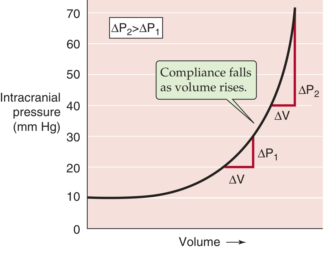
Fig: As intracranial volume increases, compliance (ΔV/ΔP) falls progressively, causing dangerous ICP surges - Medical Physiology (Boron & Boulpaep)
Classification of Cerebral Edema
There are four main types, though in clinical practice they often coexist.
1. Vasogenic Edema
Definition
An increase in extracellular fluid caused by disruption of the blood-brain barrier (BBB), allowing protein-rich fluid to pass from the intravascular compartment into the brain interstitium.
Causes
- Brain tumors (primary and metastatic)
- Brain abscess and meningitis
- Cerebral contusion / trauma
- Intracerebral hemorrhage
- Hypertensive encephalopathy
- Late phase of ischemic stroke (secondary)
- Multiple sclerosis plaques (acute attacks)
Pathophysiology
Tight junctions between cerebral endothelial cells that form the BBB are disrupted. Inflammatory mediators (cytokines, VEGF, bradykinin) and direct physical injury increase endothelial permeability. Protein-rich fluid leaks into the extracellular space. Because the brain has minimal lymphatics, resorption of this excess fluid is very slow. Vasogenic edema preferentially affects the white matter, spreading along axonal fiber tracts.
Gross Appearance
The brain is soft and swollen; gyri are flattened and sulci narrowed.
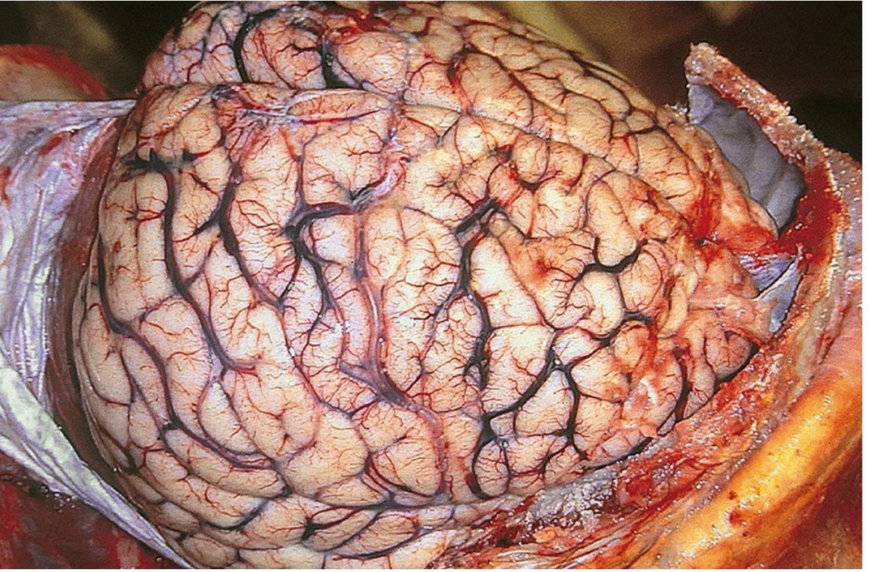
Fig: Cerebral edema - flattened gyri and narrowed sulci from brain compression against the dura mater - Robbins, Cotran & Kumar
Management
- Corticosteroids (dexamethasone 10 mg IV, then 4 mg q6h) - most effective for tumor-associated vasogenic edema; rapidly closes the BBB. Also effective in MS acute attacks (methylprednisolone 1 g/day x 3-5 days). Not indicated for stroke or hemorrhage.
- Osmotic therapy (mannitol, hypertonic saline) for acute ICP reduction
- Treat the underlying cause (resect tumor, drain abscess, antibiotics)
2. Cytotoxic Edema
Definition
An increase in intracellular fluid due to failure of cell membrane ion pumps, causing neurons, glia, and endothelial cells to swell.
Causes
- Global cerebral ischemia / hypoxia (cardiac arrest)
- Acute ischemic stroke (early phase - within hours)
- Severe hypoglycemia
- Toxin exposure (cyanide, carbon monoxide)
- Water intoxication / severe hyponatremia (also classified as osmotic edema)
- DKA-associated cerebral edema (particularly in children)
Pathophysiology
When energy metabolism fails (ATP depletion from ischemia/hypoxia), the Na+/K+-ATPase pump is disabled. Sodium and water accumulate intracellularly, causing cell swelling. Neurons, glia, and endothelial cells are all affected. The BBB remains initially intact, so this is primarily a cellular (intracellular) swelling rather than extracellular accumulation. Cytotoxic edema predominantly involves the gray matter (neuron-rich regions), while later secondary vasogenic edema involves the white matter.
As described in Goldman-Cecil Medicine, brain edema peaks between 48-72 hours after ischemic onset and can cause herniation in severe cases.
Cellular Mechanisms (AQP4)
Aquaporin-4 (AQP4) water channels, densely expressed on astrocytic endfeet surrounding capillaries, play a key role. AQP4-knockout animals are protected from cytotoxic brain swelling.
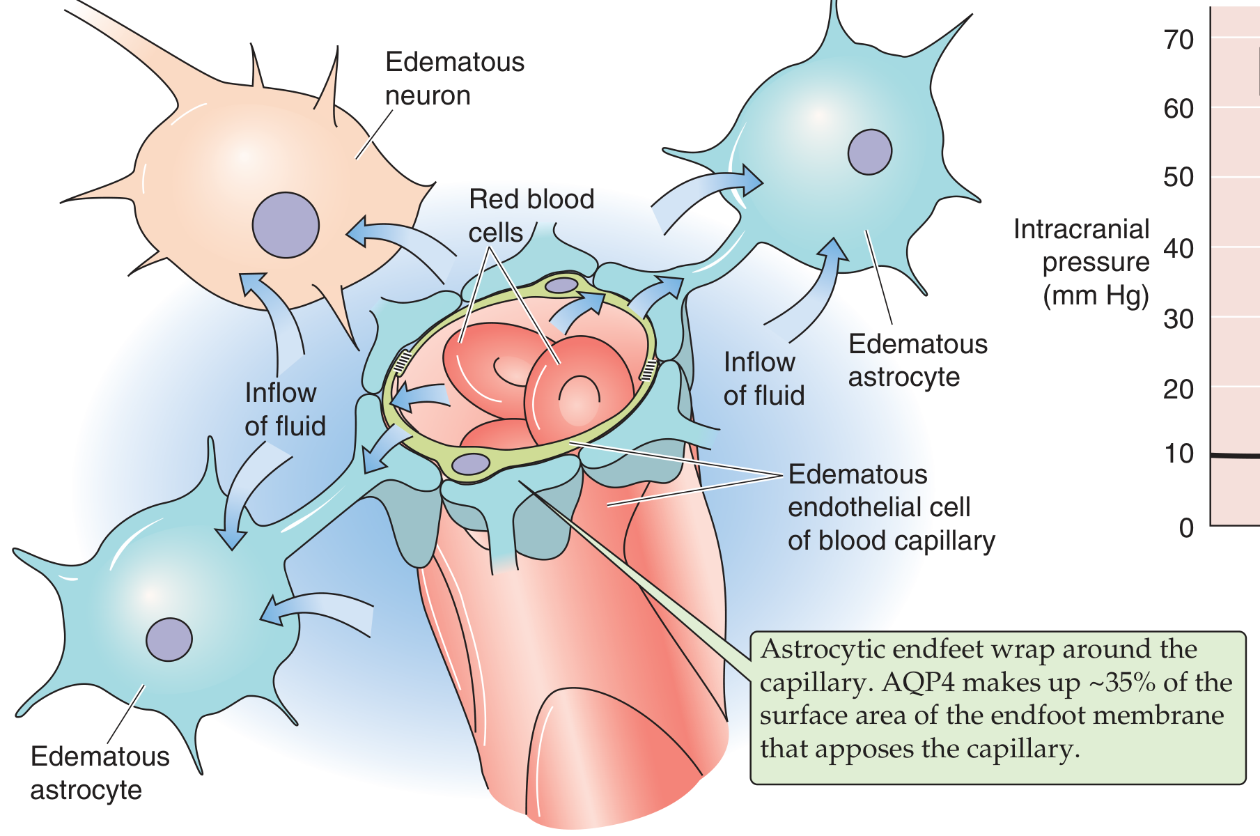
Fig: Cerebral edema - fluid flows from the vascular compartment into neurons and astrocytes. AQP4 makes up ~35% of astrocyte endfoot surface area - Medical Physiology (Boron & Boulpaep)
Management
- Osmotic therapy: Mannitol 0.25-1 g/kg IV (reduces brain tissue volume, lowers CSF production, improves cerebral blood flow, rheologic effects)
- Hypertonic saline (3% NaCl) - draws water out of cells via osmosis; particularly useful in pediatric DKA-associated edema (5-10 mL/kg over 30 min)
- Corticosteroids are NOT effective for cytotoxic edema
- Treat the underlying cause (revascularization, correct metabolic derangements)
- For ischemic stroke: no specific pharmacological agent has proven effective; decompressive craniectomy may be lifesaving in malignant hemispheric infarction
3. Osmotic (Hyponatremic) Edema
Definition
A subtype of cytotoxic/cellular edema caused by decreased plasma osmolality, driving water into brain cells along an osmotic gradient.
Causes
- Severe acute hyponatremia (Na+ < 120 mmol/L)
- SIADH
- Excessive hypotonic IV fluid administration (esp. postoperative children and premenstrual women)
- Water intoxication
- Hyperammonemia (e.g., acute liver failure)
- DKA - failure of serum Na+ to rise with therapy signals osmotic edema risk
Pathophysiology
When extracellular osmolality falls acutely, water moves into brain cells to establish osmotic equilibrium. Within 1-3 hours, the brain attempts to compensate by extruding Na+ and Cl- into the CSF, then later loses intracellular organic osmolytes (K+, glutamine, taurine, myoinositol, phosphocreatine). If hyponatremia is chronic, these adaptations largely prevent edema. Acutely, however, or in vulnerable populations (children, premenstrual women), the brain swells rapidly. AQP4 overexpression worsens swelling.
Risk factors for clinically overt cerebral edema in DKA: age <5 years, severe acidosis, severe hyperosmolality, failure of serum Na+ to rise with treatment.
Management
- Acute symptomatic hyponatremia: raise serum Na+ carefully with 3% hypertonic saline (target 1-2 mEq/L/hr initially, then slow correction)
- Fluid restriction
- In DKA: avoid high-dose insulin, avoid sodium bicarbonate, careful fluid management
- Correct the underlying cause
- Caution: Overly rapid correction of chronic hyponatremia causes osmotic demyelination syndrome (central pontine myelinolysis)
4. Interstitial (Hydrocephalic/Transependymal) Edema
Definition
Edema caused by obstruction of CSF flow, forcing CSF to seep across the ependymal lining into the periventricular white matter.
Causes
- Obstructive (non-communicating) hydrocephalus - tumor, hemorrhage, or infection blocking the foramen of Monro or cerebral aqueduct
- Communicating hydrocephalus - impaired CSF resorption
Pathophysiology
When CSF cannot drain normally, pressure builds in the ventricular system. CSF seeps through the ependymal lining into the adjacent white matter periventricular space. The edema is extracellular with a normal BBB. On CT/MRI, this appears as periventricular hypodensity ("halo" around ventricles).
Management
- Relief of CSF obstruction: ventriculostomy, ventriculoperitoneal shunt
- Treat the underlying mass or obstruction
- Acetazolamide or diuretics may modestly reduce CSF production but are not definitive
5. High-Altitude Cerebral Edema (HACE) - Special Type
Definition
HACE is a severe, life-threatening form of altitude sickness caused by hypoxia at high altitude, involving both vasogenic and cytotoxic components.
Causes
- Rapid ascent to high altitude (typically >3,500 m)
- Reduced atmospheric oxygen concentration
Pathophysiology
Two main mechanisms operate simultaneously:
- Vasogenic: Hypoxia increases cerebral blood flow → raised intravascular pressure → BBB breakdown → vasogenic edema
- Cytotoxic: Hypoxia disrupts Na+/K+-ATPase → cellular swelling
Initial compensatory hyperventilation lowers pCO2 (causing vasoconstriction) while hypoxia causes vasodilation - these initially balance. At extreme exertion and altitude, hypocapnic vasoconstriction may reduce CBF to ischemic levels. MRI shows characteristic T2 signal increase in the splenium of the corpus callosum and centrum semiovale.
Clinical Features
Progression: headache → ataxia → short-term memory loss → papilledema → coma → death
Management
- Immediate descent to lower altitude is the definitive treatment
- Supplemental oxygen
- Dexamethasone 8 mg initially, then 4 mg q6h (reduces BBB permeability, anti-inflammatory)
- Acetazolamide (carbonic anhydrase inhibitor) for prevention of acute mountain sickness
- Portable hyperbaric bag (Gamow bag) if descent impossible
General Management of Raised ICP from Cerebral Edema
| Intervention | Mechanism | Notes |
|---|---|---|
| Head elevation 30°, midline | Improves venous drainage | First-line positioning |
| Mannitol 0.25-1 g/kg IV | Osmotic: reduces brain water; ↓CSF; improves rheology; antioxidant | Monitor electrolytes; avoid prolonged use |
| Hypertonic saline (3%) | Osmotic: draws water out of brain cells | Particularly useful in cytotoxic/osmotic edema |
| Controlled hyperventilation (pCO2 35 mm Hg) | ↓pCO2 → vasoconstriction → ↓cerebral blood volume | Short-term bridge; avoid pCO2 <35 in first 24h after TBI |
| Dexamethasone | Closes BBB; ↓inflammatory permeability | Effective only for vasogenic edema (tumor, MS, abscess); contraindicated in stroke/ICH |
| Ventriculostomy/EVD | Mechanical CSF drainage | Used in hydrocephalus, post-TBI, post-operative |
| Acetazolamide | ↓CSF production | HACE prevention; marginal benefit in other types |
| Sedation/paralysis | Prevents ICP surges from Valsalva/agitation | ICU management |
| Decompressive craniectomy | Removes skull to allow brain expansion | Malignant MCA infarction; refractory TBI edema |
| Treat the cause | Revascularization, resect tumor, antibiotics | Definitive therapy |
Summary Table
| Type | Fluid Location | BBB | Key Causes | Preferential Region | Responds to Steroids? |
|---|---|---|---|---|---|
| Vasogenic | Extracellular | Disrupted | Tumor, abscess, contusion | White matter | Yes |
| Cytotoxic | Intracellular | Intact | Ischemia, hypoxia, toxins | Gray matter | No |
| Osmotic | Intracellular | Intact | Hyponatremia, DKA | Diffuse | No |
| Interstitial | Extracellular (periventricular) | Intact | Hydrocephalus | Periventricular WM | No |
| HACE | Both (vasogenic + cytotoxic) | Disrupted | High altitude/hypoxia | Corpus callosum splenium | Yes (dexamethasone) |
In practice, most serious clinical conditions (e.g., large ischemic stroke, severe TBI, malignant hypertension) involve mixed vasogenic and cytotoxic components. Understanding the dominant type guides targeted therapy - steroids are the cornerstone for vasogenic edema but are harmful in stroke and ICH.
Sources consulted:
- Robbins, Cotran & Kumar - Pathologic Basis of Disease (9th ed.), Cerebral Edema section
- Robbins & Kumar Basic Pathology (11th ed.)
- Goldman-Cecil Medicine (International Ed.), Cerebral Edema section
- Bradley and Daroff's Neurology in Clinical Practice - Brain Edema treatment & HACE
- Medical Physiology, Boron & Boulpaep - AQP4 mechanisms, ICP-volume curve
- Tintinalli's Emergency Medicine - DKA cerebral edema management
- Comprehensive Clinical Nephrology, 7th ed. - Hyponatremic cerebral edema
This is a shared conversation. Sign in to Orris to start your own chat.