
Pulses paradox
Pulsus paradoxus is an exaggerated drop in systolic blood pressure (>10 mmHg) during inspiration. It occurs because inspiration increases right heart filling and shifts the interventricular septum leftward, reducing left ventricular output. It is classically associated with cardiac tamponade, severe asthma, and constrictive pericarditis.
Searching sources...
Reading File
Searching the Web
pulsus paradoxus mechanism clinical significance
Finding Sources
Searching Images
pulsus paradoxus cardiac tamponade blood pressure waveform
Reading File
Reading File
Loading Image
Loading Image
Pulsus Paradoxus
The "Paradox" Explained
The name comes from Kussmaul's original observation: the heart sounds are audible at the precordium during inspiration, but the radial pulse disappears - pulse and heart action are dissociated. This seemed paradoxical - the heart is beating but you cannot feel the pulse. It is not a paradoxical rise in BP; it is an exaggerated fall.
Definition: A drop in systolic blood pressure of >10 mmHg during normal inspiration.
Pathophysiology - Three Parallel Mechanisms
The flowchart below (from Miller's Anesthesia) shows the three simultaneous pathways triggered by inspiration that converge to reduce left ventricular output:
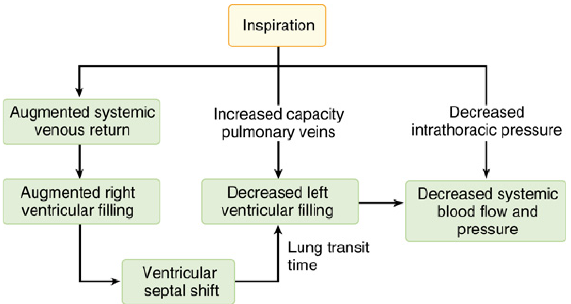
Fig. 50.68 from Miller's Anesthesia, 10e - Physiology of paradoxical pulses in cardiac tamponade
1. Ventricular Interdependence (most important in tamponade)
- Inspiration increases systemic venous return → augmented right ventricular (RV) filling
- The pericardial sac is non-compliant (especially with effusion) and acts as a fixed-volume constraint
- The distended RV pushes the interventricular septum leftward
- This reduces left ventricular (LV) filling volume and stroke volume
2. Pulmonary Vascular Pooling
- Inspiration increases the capacitance of pulmonary veins
- Blood pools in the lung parenchyma, reducing pulmonary venous return to the left atrium
- Compounded by lung transit time delay - the RV boost in output does not reach the LV for 2-3 beats
3. Decreased Intrathoracic Pressure
- Inspiration lowers intrathoracic pressure
- The aorta is partly outside the chest, so LV afterload effectively increases relative to thoracic structures
- This reduces systemic blood flow and pressure
All three pathways converge on decreased LV filling → reduced stroke volume → drop in systolic BP during inspiration.
Measurement at the Bedside
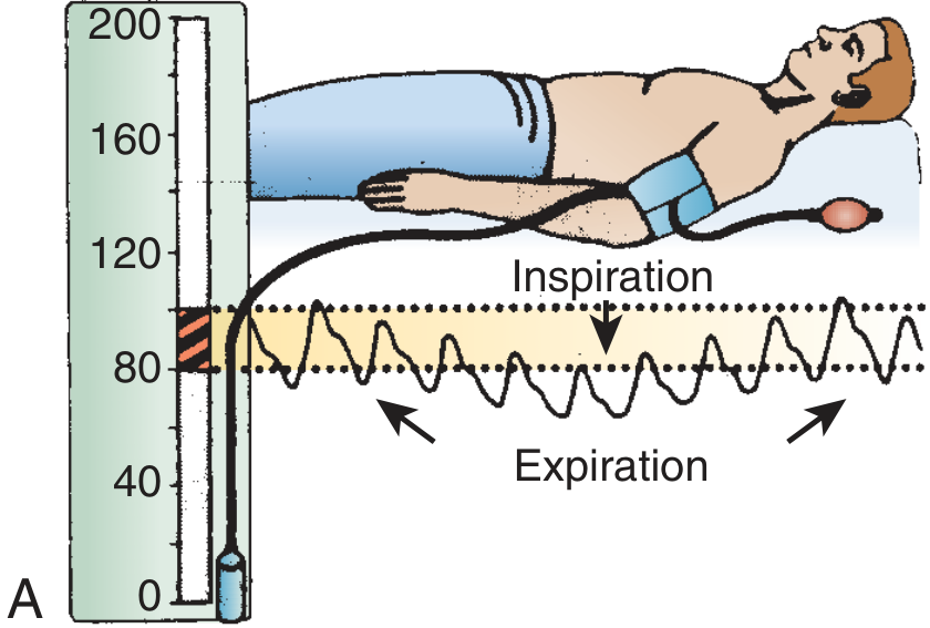
Roberts & Hedges' Clinical Procedures in Emergency Medicine - standard measurement procedure
Sphygmomanometer method:
- Patient lies at 30-45° with normal (unlabored) breathing
- Inflate cuff well above systolic pressure
- Slowly deflate until Korotkoff sounds are heard only during expiration - record this pressure
- Continue deflating until Korotkoff sounds are heard throughout the cycle - record this pressure
- The difference between the two readings = pulsus paradoxus
Alternative: Pulse oximetry plethysmographic waveform - observe loss and reappearance of the waveform during the respiratory cycle (validated in ICU settings).
| Finding | Significance |
|---|---|
| >10-12 mmHg | Abnormal (cut-off varies by source) |
| >20-30 mmHg | Strongly suggests cardiac tamponade |
| >25 mmHg (no hypotension) | Sensitive and specific for moderate-severe tamponade |
Practical caveat: Difficult to measure accurately in tachycardia, atrial fibrillation, or tachypnea. - Harrison's Principles of Internal Medicine, 22e
Clinical Causes
| Category | Conditions |
|---|---|
| Pericardial | Cardiac tamponade (classic), constrictive pericarditis (partial) |
| Respiratory | Severe asthma, COPD exacerbation, tension pneumothorax |
| Other | Massive pulmonary embolism, hypovolemic shock, large pleural effusion |
In cardiac tamponade, pulsus paradoxus has a sensitivity >80-98% for hemodynamic compromise, but sensitivity alone is not enough - absence does not exclude tamponade. - StatPearls, NCBI Bookshelf
Conditions Where Pulsus Paradoxus is Absent Despite Tamponade
This is a high-yield clinical trap. Pulsus paradoxus may be absent even with true cardiac tamponade when:
- Aortic regurgitation (AR) - elevated LVEDP prevents the LV from being further limited
- Atrial septal defect (ASD) - the shunt allows pressure equalization across the atria
- LV hypertrophy or dilatation with preexisting elevated LVEDP
- Severe pulmonary hypertension with elevated RV pressure
Constrictive Pericarditis vs. Tamponade
Pulsus paradoxus is prominent in tamponade but typically absent or less pronounced in constrictive pericarditis. In constriction, a rigid shell surrounds the heart equally, so the respiratory variation in filling is blunted - instead, Kussmaul's sign (JVP rises on inspiration) is the hallmark.
Sources: Miller's Anesthesia 10e; Roberts & Hedges' Clinical Procedures in Emergency Medicine; Harrison's Principles of Internal Medicine 22e; NCBI StatPearls; European Respiratory Journal review
Trave space
Trabes space
You likely mean Traube's space - the tympanic area over the stomach in the left lower chest, used to assess spleen size. Let me pull the full details from the textbooks.
Searching sources...
Finding Sources
Searching the Web
Traube's space anatomy percussion spleen clinical examination
Reading File
Searching Images
Traube's space percussion spleen anatomy diagram
Reading File
Searching Images
Traube space splenomegaly percussion left upper quadrant
Traube's Space
Named after: Ludwig Traube (1818-1876), German physician.
Anatomy
Traube's space (also called Traube's semilunar space) is a crescent-shaped tympanic area over the left lower thorax corresponding to the gastric air bubble. Its borders are:
| Border | Landmark |
|---|---|
| Superior | 6th rib (lower border of left lung) |
| Lateral | Left midaxillary line |
| Inferior | Left costal margin |
Anatomically it is bounded by the left lobe of the liver medially, the spleen laterally, and the lower cardiac border superiorly. The tympanic note arises from air in the gastric fundus beneath this region.
Clinical Use - Percussion for Splenomegaly
Traube's space is one of three classical percussion methods for detecting splenomegaly (alongside Nixon's and Castell's methods).
Technique:
- Patient is supine, left arm slightly abducted
- Percuss from the medial to lateral margins during normal breathing
- A resonant (tympanic) note is normal
- A dull percussion note suggests splenomegaly
The rationale: when the spleen enlarges, it occupies Traube's space and displaces or compresses the gastric air bubble, replacing tympany with dullness.
Limitations and Accuracy
Traube's space has poor operating characteristics in isolation and should not be used as the sole test:
| Metric | Value |
|---|---|
| Sensitivity | ~56% (Harrison's 22e) |
| Positive likelihood ratio | ~2.1 |
| Negative likelihood ratio | ~0.8 (poor rule-out) |
| Inter-rater reliability (kappa) | 0.19-0.41 (poor to fair) |
False positives (dullness without splenomegaly):
- Full stomach (solid gastric contents)
- Left-sided pleural effusion
- Colonic contents in the splenic flexure
- Left lower lobe consolidation/atelectasis
- Obesity
False negatives (tympany despite splenomegaly):
- Fasting state with empty stomach may still show tympany
- Massive splenomegaly can paradoxically shift gas
As a result, ultrasonography is the current preferred method for routine spleen size assessment - it is the most sensitive, specific, quick, non-invasive, and mobile modality. - Harrison's Principles of Internal Medicine, 22e
Comparison with Other Percussion Signs
| Method | Technique | Positive finding |
|---|---|---|
| Traube's space | Percuss medial to lateral, supine | Dullness throughout |
| Castell's sign | Percuss lowest interspace in anterior axillary line, ask patient to inspire | Tympany shifts to dullness on inspiration |
| Nixon's method | Patient in right lateral decubitus; percuss from posterior axillary line diagonally | Upper dullness border >8 cm above costal margin |
Of these, Castell's method (positive = tympany → dullness on inspiration) may have slightly better performance, but all three are supplemented by palpation and imaging in clinical practice.
Common Causes of Splenomegaly (when Traube's space is dull)
- Hyperplasia/hypertrophy: Hereditary spherocytosis, thalassemia, infectious mononucleosis, SLE, endocarditis
- Congestion: Portal hypertension (cirrhosis, Budd-Chiari), congestive heart failure
- Infiltration: Lymphoma, CML, myelofibrosis, polycythemia vera, amyloidosis, Gaucher's disease
Massive splenomegaly (>8 cm below costal margin) is most commonly due to: non-Hodgkin's lymphoma, CLL, hairy cell leukemia, CML, myelofibrosis, or polycythemia vera.
Sources: Harrison's Principles of Internal Medicine, 22e (p. 523); Stanford Medicine 25 - Spleen Exam; Georgetown Bedside Medicine - Splenomegaly
Content
Could you clarify what you mean by "Content"? This could refer to several things:
Asking for Preferences
What topic are you asking about?
Of trabes space
The contents of Traube's space are the structures that produce and occupy this semilunar region. The normal tympanic note comes from the gastric air bubble below, with surrounding viscera forming its walls.
Searching sources...
Finding Sources
Contents of Traube's Space
Traube's space is not a true anatomical cavity - it is a surface projection (a percussion zone) defined by its surrounding structures. What lies within and around it determines the percussion note.
Normal Contents (what fills this space)
| Structure | Position | Role |
|---|---|---|
| Gastric fundus (air bubble) | Centrally, beneath | Primary source of tympany - the gas-filled fundus of the stomach is what produces the normal resonant note |
| Left lobe of liver | Medial border | Hepatic dullness limits the space medially |
| Spleen | Lateral/posterior border | Normally tucked behind the ribs; splenic dullness bounds the space laterally |
| Lower left lung | Superior border | Lung resonance forms the upper limit at the 6th rib |
| Left hemidiaphragm | Superior, intervening | Separates the space from the pleural cavity above |
| Splenic flexure of colon | Inferior/lateral | Can contribute tympany or dullness depending on gaseous content |
| Left costal margin | Inferior bony boundary | Defines the lower limit of the space |
| Heart (lower border) | Medial/superior | Cardiac dullness can impinge medially in cardiomegaly |
Why It Is Normally Tympanic
The gastric air bubble (fundal gas) sits directly beneath this region. When you percuss, the air-filled stomach produces a hollow, drum-like (tympanic) note - the defining normal finding.
What Replaces the Tympany (causes of dullness)
When the gastric air is displaced or the space is occupied by a solid/fluid structure, dullness replaces tympany:
| Cause | Mechanism |
|---|---|
| Splenomegaly | Enlarged spleen descends and fills the space, displacing the gastric bubble |
| Full/solid stomach | Food or fluid in the fundus replaces air |
| Left pleural effusion | Fluid above the diaphragm transmits dullness downward |
| Left lower lobe consolidation/collapse | Loss of aerated lung above |
| Colonic distension | Splenic flexure gas can give variable results |
| Pericardial effusion | Enlarges the cardiac silhouette, encroaching medially |
| Subphrenic abscess/collection | Fluid beneath the diaphragm |
| Pancreatic tail mass or cyst | Posterior mass effect |
Summary
The key content of Traube's space in clinical terms is:
Gastric fundal gas - normal → tympany Spleen (enlarged) - abnormal → dullness
The space is essentially an acoustic window onto the left upper quadrant, where the stomach's air bubble serves as the natural "drum," and anything that displaces that air (most importantly an enlarged spleen) converts the note to dullness.
Source: Harrison's Principles of Internal Medicine, 22e; Georgetown Bedside Medicine Series
This is a shared conversation. Sign in to Orris to start your own chat.