
SEQs- 1- Define bruise.. Determine age of bruise - Medicolegal significance 2- Define abrasion- Types of abrasion 3- Pedestrian injuries.. types .. Primary Secondary Tertiary 4- Seat belt syndrome 5- Whiplash injury 6- Types of intracranial hemorrhage. explain extra dural hemorrhage...Lucid interval 7- Differentiate between homicidal and Suicidal cut throat 8- Qisas and Diyat For mbbs
Reading File
Now let me search the medical library for all 8 topics in parallel.
Finding Sources
Finding Sources
Finding Sources
Finding Sources
Finding Sources
Good - found the forensic pathology book. Let me now read the key sections from DiMaio's Forensic Pathology and Dikshit's Forensic Medicine book.
Reading File
Reading File
Finding Sources
Finding Sources
Finding Sources
Excellent! Now let me read the key sections from the forensic textbooks in parallel.
Reading File
Reading File
Reading File
Good. Now let me read more critical sections - bruise/contusion age, pedestrian injuries, and Qisas/Diyat from Dikshit's book.
Reading File
Reading File
Reading File
Now let me read the bruise/contusion age estimation and pedestrian injury sections, plus the cut throat differentiation.
Reading File
Reading File
Reading File
Now let me get the bruise age/colour table and Qisas/Diyat section from Dikshit's book.
Reading File
Finding Sources
Let me now get the bruise colour/age table from the forensic essentials textbook and look for Qisas/Diyat:
Finding Sources
I have enough material from the textbooks to now write a comprehensive answer. Let me compile everything into a well-structured MBBS-level response.
Forensic Medicine - SEQ Answers (MBBS Level)
1. Bruise - Definition, Age Estimation & Medicolegal Significance
Definition
A bruise (contusion) is an injury caused by blunt force that ruptures small blood vessels (capillaries, venules) beneath an intact skin surface, causing extravasation of blood into the surrounding tissues without breaking the skin. The overlying epidermis remains intact.
- Also called ecchymosis when it refers to a flat, purplish discolouration.
- Haematoma = when blood collects in a defined cavity/pocket.
Age Estimation of a Bruise (Colour Changes)
The colour of a bruise changes as haemoglobin is progressively broken down by macrophages:
| Age of Bruise | Colour |
|---|---|
| Fresh (0–1 days) | Red / Blue-red |
| 1–3 days | Blue / Purple (bluish-black) |
| 3–5 days | Green (biliverdin formation) |
| 5–7 days | Yellow (bilirubin formation) |
| 7–10 days | Brownish-yellow / Fading |
| >10–14 days | Normal skin colour (resolves) |
Important caveats:
- Colour changes are unreliable as a precise ageing method - different parts of the same bruise may show different colours simultaneously.
- Site matters: deep bruises take longer to appear on the surface. A bruise caused by a deep injury may not appear on the skin surface for 24–48 hours (delayed bruise), and when it does appear, it may be at a location distant from the original site due to gravitational tracking (e.g. subconjunctival haemorrhage in base of skull fracture - Battle's sign / raccoon eyes).
- Individual factors (age, skin tone, depth, extent of injury) all affect the timeline.
- Postmortem spectroscopy is more reliable for precise dating.
Medicolegal Significance of a Bruise
- Evidence of violence - confirms that blunt force trauma occurred.
- Indicates nature of weapon - patterned bruises (e.g., figure-8 shape of a belt buckle, tyre tread) can identify the weapon used.
- Indicates severity of force - size and depth suggest the amount of force applied.
- Determining antemortem vs postmortem injury - antemortem bruises are reddish-brown with tissue reaction; postmortem bruises are yellow, translucent, parchment-like with no vital reaction.
- Age estimation - helps establish the time of assault.
- Site of bruise vs site of impact - bruise may appear away from the site of injury (e.g., bruise tracking down the limb, "black eye" from orbital roof fracture). This is important in child abuse cases.
- Internal bruising - may indicate injuries not visible externally; always exclude in suspicious deaths.
- Defence wounds - bruises on the ulnar aspect of the forearms, dorsum of hands indicate the victim tried to defend themselves.
- Fabricated bruises - bruises may be fabricated or self-inflicted to falsely implicate another person.
- Child abuse (Non-accidental injury) - multiple bruises of varying ages in a child at unusual sites (e.g., trunk, buttocks, back) suggest repeated assault.
Source: The Essentials of Forensic Medicine and Toxicology, 36th ed.; Parikh's Textbook of Medical Jurisprudence
2. Abrasion - Definition & Types
Definition
An abrasion is an injury to the skin in which the superficial epithelial layer (epidermis) is removed by friction against a rough surface, or by compression/crushing of the superficial layers. The skin integrity is broken only at the surface; the dermis is usually spared.
- Antemortem abrasions - reddish-brown in colour; heal without scarring.
- Postmortem abrasions - yellow, translucent, parchment-like appearance; no vital reaction.
Types of Abrasions
1. Scratch / Linear Abrasion
- Caused by a sharp pointed object (e.g., fingernail, thorn, pin) drawn across the skin.
- Results in a narrow, linear mark.
- Example: scratch marks on the neck in strangulation.
2. Graze / Brush / Sliding Abrasion (Scrape Abrasion)
- Caused when the skin slides across a rough surface (or a rough surface slides across skin).
- Results in a broad area of skin loss; particles of dirt, gravel, or glass may be embedded.
- The direction of movement can sometimes be determined by heaping of epithelium at the far end.
- Example: road rash in pedestrians knocked down by vehicles; abrasions in RTA victims.
3. Pressure / Impact / Crushing Abrasion
- Caused by a blunt object pressing down and crushing the skin surface.
- May be patterned - faithfully reproduces the surface pattern of the weapon (e.g., tyre tread marks, belt buckle, cord pattern).
- Example: ligature mark grooves in hanging/strangulation; tyre tread abrasion over the body in run-over accidents.
4. Friction Abrasion
- A combination of sliding and pressure components.
- Seen in hanging (neck), strangulation (neck), or dragging of a body.
Some textbooks classify as 3 types: scrape, impact, and patterned - where "patterned abrasion" is a subset of impact abrasion that reproduces the weapon surface.
Source: DiMaio's Forensic Pathology, 3rd Ed.
3. Pedestrian Injuries - Types: Primary, Secondary, Tertiary
When a vehicle strikes a pedestrian, injuries occur in a sequential pattern depending on the speed and position of impact.
Primary (Initial/Impact) Injuries
- Injuries caused by the first direct contact between the vehicle and the pedestrian.
- Usually at the level of the bumper - hence called "bumper injuries" or "fender injuries."
- Site depends on the height of the bumper and the height of the pedestrian:
- Adults: typically the lower leg (tibia/fibula), producing a characteristic bumper fracture at knee/shin level.
- Children: because of their shorter height, impact may be at the level of the chest or abdomen - causing more severe internal injuries.
- Bumper fracture = transverse fracture of tibia/fibula at the point of impact; the level of this fracture indicates the height of the bumper, helping reconstruct the accident.
- Frequently, bumper injuries appear at different levels on the two legs or are absent on one leg - indicating the person was walking or running at the time of impact.
Secondary Injuries
- Caused when the body is thrown onto the vehicle after the primary impact.
- The body is projected upward and lands on the bonnet (hood), windscreen, or roof of the vehicle.
- Injuries include: lacerations and fractures of the pelvis, chest, abdomen, face, and head from striking the bonnet/windscreen.
- Wrap-around injuries - the body wraps around the front of the vehicle momentarily.
Tertiary Injuries
- Caused when the body hits the ground after being thrown off the vehicle.
- The body may be thrown forward (if vehicle was moving toward the person) or backward.
- Injuries depend on the surface and the body part that strikes first:
- Head injuries (if head strikes ground first) - skull fractures, brain contusions.
- Abrasions and lacerations on projecting parts (elbow, knee, shoulder, face).
- Fractures of upper limbs (defensive fall injuries).
Additional Types (in some classifications):
- Quaternary injuries - caused by running over (tyres passing over the body) - produces severe crushing, tyre tread patterned abrasions.
- Secondary secondary - injuries from being struck by parts of the vehicle during rolling under the vehicle.
Source: PC Dikshit Textbook of Forensic Medicine and Toxicology
4. Seat Belt Syndrome
Seat belt syndrome refers to the cluster of injuries caused by a lap (or shoulder) seat belt during sudden deceleration in a frontal road traffic collision.
Mechanism
In a frontal collision, the body is suddenly decelerated while the inertia continues to propel it forward. The seat belt restrains the body, but in doing so, it can transmit severe compressive and shearing forces to the structures beneath it.
Types of Injuries
A. Lap Belt Injuries
- Most commonly seen when the belt is worn above the pelvis (too high), or with submarining (pelvis slides under the belt).
- Seat belt sign = visible contusion/abrasion across the lower abdomen marking the path of the belt - a key indicator of internal injury.
- Hollow viscus injuries: perforation of duodenum, jejunum, ileum (lacerations on the anti-mesenteric side).
- Solid organ: lacerations of liver, spleen, pancreas.
- Spinal: Chance fracture (flexion-distraction fracture of the lumbar vertebrae L1–L3) - transverse fracture through vertebral body, pedicles, and posterior elements - caused by sudden forward flexion over the belt.
- Urinary bladder rupture (if bladder is full at time of impact).
B. Shoulder/Diagonal Belt Injuries (without lap belt)
- Fractures of clavicle and ribs on the side of the belt.
- Chest injuries - pulmonary contusion, pneumothorax, haemothorax.
- Carotid artery injury (dissection/intimal tear) - may cause delayed stroke.
- Rib fractures and sternal injuries.
C. Three-Point Belt (Lap + Shoulder)
- Most effective restraint; reduces mortality significantly.
- Can still cause: clavicle fractures, rib fractures, abdominal wall contusions, and the above hollow visceral injuries.
Key Features
- The "seat belt sign" (diagonal bruising across chest/lower abdomen) on examination = strong predictor of internal injury requiring investigation.
- Any child with lap belt complex should be evaluated for bowel perforation + lumbar Chance fracture.
Source: DiMaio's Forensic Pathology, 3rd Ed.; PC Dikshit Forensic Medicine
5. Whiplash Injury
Definition
Whiplash injury (also called Whiplash-Associated Disorder / WAD) is an indirect, acceleration-deceleration injury to the cervical spine caused by a sudden, forceful forward-backward (or backward-forward) movement of the head and neck, without any direct trauma to the head or neck.
Mechanism
- Most commonly occurs in rear-end collisions (tail-gating accidents).
- When a stationary vehicle is struck from behind:
- The torso is suddenly propelled forward by the seat.
- The unsupported head lags behind due to inertia - causing hyperextension of the cervical spine.
- This is followed by a rebound hyperflexion.
- This whipping motion causes strain, tears, and injuries to the cervical soft tissues.
Pathological Changes
- Anterior longitudinal ligament sprain/rupture.
- Intervertebral disc injuries - acute disc herniation.
- Facet joint capsule injuries.
- Muscle/soft-tissue tears (sternocleidomastoid, paraspinal muscles).
- In severe cases: vertebral fractures, cord injury.
- Retrolaryngeal haematoma may develop.
Clinical Features
- Neck pain and stiffness (may be delayed 6–24 hours after accident).
- Restricted range of movement of cervical spine.
- Headache (occipital to frontal radiation).
- Shoulder/arm pain (C5–C6 nerve root involvement).
- Vertigo, tinnitus, visual disturbances (in severe cases - Barré-Liéou syndrome).
- Memory/concentration difficulties.
- Sleep disturbance.
WAD Classification (Quebec Task Force)
- Grade 0: No symptoms, no signs
- Grade I: Neck pain/stiffness, no physical signs
- Grade II: Neck symptoms + musculoskeletal signs
- Grade III: Neck symptoms + neurological signs
- Grade IV: Neck symptoms + fracture/dislocation
Prognosis
Most patients recover within 6 months with conservative management (physiotherapy, analgesia, soft collar for acute phase).
Medicolegal Importance
- Frequently cited in insurance claims and compensation cases after RTAs.
- Difficult to objectively verify - can be exaggerated or fabricated.
- Requires careful clinical and radiological documentation.
Source: Scott-Brown's Otorhinolaryngology; PC Dikshit Forensic Medicine
6. Types of Intracranial Haemorrhage - Extradural Haemorrhage and Lucid Interval
Types of Intracranial Haemorrhage
| Type | Location | Usual Cause |
|---|---|---|
| Extradural (Epidural) | Between skull and dura mater | Trauma; middle meningeal artery rupture |
| Subdural | Between dura and arachnoid | Trauma (bridging vein rupture) |
| Subarachnoid | Between arachnoid and pia | Berry aneurysm rupture; trauma |
| Intracerebral | Within brain parenchyma | Trauma; hypertension; AVM |
| Intraventricular | Within ventricles | Extension from intracerebral or subarachnoid |
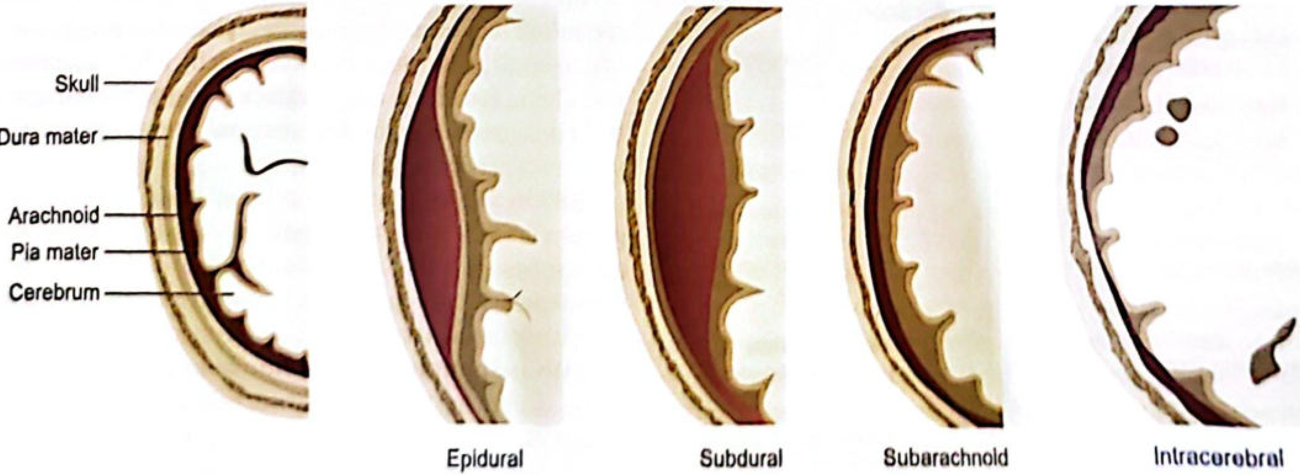
Fig: Types of intracranial haemorrhage (The Essentials of Forensic Medicine and Toxicology, 36th Ed.)
Extradural (Epidural) Haemorrhage - Detailed
Definition: Bleeding into the space between the inner surface of the skull and the outer layer of the dura mater.
Aetiology:
- In 90% of cases, caused by a fissure fracture of the temporal bone rupturing the middle meningeal artery (anterior branch) - the most commonly injured vessel.
- Less commonly: rupture of the middle meningeal vein, diploic veins, or the dural venous sinuses.
- Rarely can occur without skull fracture.
- The hematoma is almost always directly under the site of impact (cannot be contrecoup, unless skull is grossly deformed).
Location of clot:
- Most commonly temporoparietal region (where the middle meningeal artery runs in a groove on the inner skull).
- Also: fronto-temporal or parieto-occipital regions.
- Rarely: frontal, posterior fossa, or base of skull.
Nature of Clot:
- Oval or circular, rubbery, reddish-purple.
- About 10–20 cm diameter, 2–6 cm thick, weighing 30–300 g.
- The clot is adherent to the dura mater and presses it inward.
- Limited by dural attachments at suture lines (gives characteristic sharp borders).
- 100 mL is usually the minimum volume associated with fatalities.
Clinical Features - The Classic Triad:
- Initial transient loss of consciousness (from the original head injury/concussion).
- Lucid interval (period of apparent recovery).
- Rapid deterioration with deepening unconsciousness, hemiplegia, fixed dilated ipsilateral pupil (herniation), leading to death if untreated.
Lucid Interval
Definition: A period of apparent recovery of consciousness after an initial loss of consciousness following head injury, before the patient again deteriorates due to increasing intracranial pressure from the expanding extradural haematoma.
Mechanism:
- After the initial injury, the brain sustains a concussion and the patient may lose consciousness briefly.
- As the extradural clot slowly forms and expands (arterial bleeding), intracranial pressure rises gradually.
- Initially, the brain can compensate (CSF displacement, venous compression).
- During this compensation phase, the patient regains consciousness = the lucid interval.
- Once compensation is exhausted, rapid brain compression occurs with uncal herniation.
Duration:
- Classically few hours to a week (average 4–6 hours for extradural haemorrhage due to arterial source).
- The lucid interval may be shorter if the original brain injury is severe (overlapping unconsciousness).
- The lucid interval may be longer (>4 hours) in subdural haemorrhage (venous, slower bleed).
- In 25% of extradural haematoma cases, there is no initial loss of consciousness at all.
- Lucid interval is NOT seen if the brain injury itself is sufficiently severe, causing continuous unconsciousness.
Significance:
- It is a classic presentation of extradural haematoma - must be suspected in any patient who deteriorates after a head injury even if initially "well."
- A neurosurgical emergency - requires urgent CT head and burr hole/craniotomy evacuation.
- Occasionally seen in subdural haemorrhage (but the interval is longer and the history less clear-cut).
Source: The Essentials of Forensic Medicine and Toxicology, 36th Ed.; Gray's Anatomy for Students
7. Differentiation: Homicidal vs Suicidal Cut Throat
| Feature | Suicidal Cut Throat | Homicidal Cut Throat |
|---|---|---|
| Number of wounds | Usually single, or with multiple hesitation/tentative cuts nearby | Usually single deep cut; multiple wounds possible |
| Hesitation cuts | Present - multiple small, shallow, parallel cuts near the main wound (from gathering courage before the fatal cut) | Absent - no hesitation marks |
| Site | Usually over the anterior neck, often at the level of the thyroid cartilage | May be anterior or posterior; if cut from behind, wound is higher (at level of thyroid cartilage or above) |
| Direction | Usually runs horizontally or slightly upward; in a right-handed person, starts from left side and trails off to the right (deeper on left, shallower on right = "tailing") | Often deep throughout with no tailing; may be irregular |
| Depth | Deepest at the commencement (where the knife was applied) and shallows off at the other end (tailing) | Usually uniformly deep or deepest in the middle |
| Associated injuries | No defence wounds | Defence wounds commonly present on palms, dorsum of hands, forearms (victim tries to grab the blade) |
| Access | Wound must be within reach of the victim's own hand | Wound may be in areas the victim could not self-inflict |
| Position of victim | Usually found in a convenient position; instruments nearby | Evidence of struggle, disturbed scene |
| Weapon | Usually a sharp-edged weapon (razor, knife) found at the scene | Weapon may be absent (carried away by assailant) |
| Clothes | Typically clothes are lifted/undone to expose the neck | Clothes may be intact over the wound or cut through |
| Circumstantial evidence | History of depression, suicidal note, previous attempts, financial crisis | No note; evidence of struggle, motive |
| Post-mortem position | May show typical dribble of blood toward front if upright | Variable depending on position of victim |
| Bruising | Minimal or absent | May show bruising/grip marks on neck, especially if victim was restrained |
Key Memory Points:
- Hesitation cuts = suicidal hallmark.
- Defence wounds = homicidal hallmark.
- Tailing = suicidal (starts deep, ends shallow on the dominant-hand side).
- A person found with a cut throat and a razor in hand does not automatically mean suicide - examination for hesitation cuts, defence wounds, and other injuries is mandatory.
Source: Parikh's Textbook of Medical Jurisprudence, Forensic Medicine and Toxicology; DiMaio's Forensic Pathology, 3rd Ed.
8. Qisas and Diyat
These are provisions of Islamic criminal law (Hudood/Sharia) as incorporated into Pakistani legislation through the Qisas and Diyat Ordinance, 1990 (now part of Pakistan Penal Code, Chapter XVI).
Qisas (قصاص)
Definition: Qisas literally means "equality" or "retaliation." It is the right of the victim (or victim's heirs) to demand equal punishment for the offence committed against them.
Key Features:
- Applies to: Intentional (wilful) offences - murder (Qatl-i-Amd) and intentional grievous hurt.
- The punishment is "an eye for an eye" - the offender receives the same injury they inflicted on the victim.
- The heirs of the victim have the right to retaliate, pardon, or accept Diyat (blood money).
- In Qatl-i-Amd (wilful homicide): the Wali (heir of deceased) can either:
- Demand Qisas (execution).
- Pardon the offender without receiving Diyat.
- Accept Diyat (blood money) in lieu of Qisas.
- Once Qisas is waived or compounded, the state cannot impose the same punishment independently (though discretionary punishment ta'zir may apply).
- The state can demand Qisas on behalf of heirs if there are no heirs or if the heirs cannot compromise.
Diyat (دية)
Definition: Diyat means "blood money" - a monetary compensation paid to the victim or the heirs of a victim in lieu of Qisas (retaliation) for homicide or bodily harm.
Key Features:
- Diyat is fixed by the Federal Government on the basis of the value of 30,630 grams of silver (or the equivalent cash amount, revised periodically).
- Paid to the heirs of the deceased in case of murder, or to the victim in case of hurt.
- Diyat is payable in the following circumstances:
- The heirs voluntarily waive Qisas and accept blood money.
- The offence was quasi-intentional (Qatl-Shibh-i-Amd) or by mistake (Qatl-i-Khata).
- In Qatl-i-Amd, Diyat replaces Qisas by mutual agreement.
Classification of Homicide in Qisas/Diyat Law
| Category | Description | Punishment |
|---|---|---|
| Qatl-i-Amd | Wilful/intentional murder | Qisas (or Diyat if waived) |
| Qatl-Shibh-i-Amd | Quasi-intentional (intended hurt but not death) | Diyat only (no Qisas) |
| Qatl-i-Khata | Accidental killing by mistake | Diyat only |
| Qatl-bis-Sabab | Indirect cause of death | Diyat only |
Arsh (ارش)
- Compensation for injuries/hurt that has a fixed rate specified in law (e.g., loss of an eye, ear, limb).
- A percentage of Diyat (e.g., loss of one eye = 1/2 Diyat).
Daman (ضمان)
- Compensation determined by the court for injuries that do not have a fixed Arsh rate.
Medicolegal Significance of Qisas and Diyat
- The doctor's report (medicolegal examination) is critical in determining:
- Whether the injury was intentional or accidental.
- The nature and extent of hurt.
- Whether the injury caused permanent disability (affecting Arsh calculation).
- Doctors must carefully classify wounds (simple/grievous hurt, nature of weapon) as this determines whether the offence falls under Qisas or Diyat and the amount of Arsh.
- The physician must not certify under pressure from either party.
- Proper documentation of injuries, weapons, and permanence of disability is essential for court proceedings.
Source: PC Dikshit Textbook of Forensic Medicine and Toxicology; The Essentials of Forensic Medicine and Toxicology, 36th Ed.
Study Tips for MBBS:
- Bruise colour changes: "Red → Blue → Green → Yellow → Brown → Normal" - think of it as a healing rainbow over ~2 weeks.
- Lucid interval: Think "bump → okay → crash" = classic EDH.
- Suicidal cut throat: Hesitation cuts + tailing + no defence wounds.
- Homicidal cut throat: Defence wounds + no hesitation cuts + multiple/irregular wounds.
- Qisas = retaliation; Diyat = blood money.
This is a shared conversation. Sign in to Orris to start your own chat.